Original Article
Effect of ischemic preconditioning on the second choke
zone of the extended dorsal skin perforator flap
in a rat model
Liming Qing, Pangfeng Wu, Zhengbing Zhou, Fang Yu, Juyu Tang
Department of Orthopaedics, Xiangya Hospital, Central South University, # 87 Xiangya Road, Changsha 410008, Hunan, China
Received September 12, 2016; Accepted November 14, 2016; Epub March 15, 2017; Published March 30, 2017
Abstract:Background: The purpose of this study was to investigate the possible effects of ischemic preconditioning
(IP) on the second choke zone of extensive perforator flap in a rat model, and whether pedicle selection for IP influ
-enced its efficacy. Methods: A rat perforator flap model, encompassing three vascular territories, was used. In total,
60 rats were divided into one control and two IP groups (primary pedicle, secondary pedicle). On day 7, evaluation
of survival area and angiography of the flap were performed. Tissue samples were collected from the second choke
zone for histological analyses, western blot, and real-time qRT-PCR. Results: The percentage of flap survival area was greater in both IP groups than in the control group (P < 0.001). There was no significant difference between IP
groups (P > 0.05). In both IP groups, the second choke vessels dilated extensively, whereas in the control group, they dilated only slightly or remained unopened. The number of CD31-positive vessels of the second choke zone was increased in IP groups. The vascular diameter was greater in IP groups than in control group. The expression of
hypoxia inducible factor-1α and inducible nitric oxide synthase was higher in both IP groups than in control group (P
< 0.001), but there was no different between IP groups (P > 0.05). Conclusion: IP has a protective effect on ischemia
of skin flaps through effects on the second choke zone. Secondary pedicle IP was as effective as primary pedicle IP on the second choke zone of skin flaps.
Keywords: Ischemic preconditioning, choke vessels, perforator skin flap
Introduction
Perforator flaps are frequently used in plastic and reconstructive surgery to cover soft-tissue defects caused by trauma or tumor resection. Ischemic necrosis of the skin flap is a common complication and can result in significant cos-metic and functional defects [1, 2].Vascular supply to the integument is crucial to the sur-vival of surgical perforator flaps. The areas of skin perfused by blood vessels are termed angiosomes [2]. Arteriolar connections, in the form of choke vessels, exist between angio-somes [3, 4]. Choke vessels are part of both arterial and venous skin circulation, and under normal physiological conditions are small-cali-ber vessels extending between the tips of the branches of adjacent vascular trees [5]. Choke vessels play an important role in skin flap sur-vival. One perforator can safely perfuse adja-cent territory via dilation of a choke vessel in
the primary choke zone. However, the survival of the third territory is unpredictable [6]. Some angiographic studies show that the difference in the survival area of flaps is attributable to the behavior of the second choke zone [7, 8]. The behavior of choke vessels plays an important role in skin flap survival [7, 8].
Although necrosis of the distal portion of the flap can be alleviated with IP, this method may not be suitable in clinic practice because of the risk of damage to the pedicle and the extended surgical time. Preconditioning methods that do not adversely affect operation time and that increase the success of flap transfer are of interest. One promising approach performed specifically on flaps was reported by Matsumara [16], who showed that regional IP on the distal end of random pattern skin flaps increased flap survival in rats. Yildiz [10] found that secondary pedicle IP was as effective as primary pedicle IP and may be feasible in free flap transfers. We therefore hypothesized that applying different pedicle preconditioning methods would have the same effects on the second choke zone.
Local skin flaps often present with flap necrosis caused by critical disruption of the blood
sup-uth of University (Changsha, Hunan, China), and provided with free access to food and water. All manipulations and surgical proce-dures were performed in accordance with the guidelines of the China Council of Animal Care and with approval of the Central South Uni- versity Committee on Laboratory Animals.
Flap model and surgical procedures
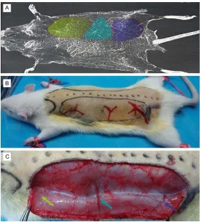
[image:2.612.89.377.69.388.2]Flap design: In this study, an extended dorsal skin perforator flap model was used. The perfo-rator flap was marked on the dorsolateral aspect. Based on previous reports and our angiographic study, this flap contains three even vascular territories, the iliolumbar artery perforator vessel, the posterior interior inter-costal artery perforator vessel, and the thora-codorsal artery perforator vessel, and two choke zones (Figure 1A). Dimensions of the Figure 1. A: A radiogram of the blood vessels showing multiple vascular
ritories supplied by three perforating arteries on the rat dorsum. The ter-ritories supplied by the iliolumbar artery perforator, the posterior intercostal artery perforator, and the thoracodorsal artery perforator are labeled Purple, Green, and Yellow, respectively. B: Design of the three-territory flap. C: The undersurface of the three-territory flap (Yellow arrow, thoracodorsal vessel; Green arrow, posterior intercostal vessel; Purple arrow, iliolumbar vessel).
ply. Hypoxia inducible factor-1α (HIF-factor-1α) and inducible ni- tric oxide synthase (iNOS) are key enzymes in regulating ge- ne expression after ischemia [17-21]. Therefore, expression of HIF-1α and iNOS in the sec-ond choke zone during isch-emic preconditioning is of great interest.
In this study, we investigated the possible effects of IP on the second choke zone of skin flaps in a rat model. This study was also undertaken to inves-tigate whether pedicle selec-tion (primary or secondary) in- fluenced the efficacy of IP on the second choke zone in ex- tensive skin perforator flaps. A third aim of this study was to analyze the expression of HIF-1α and iNOS in the second choke zone during this proc- ess.
Materials and methods
Animals
flap corresponded to anatomical landmarks, extending caudally from the caudal margin of the scapulae to 1 cm caudal to the posterior iliac crest, with the medial border at a site 5 mm lateral to the dorsal midline and extending 3.5 cm laterally. This resulted in a flap approxi-mately 3.5 cm × 10 cm (Figure 1B).
Flap elevation: All animals were anesthetized with pentobarbital sodium anesthesia (30 mg/ kg, intraperitoneal). Fur was shaved from the dorsal surface and the skin was washed with Hibitane, alcohol, and iodine. The flap was out-lined with a felt-tipped surgical marker. Flap elevation was started with an incision at the
taining the iliolumbar artery was selected as the “primary pedicle” and that with the posteri-or interiposteri-or intercostal artery perfposteri-oratposteri-or vessel as the “secondary pedicle” to be sacrificed intraoperatively. The flap was then replaced in the surgical site and secured with 4-0 monofila-ment sutures and wound clips. After anesthetic recovery, the rats were placed in a clean cage and given mash. Animals were monitored daily for signs of dehiscence and self-mutilation. Study design and experimental protocol
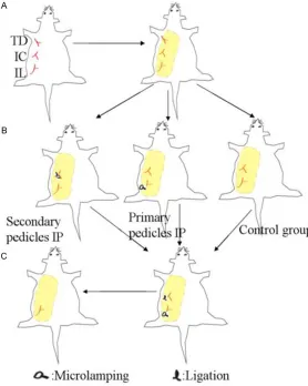
The subjects were divided into a control group (n = 20) and two experimental groups, a prima-Figure 2. Schematic view of study design in the ischemic preconditioning
and control groups. A: Elevation of the flap based on three perforating arter -ies on the rat dorsum in all groups. (TD: thoracodorsal vessel; IC: posterior
intercostal vessel; IL: iliolumbar vessel). B: The flap in the study groups were
performed with preconditioning of the pedicle comprising three
ischemia-perfusion cycles over a 1-hour period. In contrast, the flap in the control
group was perfused through a both pedicles and did not undergo IP. C: Ap-plication of global ischemia for 2-hour in both IP and control groups. (IL:
mi-croclamped; IC: ligated). The final form of an extended flap was perfused by
the iliolumbar vessel pedicle in all groups.
medial border. The periphery of the flap was incised and hemostasis was achieved. The surgical flap was then raised by sharp dissection in the plane between the pan-niculus carnosus and the deep fascia and the three blood vessels of interest were confirmed (Figure 1C). Cuta- neous blood vessels were cauterized as they were en- countered, except for the planned perforator pedicle. The thoracodorsal artery per-forator was clipped and sev-ered creating an island flap that relied solely on the ilio-lumbar artery perforator for its blood supply. After flap elevation, the anastomotic line between the thoracodor-sal artery perforator and the posterior intercostal artery perforator (the second choke zone) was marked on the flap’s surface.
[image:3.612.91.369.74.422.2]con-ry pedicles IP group (iliolumbar artecon-ry perfora-tor pedicle IP group, n = 20) and a secondary pedicles IP group (posterior intercostal artery perforator pedicle IP group, n = 20). In the con-trol group, flaps were raised based on the ilio-lumbar artery perforator pedicles and perfused for 1 h. After ligation of the posterior intercostal artery perforator pedicle, a 2-h period of global ischemia was induced in the flap using a microclamp on the pedicle. The flap was then sutured in place. In the IP groups, after flap elevation on the two perforator pedicles, three ischemic-perfusion cycles were induced by clamping the pedicles (10 min of clamping and 10 min of reperfusion). The posterior interior intercostal artery perforator vessels were then ligated and global ischemia was induced as described for the control group (Figure 2). Five rats in each group were used for angiographic examination of the flaps. Fifteen rats in each group were randomly sacrificed at postopera-tive day seven and submitted for examinations of flap viability, histology, immunohistochemis-try, and expression of HIF-1α and iNOS.
Angiography
In each group, five rats underwent whole-body angiography according to the method described by Tang [6]. Briefly, 5 g of gelatin was diluted in 100 ml of tap water heated to 40°C, and 100 mg of water-soluble red lead oxide was added. This mixture was injected into the rat’s carotid artery until the rat’s limbs turned red. After
and scar formation) and total flap area were delineated. Surface areas were calculated using Image-Pro Plus Software (version 6.0, Media Cybernetics Inc., Bethesda, MD, USA) by an investigator who was blinded to the experi-mental groups. The skin flap survival results were expressed as a percentage of the surviv-ing area relative to the total surface area of the flap.
Histology and immunohistochemistry
A sample were excised from the second choke zone and stored in 4.5% buffered formaldehy- de solution. Formaldehyde-fixed samples were processed in paraffin and stained with hema-toxylin/eosin (H&E) using standard histology protocols. Samples were examined for infiltra-tion of polymorphonuclear leukocytes (PNLs), chronic inflammatory cells, and interstitial edema. Tissue sections stained with CD31 were used to evaluate microvessel density (MVD). Rats were anesthetized and full thick-ness skin flap specimens measuring 0.5 cm × 0.5 cm were harvested from the second choke zone, fixed in 10% saline-buffered formalin, embedded in paraffin, and cut into 5-µm-thick sections. The samples were rinsed in phos-phate-buffered saline (PBS) and blocked with 1% triton and 4% non-immune horse serum for 1 h at room temperature, followed by incuba-tion with a CD31 primary antibody at a diluincuba-tion of 1:100 (mouse anti-rat PECAM-1, CD31; Chemicon, Temecula, CA, USA) at 4°C
over-Table 1. Primer sequences used for real-time quantitative RT-PCR Primer name Primer sequence
GADPH: F 5’-TGCCCCATGTTTGTGATG-3’
GADPH: R 5’-TTACGTAGGACGTGGTGGT-3’ HIF-1α: F 5’-ACCGTGCCCCTACTATGTCG-3’ HIF-1α: R 5’-GAGCCACCAGTGTCCAAAAC-3’
iNOS: F 5’-GGCAACATCAGGTCGGCCATTACTG-3’
[image:4.612.91.380.83.178.2]iNOS: R 5’-GGAACCACTCGTACTTGGGATGCTC-3’
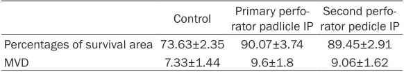
Table 2. The Table Showing Percentages of survival Area (%) And MVD Results for Each Subgroups, Respectively
Control rator padlicle IPPrimary perfo- rator pedicle IPSecond perfo-Percentages of survival area 73.63±2.35 90.07±3.74 89.45±2.91
MVD 7.33±1.44 9.6±1.8 9.06±1.62
IP: ischemic preconditioning.
injection, the integument was dissected carefully in the plane between the pannicu-lus carnosus and the deep fascia. The integument was then fixed for 24 h at 4°C. The flaps were obtained and radiographed (55 kVp, 25 mA, 20-s exposure) with a soft X-ray machine (Fuji Computerized Radiography XG-1; Fujifilm, Tokyo, Japan).
Assessment of flap survival area
[image:4.612.89.380.221.276.2]night. Slides were then rinsed in PBS and incu-bated with 1:500 followed by a biotinylated secondary antibody (anti-mouse IgG, ABC-kits, PK-6200; Vector Laboratories, Burlingame, CA, USA) for 1 h. The location of the reaction was visualized with 3,3-diaminobenzidine tetrahy-drochloride (Sigma). To assess the angiogenic response, microvessel density was estimated after CD31 staining. Briefly, three “hot spots”, or areas with the highest visible blood vessel density (marked by the vessel marker) were selected, and the blood vessels with a visible lumen were counted per high-power field (mag-nification × 100) by two pathologists blinded to the groups.
Western blotting analysis
Tissue samples from the choke zones were har-vested on postoperative day 7. These samples were homogenized in lysis buffer containing 20 mmol/L Tris-HCl, pH 7.4, 150 mmol/L NaCl, 1 mmol/L EDTA, 1 mmol/L EGTA, 1% Triton X-100, 2.5 mmol/L sodium orthovanadate, 1 µg/ml leupeptin, and 1 mmol/L phenylmethyl sulfonyl
[image:5.612.91.515.73.352.2]fluoride. The samples were centrifuged to pellet the debris and the supernatants were analyzed. A volume of each extract corresponding to 25 µg of total protein was resolved on sodium dodecyl sulfate-polyacrylamide gels and elec-trotransferred to polyvinylidene difluoride mem-branes. The membranes were blocked in phos-phate-buffered saline with 0.1% Tween-20 (PBS-T) containing 5% milk powder for 30 min at room temperature and then incubated over-night at 4°C with one of the following primary antibodies: anti-HIF-1α polyclonal antibody (Novus Biologicals, Littleton, CO) at a dilution of 1:400, anti-iNOS antibody (Santa Cruz Bio- technology, Santa Cruz, CA, USA) at a dilution of 1:1000, and anti-β-actin polyclonal antibody at a dilution of 1:1000 as a loading control. The membranes were subsequently incubated with horseradish peroxidase-conjugated anti-rabbit IgG (GE Healthcare Biosciences, Piscataway, NJ, USA) or anti-goat IgG (Santa Cruz Biote- chnology, Santa Cruz, CA, USA) for 1 h at room temperature. Immunoreactivity signal was visu-alized by Tanon Gel Imaging Systems (Shanghai, China) and analyzed by Image-Pro Plus Soft- ware.
Figure 3. Comparison of flap survival by group. A: Control Group; B: Primary pedicle perforator IP group; C: Secondary pedicle perforator IP group; D: The percentage of flap survival area was significantly greater in the ischemic pre -conditioning groups than in the control group (#, P < 0.001). The difference between the primary perforator pedicle
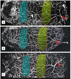
Figure 4. Representative arteriograms of each group injected with a lead ox-ide and gelatin compound. A: Control group; B: Primary perforator pedicle IP group; and C: Secondary perforator pedicle IP group. The choke arterioles
of the first choke zone (Yellow) have similar dilation and tortuous paths. The iliolumbar artery (Red arrow) has increased its territory to supply adjacent vascular territories. In both IP groups, the second choke vessels (Green) di-lated extensively, whereas those in the control group didi-lated only slightly or remained unopened.
Total RNA extraction and real-time quantitative RT-PCR
Real-time quantitative RT-PCR assay was per-formed to examine the expression of iNOS and HIF-1α. Total RNA was isolated from skin flap tissues using Trizol reagent (Invitrogen, USA) and DNA was removed using DNase I (Invitr- ogen). These RNA samples were then reverse transcribed into single-stranded cDNA using the first-strand cDNA synthesis kit (Fermentas, Lithuania). These cDNA products were further amplified using qPCR by SYBR Green RT-PCR kit (Bioteke, Beijing, China). The primers were purchased from Invitrogen (Table 1). Real-time quantitative PCR was performed with a real-time quantitative PCR machine (Stratagene, USA). The glyceraldehyde-3-phosphate dehy-drogenase (GAPDH) gene was used as an
inter-nal control. The sequences of the RT-PCR primers used in this study are listed in Table 1. Relative expression of qPCR products was deter-mined by the Delta-Delta CT method to normalize with GAPDH mRNA expression.
Statistical analysis
SPSS (SPSS Inc., Chicago, IL, USA) for Windows (version 17.0) was used for data man-agement and statistical anal-ysis. Data were expressed as mean ± SEM. Statistical anal-yses were performed using a one-way analysis of variance followed by post-hoc multiple comparisons. Values of P < 0.05 were deemed to indi-cate statistical significance.
Results
Flap survival
The percentages of surviving flap area in the control and experiment groups are shown in Table 2. Flap survival area was significantly greater in the ischemic preconditioning group than in the control group (P < 0.001; Table 2). Interestingly, the difference between the primary perforator pedicle isch-emic preconditioning group and the secondary perforator pedicle ischemic preconditioning were not significant (P = 0.582) (Figure 3). Angiography changes of the choke vessels in the extended perforator flap
On histopathological examination, the degree of PNL infiltration, chronic inflammation, and interstitial edema were similar among the groups. The vascular diameter was significantly greater in the IP groups than in the control group, but similar between two IP groups (Fi- gure 5A-C). Because the major cause of flap necrosis is generally a failure of the blood sup-ply, we evaluated the number of vessels in the second choke zone of the skin flap on postop-erative day 7. Changes in CD31 levels were a surrogate for microvessel densities in the sec-ond choke zone. The number of CD31-positive vessels was significantly different between the IP group and the control group (P < 0.05; Table 2). Blood and nutrition to the distal part of the skin flaps were supplied by choke vessels at the edges of the vascular territory. Significant enhancement in dilation of choke vessels was observed in the second choke zone (Figure 5C-E).
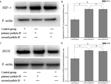
Effect of IP on hypoxia inducible factor-1α (HIF-1α) and inducible nitric oxide synthase (iNOS) expression
To test the effect of IP on expression of HIF-1α and iNOS protein in the second choke zone, we excised it and performed western blot analysis.
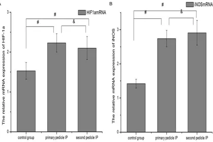
The level of HIF-1α and iNOS protein was signifi-cantly higher in the both IP groups than in the control group (P < 0.001). There was no statisti-cally significant difference between the IP groups (P > 0.05) (Figure 6). Real time qRT-PCR was used to examine the gene expression of iNOS and HIF-1α at 7 days postoperatively. The expression of iNOS and HIF-1α was higher in both preconditioning groups than the control group (P < 0.001), but there was no statistical difference between the IP groups (P > 0.05) (Figure 7).
Discussion
[image:7.612.91.527.71.289.2]The major finding of this study was that the IP affects the survival area of a perforator flap through effects on the second choke zone. To our knowledge, this is the first study to investi-gate the effects of IP on the second choke zone of skin flaps in a rat model. It is also the first to investigate whether pedicle selection (primary vs. secondary) for IP influenced the efficacy of IP on the second choke zone in the extended skin perforator flap. The expression of HIF-1α and iNOS in the second choke zone was also investigated to elucidate the underlying molec-ular events. Our results confirmed our hypoth-esis that ischemic preconditioning protects Figure 5. A-C: On histopathological examination, the degree of PNL infiltration, chronic inflammation, and interstitial edema were similar among the groups. The vascular diameter was significantly greater in the ischemic precondi -tioning groups than in the control group, but similar between IP groups. D-F: The number of CD31-positive vessels
skin flaps against ischemic necrosis through effects on the second choke zone. The positive effects of IP on the second choke zone may be attributed to upregulated expression of HIF-1α and iNOS. Interestingly, applying precondition-ing to different pedicles has the same positive effects on the extended perforator flap.
Flap loss, either partial or total, is a common fear among reconstructive surgeons. Such complications negatively alter the number of operations, duration of hospital stay, and cost of treatment. Although flap loss has various causes, ischemic reperfusion injury is one of the main causes, especially for microvascular free tissue transfers [22]. Ischemic precondi-tioning is an important technique to fight reper-fusion injury, first discovered in the canine myo-cardium by Murry [23]. Briefly, short periods of repeated ischemia/reperfusion limit the effects of the final ischemic period. This protective maneuver has received a great deal of
atten-tion and the underlying mechanisms have been investigated in various species and tissues. Wang [24] and Adanali [25] showed that capil-lary perfusion was higher after ischemic pre-conditioning in experimental muscle flaps when compared with non-preconditioned flaps. Is- chemic preconditioning has been shown to increase the survival of flaps exposed to isch-emia for up to 14 h [7-10]. Zahir [26] stated that IP may lengthen the critical ischemia time. In this study, flap survival area was obviously increased in the IP group compared with the control group.
[image:8.612.93.520.74.400.2]tomical vascular territory when the flap is ele-vated [7, 8]. Taylor and colleagues proved that at least one adjacent territory can be safely captured on vessels at the flap’s base, but the second choke zone does not dilate and the ana-tomical territory of the third perforator usually undergoes necrosis [4]. Consequently, further study into the choke vessels after extended flap elevation may further elucidate the cause of flap necrosis and shed light on strategies to prevent it. In our flap model, angiographic stud-ies showed that the difference in survival area between the groups was attributable to the behavior of the second choke zone between the posterior intercostal vessel and the iliolum-bar artery vessel territories. In both IP groups, the second choke vessels dilated extensively, whereas those in the non-preconditioning con-trol group dilated only slightly or remained unopened. The second choke zone between the posterior intercostal vessel and the iliolum-bar artery vessel territories apparently acts as a watershed in the control group, whereas the area was broken through in both IP groups.
The benefits of IP have also been demonstrat-ed experimentally by many investigators in both
free and pedicle flaps [10, 11, 14, 15]. Although the experimental results appear promising, few protocols have been translated into clinical practice. Preconditioning should be a single-stage procedure, should not injure the main pedicle, should protect the distal end of the flap, and should not unduly prolong surgical duration [10]. Ischemic preconditioning of the secondary pedicle may be more clinically valu-able for flap surgery, as it neither prolongs the operation nor increases the frequency of the procedure. Therefore, we compared the effects of applying preconditioning on different pedi-cles prior to ischemia. Interestingly, the differ-ence between the primary perforator pedicle ischemic preconditioning group and the sec-ondary perforator pedicle ischemic precondi-tioning were not significant (P > 0.05). This result shows that secondary pedicle IP is as effective as primary pedicle IP in the second choke zone and may be feasible for use in free flap transfers.
In this study, we also analyzed expression ch- anges of HIF-1α and iNOS in the second choke zone during IP. The results showed that the lev-Figure 7. Real time qRT-PCR was used to examine the gene expression of iNOS and HIF-1α. The expression of iNOS and HIF-1α was higher in both preconditioning groups than the control group (#, P < 0.001). There was no statistical
[image:9.612.97.516.73.353.2]els of HIF-1α and iNOS were significantly higher in both IP groups than in the control group, but not different between the IP groups. These results demonstrated that IP-induced HIF-1α and iNOS expression in the second choke zone plays a role in the survival of skin flaps. HIF-1α plays a pivotal role in ischemic responses and its expression is induced by the prolyl hydroxy-lase inhibitor dimethyloxalyl glycine. Many do- wnstream proteins are regulated by HIF-1α, including glucose transporter-1, vascular endo-thelial growth factor, and iNOS, which control a variety of adaptive responses to hypoxia, such as vasodilation, energy metabolism, glucose uptake, angiogenesis, erythropoiesis, cell via-bility, proliferation, and differentiation [28-30]. A number of studies show that HIF-1α could improve survival of random pattern skin flaps [31]. Recently, several studies have confirmed this role by demonstrating that intraperitoneal treatment with a HIF-1α stabilizer dimethyloxa-lylglycine or deferoxamine significantly increa- sed HIF-1α expression and enhanced skin flap survival [29, 30]. Yue [32] stated that hypoxia preconditioning effectively enhances the viabil-ity of adipose-derived stem cells by upregula-tion of HIF-1α to increase the survival rate of ischemic skin flaps. In addition, vascular den-sity in the hypoxic preconditioned group was greater than that in the control group. iNOS is one of the target genes of HIF-1α and is impli-cated in ischemic reperfusion injury (IRI) [20]. The higher expression of iNOS may play an important role in the therapeutic effect of pre-conditioning during IRI [17]. Previous studies demonstrated that iNOS plays a protective function in kidney IRI. This is consistent with the overwhelming opinion that iNOS plays a role in the protection induced by ischemic pre-conditioning against a secondary exposure to ischemia/reperfusion in the retina, heart, brain, and kidney. Some authors have also found that iNOS contributes to protective effects in skin flaps [20, 33]. Zhang et al. [20] concluded that ischemic preconditioning can enhance flap tol-erance to IRI and improve flap viability rate in a rat muscle flap model. Their study provides evi-dence that the regulation of NOS may play a role in ischemic preconditioning phenomenon. Nitric oxide (NO) is a messenger molecule regu-lating various physiological actions including vasodilation and neurotransmission [34]. Some data showed that the effect of iNOS on the skin flap was associated with NO [35]. Nitric oxide
plays an important role in the mechanism of IP, as the administration of an NO-donor prior to ischemia simulates the effect of IP, while the unspecific blocking of NO synthesis by L-NAME eliminates the protective effect of flap precon-ditioning by pre-clamping as well as by remote IP. Nitric oxide application is insufficient to pro-vide protection once NO synthesis is blocked [33]. In our flap model, the dilation of the sec-ond choke vessels resulted in improved skin flap survival. Therefore, the HIF-1α-iNOS-NO signaling pathways may be associated with be- havior of the second choke vessels. Regretfully, we did not further investigate these signaling pathways for this study. Further studies are necessary to evaluate the clinical significance and underlying molecular mechanisms of IP-improved survival of skin flaps.
Conclusion
In summary, this study provided direct evidence that ischemic preconditioning has a protective effect on ischemia of skin flaps through effects on choke vessels in the second choke zone. Ischemic preconditioning induced the second choke vessels to open and dilate extensively. Interestingly, secondary pedicle IP was as effective as primary pedicle IP in the second zone and may be feasible in free flap transfers. Our data also suggest that the expression of HIF-1α and iNOS was attributed to the develop-ing choke vessels of the second choke zone in the ischemic preconditioning process.
Acknowledgements
This research is supported in part by National Natural Science Foundation of China (No. 81472104).
Disclosure of conflict of interest
None.
Address correspondence to: Dr. Juyu Tang, Depart- ment of Orthopaedics, Xiangya Hospital, Central South University, # 87 Xiangya Road, Changsha 410008, Hunan, China. E-mail: juyutang163@163. com
References
combined free tissue transfer using the
an-terolateral thigh flap as a link. Microsurgery
2012; 32: 575-579.
[2] Hamilton K, Wolfswinkel EM, Weathers WM, Xue AS, Hatef DA, Izaddoost S, Hollier LH Jr. The delay phenomenon: a compilation of knowledge across specialties. Craniomaxillo-fac Trauma Reconstr 2014; 7: 112-118. [3] Taylor GI, Chubb DP, Ashton MW. True and
‘choke’ anastomoses between perforator an -giosomes: part i. anatomical location. Plast Reconstr Surg 2013; 132: 1447-1456. [4] Chubb DP, Taylor GI, Ashton MW. True and
‘choke’ anastomoses between perforator an -giosomes: part II. dynamic thermographic
identification. Plast Reconstr Surg 2013; 132: 1457-1464.
[5] Taylor GI, Pan WR. Angiosomes of the leg: ana-tomic study and clinical implications. Plast Re-constr Surg 1998; 102: 599-616; discussion 617-598.
[6] Zhuang Y, Hu S, Wu D, Tang M, Xu DC. A novel in vivo technique for observations of choke
vessels in a rat skin flap model. Plast Reconstr
Surg 2012; 130: 308-317.
[7] Williams BA, Currie RW, Morris SF. Impact of arteriogenesis in plastic surgery: choke vessel growth proceeds via arteriogenic mechanisms
in the rat dorsal island skin flap. Microcircula -tion 2009; 16: 235-250.
[8] Miyamoto S, Minabe T, Harii K. Effect of
recipi-ent arterial blood inflow on free flap survival
area. Plast Reconstr Surg 2008; 121: 505-513.
[9] Wang WZ. Investigation of reperfusion injury and ischemic preconditioning in microsurgery. Microsurgery 2009; 29: 72-79.
[10] Yildiz K, Karsidag S, Akcal A, Yesiloglu N, Ugur-lu K, Ozagari A, Guneren E, Bas L. Comparison
of the flap survival with ischemic precondition -ing on different pedicles under varied ischemic
intervals in a rat bilateral pedicled flap model.
Microsurgery 2014; 34: 129-135.
[11] Moosavian HR, Mirghazanfari SM, Moghad-dam KG. Effect of ischemia preconditioning
and leech therapy on cutaneous pedicle flaps
subjected to prolonged ischemia in a mouse model. Aesthetic Plast Surg 2014; 38: 1024-1029.
[12] Dacho A, Lyutenski S, Aust G, Dietz A. Ischemic
preconditioning in a rat adipocutaneous flap
model. HNO 2009; 57: 829-834.
[13] Claytor RB, Aranson NJ, Ignotz RA, Lalikos JF, Dunn RM. Remote ischemic preconditioning modulates p38 MAP kinase in rat
adipocuta-neous flaps. J Reconstr Microsurg 2007; 23: 93-98.
[14] Marian CF, Jiga LP, Ionac M. Ischemic
precon-ditioning of free muscle flaps: an experimental
study. Microsurgery 2005; 25: 524-531.
[15] Harralson T, Grossi FV, Quan EE, Tecimer T, Perez-Abadia G, Anderson G, Barker JH, Maldo-nado C. Ischemic preconditioning of skeletal muscle: duration of late-phase protection. Ann Plast Surg 2005; 55: 216-222.
[16] Matsumura H, Yoshizawa N, Vedder NB, Wata-nabe K. Preconditioning of the distal portion of
a rat random-pattern skin flap. Br J Plast Surg
2001; 54: 58-61.
[17] Yang M, Angel MF, Pang Y, Angel JJ, Wang Z, Neumeister MW, Wetter N, Zhang F. Expres-sion of inducible nitric oxide synthase in
mus-cle flaps treated with ischemic postcondition -ing. Hand (N Y) 2012; 7: 297-302.
[18] Hollenbeck ST, Senghaas A, Komatsu I, Zhang Y, Erdmann D, Klitzman B. Tissue engraftment of hypoxic-preconditioned adipose-derived
stem cells improves flap viability. Wound Re -pair Regen 2012; 20: 872-878.
[19] Civelek B, Selcuk T, Bilgen E, Demirbag E,
Cele-bioglu S. Intermittent ischaemia of skin flaps
shortens time taken to divide pedicles: an ex-perimental study in rats. Scand J Plast Recon-str Surg Hand Surg 2009; 43: 241-244. [20] Zhang F, Oswald T, Holt J, Gerzenshtein J, Lei
MP, Lineaweaver WC. Regulation of inducible nitric oxide synthase in ischemic
precondition-ing of muscle flap in a rat model. Ann Plast
Surg 2004; 52: 609-613.
[21] Rees RS, Smith DJ Jr, Adamson B, Im M, Hin-shaw D. Oxidant stress: the role of the glutathi-one redox cycle in skin preconditioning. J Surg Res 1995; 58: 395-400.
[22] Khalil AA, Aziz FA, Hall JC. Reperfusion injury. Plast Reconstr Surg 2006; 117: 1024-1033. [23] Carroll CM, Carroll SM, Overgoor ML, Tobin G,
Barker JH. Acute ischemic preconditioning of
skeletal muscle prior to flap elevation aug
-ments muscle-flap survival. Plast Reconstr Surg 1997; 100: 58-65.
[24] Wang WZ, Anderson G, Firrell JC, Tsai TM. Isch-emic preconditioning versus intermittent
re-perfusion to improve blood flow to a vascular isolated skeletal muscle flap of rats. J Trauma
1998; 45: 953-959.
[25] Adanali G, Ozer K, Siemionow M. Early and late effects of ischemic preconditioning on
micro-circulation of skeletal muscle flaps. Plast Re-constr Surg 2002; 109: 1344-1351.
[26] Zahir KS, Syed SA, Zink JR, Restifo RJ, Thom-son JG. Ischemic preconditioning improves the
survival of skin and myocutaneous flaps in a
rat model. Plast Reconstr Surg 1998; 102: 140-150; discussion 151-142.
[27] Dhar SC, Taylor GI. The delay phenomenon: the story unfolds. Plast Reconstr Surg 1999; 104: 2079-2091.
[28] Sun Y, Li QF, Zhang Y, Hu R, Jiang H. Isoflurane
preconditioning increases survival of rat skin
-pha expression. Cell Physiol Biochem 2013; 31: 579-591.
[29] Takaku M, Tomita S, Kurobe H, Kihira Y, Morim-oto A, Higashida M, Ikeda Y, Ushiyama A, Hashimoto I, Nakanishi H, Tamaki T. Systemic preconditioning by a prolyl hydroxylase
inhibi-tor promotes prevention of skin flap necrosis
via HIF-1-induced bone marrow-derived cells. PLoS One 2012; 7: e42964.
[30] Zhong X, Yan W, He X, Ni Y. Improved fat graft
viability by delayed fat flap with ischaemic
pretreatment. J Plast Reconstr Aesthet Surg 2009; 62: 526-531.
[31] Schlaudraff KU, Pepper MS, Tkatchouk EN, Eh-renburg I, Alizadeh N, Montandon D, Pittet B. Hypoxic preconditioning increases skin oxy-genation and viability but does not alter VEGF expression or vascular density. High Alt Med Biol 2008; 9: 76-88.
[32] Yue Y, Zhang P, Liu D, Yang JF, Nie C, Yang D. Hypoxia preconditioning enhances the viability of ADSCs to increase the survival rate of
isch-emic skin flaps in rats. Aesthetic Plast Surg 2013; 37: 159-170.
[33] Kuntscher MV, Juran S, Altmann J, Menke H, Gebhard MM, Germann G. Role of nitric oxide in the mechanism of preclamping and remote ischemic preconditioning of adipocutaneous
flaps in a rat model. J Reconstr Microsurg 2003; 19: 55-60.
[34] Nezami BG, Rahimpour S, Gholipour T, Ghase-mi M, Sadeghipour H, Mehr SE, EmaGhase-mi-Razavi SH, Dehpour AR. Pharmacologic
precondition-ing of random-pattern skin flap in rats usprecondition-ing
local cyclosporine and FK-506: interaction with nitric oxide system. Ann Plast Surg 2007; 59: 435-440.
[35] McDonald WS, Lo TP Jr, Thurmond M, Jones C, Cohen R, Miller A, Beasley D. Role of nitric