Original Article
Potential antidepressant role of low-dose naltrexone in
a rat model of chronic unpredictable mild stress
Xue Lin, Hui He, Ruiqin Zhang, Jingchun Xing, Wang Yang, Wenzhi Li
Department of Anesthesiology, Heilong Jiang Province Key Lab of Research on Anesthesiology and Critical Care Medicine, Second Hospital of Harbin Medical University, Harbin, Heilongjiang, China
Received July 30, 2016; Accepted February 23, 2017; Epub April 15, 2017; Published April 30, 2017
Abstract: Objectives: We aimed to investigate antidepressant effect of low-dose naltrexone (LDN) and to explore its potential mechanism of action using a rat model induced by chronic unpredictable mild stress (CUMS). Methods: The CUMS-induced rat model was established, and the antidepressant effect of LDN (0.1 mg/kg/day, given by
intra-peritoneal injection for 5 weeks) was investigated using sucrose preference test, open field test, forced swimming
test, and novelty-induced hyponeophagia test. Moreover, the molecular biological changes including beta-endorphin
(BEP), interleukin (IL)-1β, 5-hydroxy-tryptamine (5-HT), corticosterone, monoamine neurotransmitter and brain de
-rived neurotrophic factor (BDNF) were also identified. Results: Behavioral changes were observed following 5 weeks
of CUMS, including decreased sucrose preference, reduced locomotor activity, increased immobility time and
el-evated latency of feeding. However, after LDN treatment, these behavioral changes were significantly improved. In
addition, LDN could inversed reduced BEP level in hypophysis, 5-HT level and BDNF expression level in
hippocam-pus caused by CUMS. Moreover, LDN also could inversed increased levels of IL-1β and corticosterone induced by
CUMS. Conclusion: The results suggest that LDN may have therapeutic actions on depression-like behavior induced
by CUMS in rats by decreasing IL-1β in spleen and hippocampus tissue and plasma corticosterone levels and
up-regulating the BEP level in hypophysis tissue and plasma and the 5-HT, BDNF level in hippocampus tissue.
Keywords: Antidepressant effects, low-dose naltrexone, high-dose naltrexone, chronic unpredictable mild stress rat model of depression
Introduction
Depression is one of the most common psy- chiatric problems affecting more than 350 mil-lion people worldwide [1], and more than half among them would experience episodes [2].
Depression causes significant distress or
im-pairment in physical healthy, mental health and quality of life [3]. Various therapies have been considered available for treating depression, but antidepressant is usually recognized as the
first-line treatment for depression.
Recently, many synthetic chemical antidepres-sants were introduced, such as monoamine oxi-dase inhibitors, tricyclic antidepressants and selective serotonin reuptake inhibitors [4, 5]. Although these chemical antidepressants have been proved to have therapeutic responses, there are still a variety of side effects, such as psychomotor impairment and dependence
lia-bility [6, 7]. Naltrexone is clinically used to treat heroin and alcohol dependence [8, 9], which could block the pleasurable effects associated with alcohol drinking or heroin use [10]. Previous evidences showed that low-dose of naltrexone (LDN) might have a role in amelioration of psy-chiatric problems such as autism and depres-sion [11]. For example, intermediate levels of LDN (0.25 mg/kg given every other day) have
been found to benefit a subset of autistic chil -dren [12]. Kennedy et al. demonstrated that
endogenous opioid neurotransmission on
improv-ing depression is needed for further explora- tion.
Nowadays, the chronic unpredictable mild stress (CUMS) animal model has been proved as an effective animal model of depression for investigating antidepressants [17-19]. In this study, we established a CUMS rat model, and administrated LDN (0.1 mg/kg) and high-dose naltrexone (HDN) to rats to explore the poten-tial antidepressant role of naltrexone by using behavioral tests. Moreover, the potential mech-anism of action of LDN was also evaluated through detecting some molecular biological
indexes including interleukin (IL)-1β and plas -ma corticosterone.
Materials and methods
Animal and drugs
Thirty-two female Sprague-Dawley (SD) rats weighing 150-170 g were obtained from the Laboratory Animal Center (approved by the
eth-ical committee of the Second Affiliated Hospital
of Harbin Medical University, China). Rats were housed at 22-25°C with 55±2% relative humid-ity, and exposed to a 12:12 h light-dark cycle with lights on at 07:00 am, free access to food and water. All procedures were in accordance
rats were randomly divided into four matched groups (8 rats/group): (1) Control group (CON); (2) NS-CUMS group: CUMS condition with nor-mal saline; (3) LDN-CUMS group: CUMS condi-tion with low-dose naltrexone treatment (0.1 mg/kg); (4) HDN-CUMS group: CUMS condition with high-dose naltrexone (10 mg/kg).
The rats, except the CON group, were individu-ally housed and repeatedly exposed to a set of chronic unpredictable mild stressors in a ran-dom order every day for 5 weeks: cage tilt at 45°C for 24 h; inversion of the light/dark cycle;
continuous overnight flash mode; damp saw -dust for 24 h (200 mL of tap water spilled onto 100 g pad); behavior restraint in a tube for 6 h (diameter: 8 cm, length: 20 cm); fasting for 24 h; water deprivation for 24 h; high temperature at 45°C for 5 min; nip trail for 1 min [20, 21]. The rats in CON group were housed in the quiet and separate home with conventional breed-ing. All rats were administered different con-centrations of naltrexone or normal saline via intraperitoneal injection at 6:00 pm-7:00 pm during the 5-week period of CUMS (day 14 to day 49).
After establishing models, animals were
reori-ented to the specific pathogen-free animal
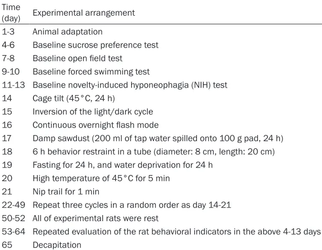
[image:2.612.91.409.97.344.2]house for 3 days. Behavior test was handled on the fourth day (in Table 1).
Table 1. Schematic representation of the experimental procedure and behavioral test
Time
(day) Experimental arrangement 1-3 Animal adaptation
4-6 Baseline sucrose preference test 7-8 Baseline open field test
9-10 Baseline forced swimming test
11-13 Baseline novelty-induced hyponeophagia (NIH) test 14 Cage tilt (45°C, 24 h)
15 Inversion of the light/dark cycle 16 Continuous overnight flash mode
17 Damp sawdust (200 ml of tap water spilled onto 100 g pad, 24 h) 18 6 h behavior restraint in a tube (diameter: 8 cm, length: 20 cm) 19 Fasting for 24 h, and water deprivation for 24 h
20 High temperature of 45°C for 5 min 21 Nip trail for 1 min
22-49 Repeat three cycles in a random order as day 14-21 50-52 All of experimental rats were rest
53-64 Repeated evaluation of the rat behavioral indicators in the above 4-13 days
65 Decapitation
with the “Principles of Laboratory Animal Care” and the PR China legislation for the use and care of laboratory animals. Moreover, the suffer-ing of animals was minimized as much as possible.
Naltrexone was obtai- ned from Sigma-Ald- rich (St Louis, MO), and was dissolved in normal saline before experiment.
Chronic unpredict-able mild stress rat model of depression
Behavioral tests
Rats were housed in the testing chamber 30 min before each test. All tests were performed between 7:00 and 12:00 am.
Sucrose preference test
The sucrose preference test was performed as employed previously [21, 22]. Rats were culti-vated to adapt to sucrose solution (1%, w/v) 2 days before the test. During adaptation period, 2 bottles of sucrose solution were placed in each cage for 24 h, and then one bottle of sucrose solution was substituted with tap-water for 24 h. After the adaptation period, the rats was expropriated of food and water for 24 h, and then rats were given free access to a bottle of 1% sucrose solution and a bottle of tap-water for 1 h in the sucrose preference test. At the end of the test, the bottles contain-ing the remaincontain-ing sucrose solution and tap-water were weighed, and the sucrose prefer-ence index was calculated as follows: sucrose preference index (%) = sucrose consumption/ (sucrose consumption + tap-water consump- tion).
Open field test
The open field test was performed according to
previous studies [22, 23]. Briefly, the test was
performed in a gray square box (100 cm × 100
cm × 40 cm), the floor of which was divided into
25 equal squares, and was illuminated by a
100 W lamp above the center of the open field. Each rat was placed in the center of open field
and observed for 5 min. The numbers of the locomotion, rearing and grooming were record-ed using a video camera locatrecord-ed 155-165 cm
above the open field. Additionally, the arena
was cleaned with 70% alcohol after each test. Novelty-induced hyponeophagia (NIH) test
Hyponeophagia was defined as feeding inhibi -tion produced by exposing to novelty, and the NIH test was performed according to previous studies [20, 24]. After fasting for 48 h, rats were placed into a new cage, in which there were thirty pieces of rat food particles in the
cage center. The latency of feeding defined as
time elapsed until rats began to eat was manu-ally recorded by competent observers.
Forced swimming test
Forced swimming test was carried out on rats
as described previously with minor modifica -tions [25]. Briefly, each rat was placed in aplas
-tic drum (40 cm tall, 25 cm in diameter) filled to
21.5±0.5 cm with 24±0.5°C water. Rats were forced to swim in the drum for 5 min, and each rat’s behavior was recorded using a video cam-era above the drum in a dim light. Immobility
time referred to the time spent by rat floating without floundering in the water except for
small movement necessary to keep its head above the water. In addition, the drum would be cleaned after each test.
Preparation of blood and tissue samples
Eight rats from each group were deeply anes-thetized with chloral hydrate (10%, 0.3 mL/100 g, i.p.), and the samples of blood and tissue were collected between 8:00 and 10:00 am. The collected blood samples were centrifuged immediately at 1000 × g for 15 min at 4°C to separate blood plasma.
In addition, the hypothalamus, hypophysis, hip-pocampus and spleen of rats were dissected onto a frosted glass plate on top of crushed ice. Tissue samples were snap-frozen in liquid nitro-gen and stored at -80°C until use.
Measurement of beta-endorphin (BEP) in plasma and brain tissue
The BEP levels in plasma (containing 75% EDTA and aprotinin), hypothalamus and hypophy-
sis were quantified by measuring the radioac -tivity of precipitate using a gamma-counter (GC-2010, USTC Chuangxin Co., Ltd, ZONKIA Bra- nch) according to the instructions of commer-cially available radioimmunoassay kits (BNIBT, Beijing, China).
Measurement of interleukin (IL)-1β level
The spleen and hippocampus were
homoge-nized in 5 μL/mg ice-cold phosphate-buffered
saline (PBS) (0.02 mol/L, pH = 7.0-7.2) and cen-trifuged at 1500 × g for 15 min at 4°C, then the
supernatant was collected. The levels of IL-1β
Measurement of corticosterone in plasma The levels of corticosterone in plasma were measured using high performance liquid chro-matography (HPLC) method as reported previ-ously [26]. Before measurement, the plasma was extracted with 5 mL ethyl acetate, and vortex-mixed for 30 seconds. Then, the extract was washed with 1 mL of 0.1 M sodium hydrox-ide and 1 mL of water, transferred to clean tubes, and evaporated to dryness. Finally, the residue was re-dissolved in mobile phase (ace-tonitrile-water-acetic acid-TEA, 22:78:0.1:0.03, v/v) and injected into a HPLC (ShimadzuLC20A, Japan) equipped with a 25 cm C18 column, and an ultraviolet detector at 254 nm.
Measurement of monoamine neurotransmitter The levels of 5-hydroxy-tryptamine (5-HT), nor-epinephrine (NE) and dopamine (DA) in rat’s unilateral hippocampus tissue were detected as described previously [27]. Briefly, the brain
tissue were weighed, homogenized in ice-cold 0.1 Nperchloric acid (0.5 mL/100 mg tissue) containing 10-7 M ascorbic acid and centrifuged
at 5200 × g for 30 min at 4°C before detection. The 200 μL supernatant obtained through a
Immunohistochemistry analysis of brain de-rived neurotrophic factor (BDNF)
Another 3 rats in each group were sacrificed
and perfused intracardially with heparinized PBS followed by 0.1 mol/L PBS (pH = 7.4) con-taining 4% paraformaldehyde. The coronal hip-pocampus was dissected, and then cut into 5
μm thick hippocampus sections after fixing
with a solution containing 4%
paraformalde-hyde for 24 h and embedding in paraffin. After
dewaxed, rehydrated and blocked with 5% bovine serum albumin for 30 min, the sections were incubated with rabbit anti-BDNF antibody (1:50 dilution, BOSTER, Wuhan, China) over-night at 4°C, and subsequently reacted with the biotinylated secondary antibody for 30 min at room temperature. Diaminobenzidine was used as a chromogen, and the hippocampal CA3 region of each brain section was observed and pictured with microscope (Nikon H600L, Japan). The expression of BDNF proteins was
quantified and statically analysed followed the
[image:4.612.94.522.84.197.2]reference reported [28]. The staining density was measured by detecting the average gray value and integrated optical density (IOD) using the Image-Pro Plus 6.0.
Table 2. Baseline values of the tests conducted from day 4 to 13
Indexes CON NS-CUMS LDN-CUMS HDN-CUMS F P
N Mean ± SD N Mean ± SD N Mean ± SD N Mean ± SD
Locomotor activity 8 108.63±16.52 8 111.63±21.62 8 107.38±22.97 8 109.88±18.92 0.065 0.978 Rearing 8 34.88±6.92 8 35.25±7.48 8 31.50±7.09 8 28.75±4.53 1.725 0.185 Grooming 8 3.50±2.07 8 3.75±1.91 8 3.88±2.10 8 4.63±2.07 0.452 0.718 Rearing and grooming 8 38.38±7.19 8 39.00±7.37 8 35.38±8.53 8 33.38±5.07 1.084 0.372 Sucrose preference index 8 0.80±0.08 8 0.81±0.08 8 0.80±0.10 8 0.82±0.07 0.082 0.969 Immobility time 8 155.63±11.30 8 160.75±21.20 8 165.75±18.53 8 159.38±22.57 0.392 0.759 Latency of feeding 8 131.88±49.66 8 135.00±38.55 8 139.50±42.18 8 133.63±32.35 0.050 0.985 SD: Standard Deviation; N: Number of cases. CON: control group. NS-CUMS: Normal saline-chronic unpredictable mild stress group. LDN-CUMS: Low-dose naltrexone-chronic unpredictable mild stress group. HDN-CUMS: High-dose naltrexone-chronic unpredictable mild stress group.
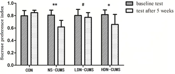
Figure 1. Effects of low-dose naltrexone on sucrose preference index.
*P<0.05, **P<0.01 vs. CON group; #P<0.05 vs. NS-CUMS group.
0.22 μm filter was reacted
with the benzylamine deriva- tization reagent solution for 2 min at 24°C, and followed with the diphenyl ether de- rivatization reagent solution for 20 min at 50°C. Finally, 20
μL sample was injected into a HPLC with a fluorescence
[image:4.612.92.376.240.360.2]Statistical analyses
The experimental data were analyzed by SPSS software (version 13.01S; Beijing Stats Data Mining Co. Ltd., Beijing, China). All the data were reported as means ± SD (standard devia-tions). The differences about behavioral data among groups were analyzed by repeated mea-surement ANOVA (analysis of variance) and bio-chemical data were analyzed by one-way ANOVA (Furthermore, homogeneity of variance assumptions was checked using Levene test. If P<0.1, we defined homogeneity of variance is satisfied. Then, Tukey’s HSD (Honestly Signi-ficant Difference) test would be used to assess
differences among groups. Otherwise, Dun- nett’s T3 would be carried out to analyze differ-ences among groups.
Results
Overall values comparisons for analyzing LDN anti-depression effects
Baseline values of the tests conducted from day 4 to 13 were shown in Table 2. The differ-ences among groups were analyzed using
ANOVA, and no significant difference were
found for all included indexed in the tests among 4 groups. Therefore, it is available for next step study.
Sucrose preference index increased in LDN-CUMS rats
As shown in Figure 1, rats in NS-CUMS and
HDN-CUMS groups showed significantly lower
sucrose preference index than that in CON group (P<0.01; P<0.05). Meanwhile, LDN
treat-ment significantly increased the sucrose prefer -ence index compared with the NS-CUMS group (P<0.05).
Unsignificant difference found between LDN-CUMS and CON rats in the open field test
The activity of locomotion, rearing and
groom-ing were observed in the open field test (Figure 2). Compared with CON group, locomotor
activ-ity was significantly decreased in NS-CUMS and
HDN-CUMS groups (P<0.05). Both rearing and total number of rearing and grooming were
sig-nificantly reduced after HDN treatment com -pared with CON group (P<0.01). Moreover, no
significant differences on performance in the open field test were found between LDN-CUMS
and CON group.
Unsignificant difference found between
LDN-CUMS and CON rats in immobility time and latency of feeding test
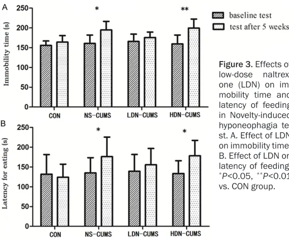
As shown in Figure 3A, the immobility time in
NS-CUMS and HDN-CUMS groups were signifi -Figure 2. Effects of low-dose naltrexone on chronic unpredictable mild stress-induced rat model on locomotor activ-ity, rearing, grooming, and the total number of rearing and grooming. (A) Effects of low-dose naltrexone on locomotor
[image:5.612.86.517.72.288.2]cantly longer than that in CON group (P<0.05; P<0.01). Compared with CON group, latency of
feeding was significantly increased in NS-CUMS
group and HDN-CUMS group (P<0.05) (Figure 3B). In addition, no significant differences were
found both in forcing swimming and novel feed tests between LDN-CUMS group and CON group.
BEP and 5-HT up-expression
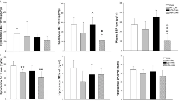
As shown in Figure 4A, compared with CON group, the BEP concentrations in hypophysis
and plasma were significantly decreased in
HDN-CUMS group (P<0.05). In addition, com-pared with NS-CUMS group, hypophysial BEP
concentrations were significantly increased in
LDN-CUMS group (P<0.05). As for BEP
concen-tration in hypothalamus, no significant differ -ences were found among the four groups. As shown in Figure 4B, the levels of 5-HT in
hip-pocampus tissue were significantly lower in
both NS-CUMS group and HDN-CUMS group (P<0.01) than those in CON group. Additionally,
no significant differences were found among
the four groups with regard to DA and NE levels in hippocampus tissue.
IL-1β down-expression in the spleen and hip -pocampus
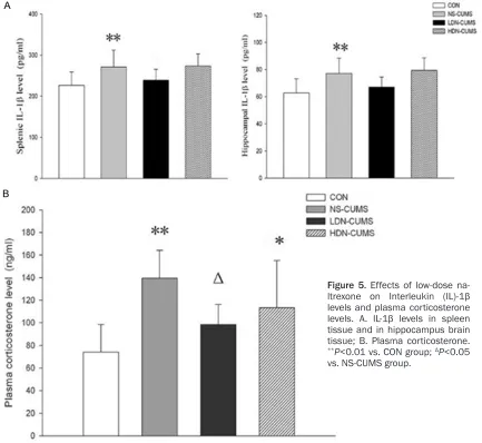
As shown in Figure 5A, the IL-1β levels both in spleen and hippocampus were significantly
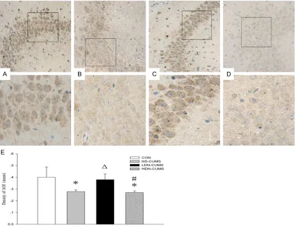
MS and HDN-CUMS groups (P<0.05). The BDNF expression levels of LDN-CUMS group were elevated compared with NS-CUMS group (P<0.05). Furthermore, the expression of BDNF
was significantly decreased in HDN-CUMS
group compared with LDN-CUMS group (P< 0.05).
Discussion
Depression is a serious public mental disease [29]. Naltrexone has been widely used in treat-ment of alcohol dependence and heroin depen-dence [30, 31]. However, the research focusing on antidepressant effects of LDN has rarely been reported. In this study, we established a CUMS-induced rat model, and studied the anti-depressant effects of LDN using 4 groups’ rats which showed similar baseline value during behavioral tests before the CUMS procedures. We found that LDN would have a therapeutic effect on depression behavior in the rat model by improving sucrose preference, decreasing locomotor activity, reducing immobility time and holding back prolongation of latency of feeding. Further data showed that antidepres-sant effects might be mediated by decreasing plasma corticosterone level, up-regulating BEP level in hypophysis, 5-HT level in hippocampus, and BDNF expression level, and inhibiting the
increase of IL-1β caused by CUMS.
[image:6.612.91.375.70.309.2]The CUMS-induced rat model is considered as an effective animal model of depression Figure 3. Effects of
low-dose naltrex-one (LDN) on im-mobility time and latency of feeding in Novelty-induced hyponeophagia te- st. A. Effect of LDN on immobility time; B. Effect of LDN on latency of feeding.
*P<0.05, **P<0.01
vs. CON group.
increased in NS-CUMS group (P<0.01) than those in CON group. As shown in Figure 5B, the levels of plasma
corticos-terone were significantly high -er in NS-CUMS and HDN-CUMS groups than that in CON group (P<0.05). Com- pared with NS-CUMS group, plasma corticosterone level in LDN-CUMS group were
sig-nificantly lower (P<0.05) than that in NS-CUMS group (P< 0.05).
BDNF up-expression in rat hippocampus
As shown in Figure 6, com-pared with CON group, the expression of BDNF was
NS-CU-Figure 4. Effects of low-dose naltrexone on β-Endorphin (BEP) concentrations and levels of 5-hydroxytrypytamine(5-HT), norepinephrine (NE) and dopamine (DA). A. BEP in hypothalamus brain tissue, hypophysis brain tissue and plasma; B. 5-HT, NE and DA in hippocampal brain tissue. *P<0.05, **P<0.01 vs. CON group; ΔP<0.05
[image:7.792.97.702.72.414.2]because the rat exposing to CUMS can exhibit a series of depression-like symptoms [18]. CUMS for 5-weeks could induce behavior changes such as anhedonia, (i.e., lessened intake of a delicious sweet solution) [21], psy-chomotor retardation (i.e., decreased locomo-tor activity) [32], refraclocomo-tory loss of interests (i.e., decreased rearing and grooming, which is driving by the instinct interests of rats to expl- ore a novel environment) [17, 33], recurrent thoughts of death (i.e., dramatically increased immobility time) [34], and anxiety-like behavior (i.e., increased latency for feeding) [20, 24]. CUMS caused depression-like symptoms by inducing changes in behavior, including decre- ased sucrose preference, reduced locomotor activity and total number of rearing and groom-ing, increased immobility time and elevated latency of feeding, which indicated that a
CUMS-induced rat model was successfully established in our research.
Recent studies have demonstrated that inflam
-matory mediators (i.e., IL-1β) may play a critical
role in the pathophysiology of depression [35].
IL-1β is considered as an inflammatory bio -marker for depression, and the peripheral
lev-els of IL-1β are increased in depression patients
[36, 37]. In the present study, CUMS markedly
increased IL-1β levels in the spleen and hippo
-campus. Furthermore, LDN treatment signifi -cantly reversed the changes and reduced the
IL-1β levels. The above experimental data sug -gested that LDN could inhibit the increase of
IL-1β levels induced by CUMS.
Meanwhile, increasing evidences have showed
[image:8.612.90.523.74.472.2]that inflammation could lead to depression by
Figure 5. Effects of low-dose
na-ltrexone on Interleukin (IL)-1β
levels and plasma corticosterone
levels. A. IL-1β levels in spleen
tissue and in hippocampus brain tissue; B. Plasma corticosterone.
**P<0.01 vs. CON group; ΔP<0.05
affecting the hypothalamic-pituitary-adrenal (HPA) axis activation, modulating neurotrans-mitters, and alternating of neuroplasticity [38]. It has been found that animals with exogenous
administration of IL-1β into brain could exhibit
depressive-like symptoms, and this could be
attenuated by pretreatment with IL-1β receptor
antagonist [39, 40]. Recent researches have
shown that IL-1β could activate HPA axis, which
is accompanied with increase of plasma levels of ACTH and corticosterone [41, 42]. Moreover, it has been reported that mice with deletion of the IL-1 receptor type I did not increase CUMS-induced plasma corticosterone level, and the obstruction of endogenous corticoste-rone release eliminated CUMS-induced depres-sion [43]. In addition, previous data suggested that naltrexone could participate in regulating
chronic physical or psychological stress through modifying corticosterone plasma levels [44]. In our study, we found that LDN administration
significantly reversed the increase of IL-1β lev -els and plasma corticosterone level induced by CUMS. Thus, the potential mechanism of LDN as an antidepressant in the CUMS-induced model may be related to the decreased plasma
corticosterone and IL-1β levels in CUMS model.
[image:9.612.91.524.71.403.2]BEP, a neuromodulator and neurotransmitter in the central nervous system, has been demon-strated to inhibit the generation of stress hor-mones, produces analgesia and a feeling of cheerfulness [43]. Accumulating evidences suggest that a low level of central BEP is relat-ed with the course of stress-relatrelat-ed psycho-disturbance, depression and posttraumatic Figure 6. Effect of low-dose naltrexone on expression of brain derived neurotrophic factor (BDNF) in hippocampus brain tissue of CUMS rat. A. CON group; B. NS-CUMS group; C. LDN-CUMS; D. HDN-CUMS group. BDNF levels were determined by immunohistochemical assay and the cells in brown were BDNF positive. The images above were
magnified 400 times, and the images below were magnified 1000 times. E. Comparison of the mean density of
AOI (area of interesting) of BDNF. Immunohistochemical analysis of BDNF proteins was performed on sections of rat brain of CON group, NS-CMS group, LDN-CUMS and HDN-CUMS group. *P<0.05 vs. CON group; ΔP<0.05 vs.
stress disorder [45, 46]. Moreover, previous study has put forward that naltrexone could elevate BEP expression levels in autistic chil-dren [47]. Our results inferred that BEP expres-sion levels might be regulated after the LDN treatment on depression. In addition, several studies showed that monoamine neurotrans-mitters (NE, DA and 5-HT) are also involved in the pathogenesis of depression and play impor-tant roles in mediating the effects of antide-pressant [48]. Moreover, IL-1 may reduce 5-HT level by lowering the availability of its precursor
tryptophan [49]. In our study, IL-1β levels decreased and 5-HT was significantly increased
in CUMS rats after LDN treatment. Thus, we suggested that up-regulation of 5-HT levels
might be related with inversed IL-1β levels.
Ample evidence indicates that IL-1β could
re-gulate hippocampal neurogenesis via the inhi-bition of the expression level of BDNF, which is involved in synaptic plasticity and related with cognitive dysfunction by CUMS induced-depression [43, 50], and up-regulation of BDNF expression plays a critical role in antidepres-sant treatment [51]. In line with the previous reports, our results indicated that rats exposed to CUMS exhibited a reduced expression of BDNF proteins in the hippocampus [52, 38]. After LDN treatment, BDNF levels in CUMS-induced rats were similar with that in healthy control group. These results indicated that the mechanism of the antidepressant effect of LDN may be also related to the increased BDNF expression in the hippocampus.
However, several limitations in this present study should be reminded. Firstly, although the experiment evidence has demonstrated the effect of naltrexone on up-regulation of BEP was dose-dependent, we did not explore the best dose of naltrexone on regulating BEP lev-els. Further experiment is needed to explore the relationship between naltrexone and BEP levels before clinical use. Secondly, positive control group was not established in this study. Although change of neurotransmitter concen-tration has been observed in previous study on traditionally antidepressants, including amitrip-tyline and desipramine [15, 53], intuitive para- meters to evaluate LDN antidepressant effect is still needed. In addition, neurobiological results on unstressed rats about how naltrex-one works will be provided in our further study.
Conclusions
In conclusion, it appears that LDN has a thera-peutic effect on depression behavior in CUMS-induced rat model, and mechanisms underly- ing the antidepressant-like effects of LDN might be associated with the decrease in plas-ma corticosterone levels, up-regulation of BEP in hypophysis, 5-HT level in hippocampus, and BDNF expression by inhibiting the increase of
IL-1β induced by CUMS. However, the exact tar -get of the antidepressant effect of LDN remains unknown, and our group will try to resolve the above limitations in future studies.
Acknowledgements
This work was supported by the Department
of Anesthesiology of Second Affiliated
Hospi-tal of Harbin Medical University and Heilong Jiang Province Key Laboratory of Research on Anesthesiology and Critical Care Medicine.
Disclosure of conflict of interest
None.
Address correspondence to: Wenzhi Li, Department of Anesthesiology, Heilong Jiang Province Key Lab of Research on Anesthesiology and Critical Care Medicine, Second Hospital of Harbin Medical Uni- versity, 246 Xuefu Road, Harbin 150081, Heilong- jiang, China. Tel: 451-86605029; Fax: 0086-451-86605028; E-mail: [email protected]
References
[1] Organization WH. Depression Fact Sheet. 2013.
[2] Eaton WW, Shao H, Nestadt G, Lee BH, Bien-venu OJ and Zandi P. Population-based study
of first onset and chronicity in major depres -sive disorder. Arch Gen Psychiatry 2008; 65: 513-520.
[3] Depression. In: Preedy V, Watson R, editors. Handbook of disease burdens and quality of life measures. New York: Springer; 2010. pp. 4188-4188.
[4] Youdim MB, Edmondson D and Tipton KF. The therapeutic potential of monoamine oxidase inhibitors. Nat Rev Neurosci 2006; 7: 295-309.
[5] Anderson IM. Selective serotonin reuptake in-hibitors versus tricyclic antidepressants: a
[6] Dai Y, Li Z, Xue L, Dou C, Zhou Y, Zhang L and Qin X. Metabolomics study on the anti-depres-sion effect of xiaoyaosan on rat model of chronic unpredictable mild stress. J Ethno-pharmacol 2010; 128: 482-489.
[7] Su GY, Yang JY, Wang F, Ma J, Zhang K, Dong YX, Song SJ, Lu XM and Wu CF. Antidepressant-like effects of Xiaochaihutang in a rat model of chronic unpredictable mild stress. J Ethno-pharmacol 2014; 152: 217-226.
[8] Pettinati HM, Oslin DW, Kampman KM, Dun-don WD, Xie H, Gallis TL, Dackis CA and O’Brien CP. A double-blind, placebo-controlled trial combining sertraline and naltrexone for treat-ing co-occurrtreat-ing depression and alcohol de-pendence. Am J Psych 2010; 167: 668-675. [9] Ritter AJ. Naltrexone in the treatment of heroin
dependence: relationship with depression and risk of overdose. Australas Psych 2002; 36: 224-228.
[10] Berg B, Pettinati H and Volpicelli J. A
risk-bene-fit assessment of naltrexone in the treatment
of alcohol dependence. Drug Saf 1996; 15: 274-282.
[11] Brown N and Panksepp J. Low-dose naltrexone for disease prevention and quality of life. Med Hypotheses 2009; 72: 333-337.
[12] Bouvard MP, Leboyer M, Launay JM, Recasens C, Plumet MH, Waller-Perotte D, Tabuteau F, Bondoux D, Dugas M, Lensing P, et al. Low-dose naltrexone effects on plasma chemistries and clinical symptoms in autism: a double-blind, placebo-controlled study. Psychiatry Res 1995; 58: 191-201.
[13] Kennedy SE, Koeppe RA, Young EA and Zubie-ta JK. Dysregulation of endogenous opioid emotion regulation circuitry in major depres-sion in women. Arch Gen Psychiatry 2006; 63: 1199-1208.
[14] Weerts EM, Kim YK, Wand GS, Dannals RF, Lee JS, Frost JJ and McCaul ME. Differences in delta- and mu-opioid receptor blockade mea-sured by positron emission tomography in nal-trexone-treated recently abstinent alcohol-de-pendent subjects. Neuropsychopharmacology 2008; 33: 653-665.
[15] Chen B, Dowlatshahi D, MacQueen GM, Wang JF and Young LT. Increased hippocampal BDNF immunoreactivity in subjects treated with anti-depressant medication. Biol Psychiatry 2001; 50: 260-265.
[16] Donahue RN, McLaughlin PJ and Zagon IS. Low-dose naltrexone suppresses ovarian can-cer and exhibits enhanced inhibition in combi-nation with cisplatin. Exp Biol Med (Maywood) 2011; 236: 883-895.
[17] D’Aquila PS, Peana AT, Carboni V and Serra G. Exploratory behaviour and grooming after re-peated restraint and chronic mild stress: effect
of desipramine. Eur J Pharmacol 2000; 399: 43-47.
[18] Mineur YS, Belzung C and Crusio WE. Effects of unpredictable chronic mild stress on anxiety and depression-like behavior in mice. Behav Brain Res 2006; 175: 43-50.
[19] Willner P. Validity, reliability and utility of the chronic mild stress model of depression: a 10-year review and evaluation. Psychopharmacol-ogy (Berl) 1997; 134: 319-329.
[20] Dang H, Chen Y, Liu X, Wang Q, Wang L, Jia W and Wang Y. Antidepressant effects of ginseng total saponins in the forced swimming test and chronic mild stress models of depression. Prog Neuropsychopharmacol Biol Psychiatry 2009; 33: 1417-1424.
[21] Willner P, Towell A, Sampson D, Sophokleous S and Muscat R. Reduction of sucrose prefer-ence by chronic unpredictable mild stress, and its restoration by a tricyclic antidepressant. Psychopharmacology (Berl) 1987; 93: 358-364.
[22] Luo DD, An SC and Zhang X. Involvement of hippocampal serotonin and neuropeptide Y in depression induced by chronic unpredicted mild stress. Brain Res Bull 2008; 77: 8-12. [23] Blokland A, Lieben C and Deutz NE. Anxiogenic
and depressive-like effects, but no cognitive
deficits, after repeated moderate tryptophan
depletion in the rat. J Psychopharmacol 2002; 16: 39-49.
[24] Dulawa SC and Hen R. Recent advances in animal models of chronic antidepressant ef-fects: the novelty-induced hypophagia test. Neurosci Biobehav Rev 2005; 29: 771-783. [25] Porsolt RD, Le Pichon M and Jalfre M.
Depres-sion: a new animal model sensitive to antide-pressant treatments. Nature 1977; 266: 730-732.
[26] Ling S and Jamali F. Effect of cannulation sur-gery and restraint stress on the plasma corti-costerone concentration in the rat: application of an improved corticosterone HPLC assay. J Pharm Pharm Sci 2003; 6: 246-251.
[27] Yoshitake T, Kehr J, Todoroki K, Nohta H and Yamaguchi M. Derivatization chemistries for determination of serotonin, norepinephrine and dopamine in brain microdialysis samples
by liquid chromatography with fluorescence
detection. Biomed Chromatogr 2006; 20: 267-281.
[28] Li S, Wang C, Wang W, Dong H, Hou P, Tang Y. Chronic mild stress impairs cognition in mice: from brain homeostasis to behavior. Life Sci 2008; 82: 934-942.
[30] Krystal JH, Cramer JA, Krol WF, Kirk GF and Rosenheck RA. Naltrexone in the treatment of alcohol dependence. N Engl J Med 2001; 345: 1734-1739.
[31] Kirchmayer U, Davoli M and Verster A. Nal-trexoneaintenance treatment for opioid depen-dence. Cochrane Database Syst Rev 2003; CD001333.
[32] Anisman H and Matheson K. Stress, depres-sion, and anhedonia: caveats concerning ani-mal models. Neurosci Biobehav Rev 2005; 29: 525-546.
[33] Katz RJ, Roth KA and Carroll BJ. Acute and
chronic stress effects on open field activity in
the rat: implications for a model of depression. Neurosci Biobehav Rev 1981; 5: 247-251. [34] Bielajew C, Konkle AT, Kentner AC, Baker SL,
Stewart A, Hutchins AA, Santa-Maria Barbagal-lo L and Fouriezos G. Strain and gender
spe-cific effects in the forced swim test: effects of
previous stress exposure. Stress 2003; 6: 269-280.
[35] Serafini G, Pompili M, Elena Seretti M, Stefani
H, Palermo M, Coryell W and Girardi P. The role
of inflammatory cytokines in suicidal behavior:
a systematic review. Eur Neuropsychopharma-col 2013; 23: 1672-1686.
[36] Dowlati Y, Herrmann N, Swardfager W, Liu H, Sham L, Reim EK and Lanctot KL. A meta-anal-ysis of cytokines in major depression. Biol Psy-chiatry 2010; 67: 446-457.
[37] Han QQ and Yu J. Inflammation: a mechanism
of depression? Neurosci Bull 2014; 30: 515-523.
[38] Gronli J, Bramham C, Murison R, Kanhema T, Fiske E, Bjorvatn B, Ursin R and Portas CM. Chronic mild stress inhibits BDNF protein ex-pression and CREB activation in the dentate gyrus but not in the hippocampus proper. Phar-macol Biochem Behav 2006; 85: 842-849. [39] Koo JW and Duman RS. IL-1beta is an
essen-tial mediator of the antineurogenic and anhe-donic effects of stress. Proc Natl Acad Sci U S A 2008; 105: 751-756.
[40] O’Connor KA, Johnson JD, Hansen MK, Wiesel-er Frank JL, Maksimova E, Watkins LR and
Maier SF. Peripheral and central proinflamma -tory cytokine response to a severe acute stressor. Brain Res 2003; 991: 123-132. [41] Haddad JJ, Saade NE and Safieh-Garabedian
B. Cytokines and neuro-immune-endocrine in-teractions: a role for the hypothalamic-pitu-itary-adrenal revolving axis. J Neuroimmunol 2002; 133: 1-19.
[42] Gadek-Michalska A, Tadeusz J, Rachwalska P, Spyrka J and Bugajski J. Brain nitric oxide syn-thases in the interleukin-1beta-inducedactiva-tion of hypothalamic-pituitary-adrenal axis. Pharmacol Rep 2012; 64: 1455-1465.
[43] Goshen I, Kreisel T, Ben-Menachem-Zidon O, Licht T, Weidenfeld J, Ben-Hur T and Yirmiya R. Brain interleukin-1 mediates chronic stress-in-duced depression in mice via adrenocortical activation and hippocampal neurogenesis sup-pression. Mol Psychiatry 2008; 13: 717-728. [44] Retana-Marquez S, Bonilla-Jaime H,
Vazquez-Palacios G and Martinez-Garcia R. Naltrexone effects on male sexual behavior, corticoste-rone, and testosterone in stressed male rats. Physiol Behav 2009; 96: 333-342.
[45] Darko DF, Risch SC, Gillin JC and Golshan S.
Association of beta-endorphin with specific
clinical symptoms of depression. Am J Psychia-try 1992; 149: 1162-1167.
[46] Akil H, Watson SJ, Young E, Lewis ME, Khacha-turian H and Walker JM. Endogenous opioids: biology and function. Annu Rev Neurosci 1984; 7: 223-255.
[47] Ernst M, Devi L, Silva RR, Gonzalez NM, Small AM, Malone RP and Campbell M. Plasma beta-endorphin levels, naltrexone, and haloperidol in autistic children. Psychopharmacol Bull 1993; 29: 221-227.
[48] Savegnago L, Jesse CR, Pinto LG, Rocha JB, Nogueira CW and Zeni G. Monoaminergic agents modulate antidepressant-like effect caused by diphenyl diselenide in rats. Prog Neuropsychopharmacol Biol Psychiatry 2007; 31: 1261-1269.
[49] Stone TW and Darlington LG. Endogenous kyn-urenines as targets for drug discovery and de-velopment. Nat Rev Drug Discov 2002; 1: 609-620.
[50] Barrientos RM, Sprunger DB, Campeau S, Wat-kins LR, Rudy JW and Maier SF. BDNF mRNA expression in rat hippocampus following con-textual learning is blocked by intrahippocam-pal IL-1beta administration. J Neuroimmunol 2004; 155: 119-126.
[51] Duman RS and Monteggia LM. A neurotrophic model for stress-related mood disorders. Biol Psychiatry 2006; 59: 1116-1127.
[52] Bland ST, Tamlyn JP, Barrientos RM, Green-wood BN, Watkins LR, Campeau S, Day HE and
Maier SF. Expression of fibroblast growth fac -tor-2 and brain-derived neurotrophic factor mRNA in the medial prefrontal cortex and hip-pocampus after uncontrollable or controllable stress. Neuroscience 2007; 144: 1219-1228. [53] Charney DS, Heninger GR and Sternberg DE.