Original Article
Different brain activation patterns in Uygur and
Chinese speakers during verb generation
task: a BOLD-fMRI study
Yanling Xi1, Wei Yan2, Kuerbannaimu Kaheman1, Jian Wang3, Chunhui Jiang3, Haixia Huang1, Baolan Wang1
Departments of 1Rehabilitation Medicine, 3Radiology, The First Affiliation Hospital of Xinjiang Medical University, Urumqi 830001, Xinjiang, China; 2Department of Neurology, Kashgar Region’s First People’s Hospital, Kashgar 844000, Xinjiang, China
Received September 16, 2015; Accepted February 26, 2016; Epub April 15, 2016; Published April 30, 2016
Abstract: Objective: This study is to investigate and compare the activated brain regions in Uyghur and Chinese speakers during the verb generation task with the blood oxygenation level dependent-functional magnetic reso-nance imaging (BOLD-fMRI). Method: Totally 15 Uyghur and 15 Chinese native speakers were included in this study. These subjects were asked to perform the verb generation task, and meanwhile the activated brain regions were analyzed and compared with BOLD-fMRI. Results: Uyghur and the Chinese speakers exhibited significant activation in multiple brain regions during the verb generation task. The dominant hemisphere for both Uyghur and Chinese speakers is the left cerebral hemisphere. However, in the Uyghur speakers, the differentially activated brain regions during the verb generation task mainly included the left inferior temporal gyrus (BA37), left inferior parietal lobule, left fusiform gyrus, and left parahippocampal gyrus, which were significantly less activated in the Chinese speakers. On the other hand, in the Chinese speakers, significant differential activation was observed in the right superior temporal gyrus (BA38) during the verb generation task, whereas the Uyghur speakers exhibited weak activation in this region. Conclusion: Differential brain activation patterns are observed for the Uyghur and Chinese speakers during the verb generation task. Compared with the Uyghur language, processing of Chinese characters may involve the right hemisphere more extensively.
Keywords: Brain language function, Uyghur, Chinese, verb generation task, blood oxygenation level dependent-functional magnetic resonance imaging (BOLD-fMRI)
Introduction
Language is a unique and sophisticated capac-ity of humans, and the brain processing mecha-nism of language has become a research focus in recent years. In China, previous related stud-ies mainly focus on the Chinese language, while researches concerning minority languages have been rarely reported [1-3]. Specifically, up to now there are only two reports on the seman-tic processing in the brain functional regions regarding the Uyghur language [4, 5]. However, the motor speech brain regions for Uyghur, and the difference in the functional brain regions between Uyghur and Chinese have not yet been reported.
Uyghur and Chinese are in fact substantially dif-ferent languages. Uyghur belongs to the Altaic
each specific language might have its own cor-tical representation [4].
In this study, the blood oxygenation level depen-dent-functional magnetic resonance imaging (BOLD-fMRI) technology was used to investi-gate the functional brain regions in Uyghur and Chinese speakers during word task. These sub-jects were asked to perform the verb genera-tion task, and meanwhile the activated brain regions between the two groups were analyzed and compared.
Materials and methods
Subjects
Totally 15 Uyghur native speakers were includ-ed in this study, who were undergraduate fresh-men in the Xinjiang Medical University. Another 15 Chinese native speakers, junior undergrad-uate students from the same university, also participated in the study. All these subjects pre-sented with normal uncorrected or corrected visual acuity, with no history of any mental dis-orders or organic brain diseases. Before the investigation, all the subjects underwent a detailed language function test, including the language fluency, spontaneous oral expression, listening comprehension, naming, repeating, reading, and reading comprehension. In addi-tion, handedness was assessed using the handedness rating scale (standard Chinese version). Prior written and informed consent were obtained from each patient and the study was approved by the ethics review board of the First Affiliation Hospital of Xinjiang Medical University.
shown for the Uyghur subjects, whereas Chinese was shown for the Chinese subjects. The contents (meaning of the words in the task) were identical for both groups. During the stim-ulating period (30 s), the tasks (i.e., noun pre-sentation and verb generation) were repeated for six times, followed by a resting period for 30 s. The stimulating and resting cycles (60 s) were repeated for nine times.
fMRI scanning
The fMRI scanning was performed on each ject during the verb generation task. The sub-jects were asked to gaze at the screen center and keep their heads still throughout the detec-tion. Initial scanning signal (18 s) before the task was collected as baseline, followed by nine stimulating and resting cycles (block sequences), with a total scanning time of 9 min and 18 s.
A GE signal 3.0T MRI scanning system was used for the image acquisition, with an 8-chan-nel cardiac coil. The craniocerebral axial T1-weighted images were first obtained using the 3D thin-slice scanning sequence, with the following scanning parameters: TR, 550 ms; TE, 67 ms; section thickness, 1.0 mm; with zero interval; field of view (FOV), 240 mm × 240 mm; and matrix, 320 mm × 192 mm. A total of 136 slices were obtained from the skull base to the parietal layer. Then, the gradient-echo echo pla-nar imaging (GRE-EPI) sequence was used for the BOLD data acquisition, with the following scanning parameters: TR, 2000 ms; TE, 30 ms; section thickness, 5.0 mm; with zero interval; flip angle, 90°; FOV, 240 mm × 240 mm; and matrix, 960 mm × 960 mm. A total of 25 slices were obtained from the skull base to the
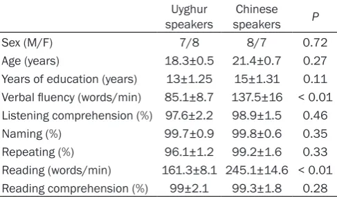
pari-Table 1. General information and language function test
of Uyghur and Chinese speakers Uyghur
speakers speakersChinese P
Sex (M/F) 7/8 8/7 0.72
Age (years) 18.3±0.5 21.4±0.7 0.27 Years of education (years) 13±1.25 15±1.31 0.11 Verbal fluency (words/min) 85.1±8.7 137.5±16 < 0.01 Listening comprehension (%) 97.6±2.2 98.9±1.5 0.46
Naming (%) 99.7±0.9 99.8±0.6 0.35
Repeating (%) 96.1±1.2 99.2±1.6 0.33 Reading (words/min) 161.3±8.1 245.1±14.6 < 0.01 Reading comprehension (%) 99±2.1 99.3±1.8 0.28
Verb generation task
[image:2.629.100.344.106.249.2]etal layer, and 279 frames were collected for each slice.
Data processing and statistical analysis
Image processing and analysis was conducted using the statistical parametric mapping (SPM) software (SPM5) (Welcome Department of Imaging Neuroscience; http://www.fil.ion.ucl. ac.uk). After the time alignment and head movement calibration, the data was standard-ized to the standard template of the Montreal Neurological Institute (MNI) using the SPM5 software. Each pixel was re-sampled to 3 mm × 3 mm × 3 mm. General linear model (GLM) was used to estimate the parameters for the image time series. SPM was obtained from the ran-dom effect analysis. Statistical tests were per-formed based on these parameters to obtain the specific t-value for each voxel. Then the map of activated parameters was superim-posed on the T1 template of the standard brains from MNI (avg152) to obtain a two-dimensional activation image. The SPM plugin xjview (http://www.nitrc.org/projects/xjview/) was used to obtain the spatial coordinates for each activated region, the corresponding
func-tion posifunc-tioning on the standard brains from MNI, and the size of activated voxels.
The SPSS17.0 software was used for the statis-tical analysis. After the normality test, two-sam-ple t-test was used for the data within the same group. For the image analysis with the SPM5 software, the influence of head movement (for individual analysis) and age, sex, and mean whole-brain signals (for group analysis) were removed. The statistical threshold was adjust-ed to P ≤ 0.05, with voxels (cluster size) ≥ 10 after false discovery rate (FDR) correction. P < 0.05 was considered statistically significant.
Results
General information and language function test of Uyghur and Chinese speakers
[image:3.629.103.529.94.382.2]The baseline characteristics of the Uyghur and Chinese speakers and the results from the lan-guage function test were shown in Table 1. Statistical analysis indicated significant differ-ences in the speech fluency and reading between these two groups (P < 0.05). However, due to the substantial different characteristics between the Uyghur and Chinese languages,
Table 2. Activated brain regions in Uyghur speakers during verb generation task
Differently activated brain regions Cluster p (cor) Cluster equivk Cluster p (unc) (FDR-cor) Voxel TVoxel p Voxel p (unc) {mm MNI}x, y, z Left IFG, pars triangularis/BA 48 0.006 25 0.114 0.000 7.31 0.000 57, 21, 12
Left IFG, pars opercularis/BA 44 0.000 5.62 0.000 54, 24, 33
BA 48 0.003 39 0.054 0.000 7.3 0.000 -51, -9, 24
Right precuneus/BA 7 0.006 26 0.108 0.000 6.47 0.001 -12, -60, 63
Right precuneus_ 0.000 5.75 0.000 -12, -57, 51
Right IFG, pars orbitalis/BA 47 0.006 24 0.121 0.000 6.25 0.000 -27, 36, -6 Right precentral gyrus 0.012 14 0.229 0.000 6.2 0.000 -42, -15, 60 Right IFG, pars opercularis 0.003 41 0.049 0.000 5.94 0.000 -54, 15, 27
Left calcarine 0.007 21 0.145 0.000 5.87 0.000 12, -84, 9
Right superior medial frontal gyrus 0.012 14 0.229 0.000 5.71 0.000 -12, 39, 45 Left middle frontal gyrus 0.01 17 0.187 0.000 5.61 0.000 27, 42, 15 Right middle frontal gyrus 0.016 10 0.308 0.000 5.52 0.000 -45, 6, 54
Right superior occipital gyrus 0.000 4.4 0.000 -30, -75, 18
Right superior frontal gyrus 0.006 25 0.114 0.000 5.15 0.000 -27, -9, 57
Right SMA 0.000 5.1 0.000 -12, -15, 60
Right SMA 0.000 4.84 0.000 -12, -18, 69
Right middle frontal gyrus 0.007 22 0.137 0.000 5.07 0.000 -48, 33, 24
Right IFG, pars triangularis 0.000 5.03 0.000 -45, 30, 9
Right inferior parietal gyrus 0.013 12 0.265 0.000 4.93 0.000 -45, -36, 51 Right IFG, pars triangularis/BA 45 0.016 10 0.308 0.000 4.78 0.000 -45, 21, 12
we supposed that these two indicators in the language function test were not comparable between the subject groups. Moreover, no sig-nificant differences were observed in the gen-der, age, years of education, and the rest indi-cators in the language function test between the Uyghur and Chinese speakers (all P > 0.05). These results suggest that these Uyghur and Chinese speakers are suitable for the following investigation.
Brain activation in Uyghur and Chinese speak-ers during verb generation task
The Uyghur and Chinese speakers were sub-jected to the verb generation task, and mean-while the activated brain regions in these sub-jects were detected with BOLD-fMRI. Our
results showed that, the Uyghur and Chinese speakers exhibited significant activation in mul-tiple brain regions during the verb generation task. For these right-handed subjects, the dis-tribution of the activated brain regions exhibit-ed an obvious tendency to the left-sidexhibit-edness. However, in the Uyghur speakers, the brain regions differentially activated during the verb generation task were mainly located in the local brain regions, including the left inferior tempo-ral gyrus (BA37), left inferior parietal lobule, left fusiform gyrus, and left parahippocampal gyrus, which were significantly less activated in the Chinese speakers (P < 0.05) (Tables 2, 4, 6
and Figure 1A-C). On the other hand, in the
[image:4.629.104.530.93.357.2]Chinese speakers, significantly differential acti-vation was observed in the right superior tem-poral gyrus (BA38) during the verb generation
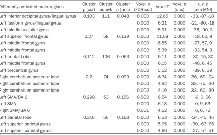
Table 3. Activated brain regions in Chinese speakers during verb generation task
Differently activated brain regions Cluster p (cor) Cluster equivk Cluster p (unc) (FDR-cor) Voxel TVoxel p Voxel p (unc) {mm MNI}x, y, z Left inferior occipital gyrus/lingual gyrus 0.103 111 0.048 0.000 12.65 0.000 -33, -87,-18 Left fusiform gyrus/lingual gyrus 0.000 6.21 0.000 -21, -90, -18
Left middle occipital gyrus 0.000 5.91 0.000 -36, -90, 3
Left superior frontal gyrus 0.27 58 0.139 0.000 11.08 0.000 -18, 60, 9
Left middle frontal gyrus 0.000 6.85 0.000 -27, 57, 9
Left middle frontal gyrus 0.000 5.39 0.000 -33, 54, 3
Left frontal Lobe 0.112 106 0.053 0.000 9.11 0.000 -30, 15, 30
Left middle frontal gyrus 0.000 6.15 0.000 -48, 6, 45
Left precentral gyrus 0.000 5.52 0.000 -39, 6, 39
Right cerebellum posterior lobe 0.2 74 0.099 0.000 6.74 0.000 36, -69, -24
Right cerebellum posterior lobe 0.000 4.82 0.000 33, -75, -30
Right cerebellum posterior lobe 0.002 4.19 0.000 33, -60, -30
Left SMA/BA 6 0.298 53 0.156 0.000 6.54 0.000 -9, 0, 66
Left SMA 0.000 6.18 0.000 0, 9, 63
Right SMA/BA 6 0.001 4.52 0.000 6, 6, 72
Left parietal lobe 0.316 50 0.168 0.000 6.53 0.000 -24, -45, 45
Left superior parietal gyrus 0.000 5.05 0.000 -30, -63, 66
Left superior parietal gyrus 0.000 4.66 0.000 -27, -57, 51
Statistical level: P < 0.05 (PDR correction) voxel > 50.
Table 4. Differentially activated brain regions in Uyghur speakers during verb generation task
Differentially activated brain regions
(Uyghur > Chinese) Cluster p (cor) Cluster equivk Cluster p (unc) (FDR-cor) Voxel TVoxel p Voxel p (unc) {mm MNI}x, y, z Left inferior parietal lobule 0.147 10 0.001 0.000 5.33 0.000 -54, -36, -24 Left inferior temporal gyrus (BA37) 0.098 11 0.001 0.001 4.59 0.000 -45, -39, 24 Left fusiform gyrus 0.098 11 0.001 0.001 3.83 0.000 -27, -30, -12
Left parahippocampal gyrus 0.001 3.65 0.001 -30, -21, -15
[image:4.629.94.531.403.480.2]task, whereas the Uyghur speakers exhibited weak activation in this region (P < 0.05) (Tables 3, 5, 6 and Figure 1D). Taken together, these results suggest that, differential brain activa-tion patterns could be observed in the Uyghur and Chinese speakers during the verb genera-tion task.
Discussion
In the present study, our results demonstrated that the Uyghur and Chinese speakers exhibit-ed significant activation in multiple brain regions during the verb generation task, which was in line with the previous findings [7]. For these right-handed subjects, the functional brain regions for language were mainly located in the left side, which was also consistent with the results from a recent study [8]. However, the activated brain regions during the verb gen-eration task were not identical for these Uyghur and Chinese speakers, which may be related to the different characteristics of the Uyghur and Chinese languages.
It has been shown that the formation of lan-guage regions in the brain is the result of long-term adaptable contention in the language environment [9], which is not only related to the brain neural layout, but also associated with the competition of brain regions in the lan-guage training. Lanlan-guage is a comprehensive capability of humans, involving listening, speak-ing, readspeak-ing, and writing. Therefore, the compe-tition of the brain regions must be more com-plex, and the multipolarity of language centers is an inevitable result. Our results
demonstrat-ed that the Uyghur speakers displaydemonstrat-ed signifi-cantly differential activation in the local brain regions, such as the left inferior temporal gyrus (BA37), left inferior parietal lobule, left fusiform gyrus, and left parahippocampal gyrus, where-as relatively weak activation in these regions was observed in the Chinese speakers. It has been shown that the temporal lobe region is related to the extraction of phonetic elements, and associated with the language comprehen-sion and generation [7]. Based on these find-ings, the brain region dealing with phonetics is likely to be located in the left superior temporal lobe, and the region dealing with semantics might be in the left middle temporal gyrus and inferior temporal gyrus [10]. The rear part of the left temporal region (BA37) may be the connection between the regions dealing with phonetics and semantics in word generation [11, 12].
[image:5.629.97.535.93.137.2]At present, it has been widely accepted that the left fusiform gyrus covers the visual word form area [13]. This brain region can be activated in lexical and semantic tasks, which is primarily related to the semantic processing in visual perception [14-16]. Moreover, the left inferior temporal gyrus, left fusiform gyrus, and left parahippocampal gyrus, as well as the right hemisphere mirror regions, have been shown to be related to semantic rendering and lexical retrieval [12, 17]. Our results showed that the left inferior parietal lobule was activated in the subjects during the verb generation task. This phenomenon might be explained that the verb generation task required in-depth semantic analysis of the noun, and comparison and
Table 5. Differentially activated brain regions in Chinese speakers during verb generation task
Differentially activated brain regions
(Chinese > Uyghur) cluster p (cor) cluster equivk p (unc)cluster (FDR-cor) voxel Tvoxel p voxel p (unc) {mm MNI}x, y, z Right superior temporal gyrus (BA38) 0.014 16 0.000 0.000 6.37 0.000 39, 15, -33
Statistical level: P < 0.05 (PDR correction) voxel ≥ 10.
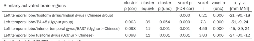
Table 6. Similarly activated brain regions in Uyghur and Chinese speakers during the verb generation
task
Similarly activated brain regions cluster p (cor) cluster equivk p (unc)cluster (FDR-cor)voxel p voxel T voxel p (unc) {mm MNI}x, y, z
Left temporal lobe/fusiform gyrus/lingual gyrus ( Chinese group) 0.000 6.21 0.000 -21, -90, -18 Left temporal lobe/BA 48 (Uyghur group) 0.003 39 0.054 0.000 7.3 0.000 -51, -9, 24 Left temporal lobe/inferior temporal gyrus/BA37 (Uyghur > Chinese) 0.098 11 0.001 0.001 4.59 0.000 -45, -39, 24 Left temporal lobe fusiform gyrus (Uyghur > Chinese) 0.098 11 0.001 0.001 3.83 0.000 -27, -30, -12
[image:5.629.95.534.193.264.2]selection of related verbs. Further studies are still needed to elucidate the detailed mechanisms.
In this study, our results showed that the Chinese speakers displayed significantly differ-ential activation in the right superior temporal gyrus (BA38), whereas the Uyghur speakers displayed weak activation in this region. In line with this, Tan et al. [4] have found that the word association task activates the right BA38 region, suggesting that the right hemisphere is more intensively involved in the processing of
Chinese characters. The right hemisphere has the functions of periodic memory and visual-spatial detection [18-22], which are needed in the reading of Chinese characters constituted by a variety of strokes. The recognition of Chinese characters not only activates the visu-al-spelling system, but also involves the pho-netic and semantic processing [23-28]. The left hemisphere is usually involved in logical analy-sis, as well as phonetic and semantic process-ing. Tan et al. [29] have found that the left mid-dle frontal gyrus is involved in the visuospatial processing of Chinese characters, and the
[image:6.629.101.530.84.492.2]gration of phonetic and semantic analysis. Moreover, it has been demonstrated that the upper temporal lobe, particularly the left supe-rior temporal gyrus, is related to the extraction of phonetic features, and the right superior temporal gyrus may be associated with the rec-ognition of the unique tone and pitch of Chinese characters [30]. Taken together, the processing of Chinese characters is actually associated with a wide range of neural activity in the cere-bral cortex.
In conclusion, our results showed that, the dominant hemisphere for these right-handed Uyghur and Chinese speakers is the left cere-bral hemisphere. However, in the Uyghur speak-ers, the brain regions differentially activated during the verb generation task were mainly located in the local brain regions, including the left inferior temporal gyrus (BA37), left inferior parietal lobule, left fusiform gyrus, and left parahippocampal gyrus, while in the Chinese speakers, significantly differential activation was observed in the right superior temporal gyrus (BA38). Differential brain activation pat-terns were observed for the Uyghur and Chinese speakers during the verb generation task. Compared with the Uyghur language, process-ing of Chinese characters may involve the right hemisphere more extensively. In China, there is a Uyghur population of nearly ten million living in Xinjiang. This study might contribute to the investigation of the dominant language hemi-sphere and functional language regions in Uyghur native speakers, and the understanding of the universality and particularity of language processing mechanism in the brain. Moreover, our findings provide valuable information for the evaluation and treatment of language dys-function after brain injuries in clinic.
Acknowledgements
This study was supported by the Natio- nal Natural Science Foundation of China (81260181) and the Natural Science Found- ation of the Xinjiang Uygur Autonomous Region (2010211B18).
Disclosure of conflict of interest
None.
Address correspondence to: Baolan Wang, De- partment of Rehabilitation Medicine, The First
Affiliation Hospital of Xinjiang Medical University, No. 392, Liyushan Road, Urumqi 830001, Xinjiang, China. Tel: 86-0991 4366 055; Fax: 86-0991 4366 055; E-mail: [email protected]
References
[1] Xi Y, Yang J, Abudusalamu R, Kaheman K and Wang B. The standardization of a Uighur Apha-sia battery. Chinese Journal of Physical Medi-cine and Rehabilitation 2015; 37: 509-512. [2] Xi Y, Jiang C, Zhang J, Kaheman K and Zhang X.
The cortical organization in language tasks of Mandarin and Uyghur speakers. Chinese Jour-nal of Physical Medicine and Rehabilitation 2013; 35: 847-851.
[3] Xi Y, Zhou J, Abudusalamu R, Kaheman K and Wang B. Reliability of aphasia battery of Ui-ghur. Chinese Journal of Rehabilitation 2015; 30: 90-93.
[4] Tan LH, Spinks JA, Gao JH, Liu HL, Perfetti CA, Xiong J, Stofer KA, Pu Y, Liu Y and Fox PT. Brain activation in the processing of Chinese charac-ters and words: a functional MRI study. Hum Brain Mapp 2000; 10: 16-27.
[5] Xiong X, Du P, Yang L, Li Q and Wand W. Study on functional MRI of two language tasks in nor-mal subjects. Medical Journal of National De-fense Forces in Southwest China 2009; 19: 561-563.
[6] Vaid J and Hull R. Re-envisioning the bilingual brain using functional neuroimaging: Method-ological and interpretive issues. Advances in the Neurolinguistic Study of Bilingualism 2002; 315-355.
[7] Indefrey P and Levelt WJ. The spatial and tem-poral signatures of word production compo-nents. Cognition 2004; 92: 101-144.
[8] Wu H, Qiu W, Kang Z, Xie C, Wan G, Yang Q and Chen S. Blood oxygen level and the pathogenic mechanism of expressive aphasia after stroke. Chinese Journal of Physical Medicine and Re-habilitation 2014; 36: 407-412.
[9] Zhao X. Magnetic Resonance Imaging. Beijing: Science Publishing House 2004.
[10] Del Prato P and Pylkkanen L. MEG evidence for conceptual combination but not numeral quantification in the left anterior temporal lobe during language production. Front Psychol 2014; 5: 524.
[11] Martin RC. Language processing: functional organization and neuroanatomical basis. Annu Rev Psychol 2003; 54: 55-89.
[13] Caspers J, Zilles K, Amunts K, Laird AR, Fox PT and Eickhoff SB. Functional characterization and differential coactivation patterns of two cytoarchitectonic visual areas on the human posterior fusiform gyrus. Hum Brain Mapp 2014; 35: 2754-2767.
[14] Luders H, Lesser RP, Hahn J, Dinner DS, Morris HH, Wyllie E and Godoy J. Basal temporal lan-guage area. Brain 1991; 114: 743-754. [15] Horwitz B, Rumsey JM and Donohue BC.
Func-tional connectivity of the angular gyrus in nor-mal reading and dyslexia. Proc Natl Acad Sci U S A 1998; 95: 8939-8944.
[16] Murtha S, Chertkow H, Beauregard M and Ev-ans A. The neural substrate of picture naming. J Cogn Neurosci 1999; 11: 399-423.
[17] Burnstine TH, Lesser RP, Hart J Jr, Uematsu S, Zinreich SJ, Krauss GL, Fisher RS, Vining EP and Gordon B. Characterization of the basal temporal language area in patients with left temporal lobe epilepsy. Neurology 1990; 40: 966-970.
[18] Bryden M. Laterality functional asymmetry in the intact brain. Elsevier; 2012.
[19] Ellis AW, Young AW and Anderson C. Modes of word recognition in the left and right cerebral hemispheres. Brain Lang 1988; 35: 254-273. [20] Jonides J, Smith EE, Koeppe RA, Awh E,
Minoshima S and Mintun MA. Spatial working memory in humans as revealed by PET. Nature 1993; 363: 623-625.
[21] Kosslyn SM, Alpert NM, Thompson WL, Maljkovic V, Weise SB, Chabris CF, Hamilton SE, Rauch SL and Buonanno FS. Visual Mental Imagery Activates Topographically Organized Visual Cortex: PET Investigations. J Cogn Neu-rosci 1993; 5: 263-287.
[22] McCarthy G, Blamire AM, Puce A, Nobre AC, Bloch G, Hyder F, Goldman-Rakic P and Shul-man RG. Functional magnetic resonance imag-ing of human prefrontal cortex activation dur-ing a spatial workdur-ing memory task. Proc Natl Acad Sci U S A 1994; 91: 8690-8694.
[23] Chua FK. Phonological recoding in Chinese logograph recognition. Journal of Experimental Psychology: Learning, Memory, and Cognition 1999; 25: 876.
[24] Perfetti CA and Zhang S. Very early phonologi-cal activation in Chinese reading. Journal of Experimental Psychology: Learning, Memory, and Cognition 1995; 21: 24.
[25] Pollatsek A, Tan LH and Rayner K. The role of phonological codes in integrating information across saccadic eye movements in Chinese character identification. J Exp Psychol Hum Percept Perform 2000; 26: 607-633.
[26] Tan LH, Hoosain R and Siok WW. Activation of phonological codes before access to character meaning in written Chinese. Journal of Experi-mental Psychology: Learning, Memory, and Cognition 1996; 22: 865.
[27] Weekes BS, Chen MJ and Lin YB. Differential effects of phonological priming on Chinese character recognition. Reading and Writing 1998; 10: 201-221.
[28] Ziegler JC, Tan LH, Perry C and Montant M. Phonology matters: The phonological frequen-cy effect in written Chinese. Psychol Sci 2000; 11: 234-238.
[29] Tan XJ, Ma LF, Yu W, Zhang ZQ, Wang XY and Weng XC. Frequency effect in reading aloud ir-regular Chinese characters: an fMRI study. Chi-nese Journal of Medical Imaging Technology 2004; 11.