Original Article
Metformin suppresses gastric tumorigenesis
by regulating chemokine CXCR4
Weiqiang Wu1, Heng Xi2, Feng Gao1, Feng Song1, Yong Zhao1, Ming Xu1, Xin Zhang1, Zengqiang Yang1
1Department of Colorectal Surgery, Lanzhou General Hospital of Lanzhou Command PLA, Lanzhou, China; 2Department of Pharmacy, The Third People’s Hospital of Chengdu, Chengdu, China
Received February 24, 2016; Accepted June 4, 2016; Epub November 15, 2016; Published November 30, 2016
Abstract: Objective: To investigate the role of metformin in the treatment of gastric cancer. Methods: Invitro, fol-lowing metformin exposure, the proliferation of MFC cells was determined using MTT assay, and the mRNA and protein levels of CXCR4 were detected by real-time quantitative PCR and western blot, respectively. In vivo, thirty-six mice were randomly divided into two groups, the experimental group and the control group (n = 18), followed by i.p. injection of gastric cancer cells. The experimental group was treated with metformin (10 mg/kg), while the control group was injected i.v. with saline. The tumor volume was measured. Results: Results from real-time quantitative PCR and western blot showed that both CXCR4 mRNA and protein levels were significantly suppressed by metfor-min, which resulted in reduced gastric cancer cell viability in vitro. Moreover, metformin inhibited tumor growth in vivo. Conclusion: Our findings suggest that metformin inhibits gastric tumor cell growth via decreasing CXCR4 both
in vitro and in vivo.
Keywords: Metformin, gastric cancer, CXCR4
Introduction
Gastric cancer is now the second-leading cause of cancer-related deaths worldwide, and the prognosis of advanced gastric cancer is poor [1, 2]. Apart from potentially curative surgery, chemotherapy and radiochemotherapy may be applied at advanced stages of gastric can-cer but neither of these can be curative [3, 4]. Thus, there is a strong demand for new effec-tive approaches to advanced gastric cancer. Metformin is a biguanide family member com-monly used in the treatment of type 2 diabetes. It increases liver and peripheral tissue sensi- tivity to insulin and reduces hepatic glucose production [5]. According to a recent epidemio-logic survey, metformin inhibits tumorigenesis. However, the molecular mechanism underlying the suppression of cancer growth by metformin remains relatively unknown.
Recent data suggest that certain chemokines and their receptors, in particular stromal cell-derived factor (SDF)-1 and CXC chemokine re- ceptor 4 (CXCR4), play an important role in the behavior of cancer cells and modulate cell mi-
gration, proliferation, and survival. It has been shown that neutralizing the interaction of SDF- 1 and CXCR4 significantly impairs the metas- tasis of breast cancer cells to regional lymph nodes and lung in vivo, suggesting that chemo-kines and their receptors have a critical role in determining the metastatic destination of tumor cells [6].
Here, we showed that metformin inhibits gas-tric cancer cell growth by decreasing CXCR4 expression.
Materials and methods
Animals
Cell line
Mouse gastric cancer cells MFC were obtain- ed from Chinese Academy of Sciences Typical Culture Preservation Commission Cell Bank (Shanghai, China). MFC cells were routinely cultured in RPMI-1640 (GIBCO) supplemented with 10% fetal bovine serum (Hyclone Labo- ratories) at 37°C in an incubator with 5% CO2. Culture medium was changed every 1-2 days.
Real-time PCR
Total RNA was isolated from MFC cells using Trizol reagent (Invitrogen), and was reverse-transcribed to cDNA. The forward primer of CXCR4 gene was 5’-GCCTGAGCTACAGATGCCC- A-3’, and the reversed primer was 5’-TTCGGG- TCAATGCACTTGT-3’. The glyceraldehydes phos-phate dehydrogenase (GAPDH) gene was used as internal reference, and the primers were: 5’-GAAGGTGAAGGTCGGAGTC-3’ and 5’-GAAGA- TGGTGATGGGATTTC-3’. The real time-PCR (RT- PCR) was performed on ABI Prism 7700 PCR instrument (Applera Corporation Norwalk). The amplification conditions were 95°C for 5 min; 35 cycles of 94°C for 1 min, 60°C for 1 min, and 72°C for 1 min; and a terminal extension step at 72°C for 10 min. After the reaction, 5 μL DNA was analyzed by agarose gel electrophore-sis, and the gel image was scanned and the gray scale was measured. The ratio of CXCR4 and GAPDH was expressed as their relative expression.
Western blotting
The protein was extracted from MFC cells, and the protein concentration was measured using
Brandford method. Samples were separated using SDS-polyacrylamide gel electrophoresis (SDS-PAGE), and transferred to polyvinylidene difluoride (PVDF) membrane. After blocking with 5% defatted milk, the membrane was incu-bated with mouse CXCR4 monoclonal anti-body (1:1000) at 4°C overnight, followed by incubation with HRP-labeled goat anti-mouse IgG (1:5000) at 37°C for 45 min. Protein bands were visualized with ECL development. The images were analyzed by measuring the gray scale, and the ratio of CXCR4 and GAPDH was expressed as their relative protein expression.
MTT assay
Cell growth inhibition was determined by mo- dified microculture tetrazolium (MTT) assay based on the enzymatic reduction of 3-(4, 5-dimethylthiazol-2-yl)-2,5-diphenyl tetrazolium bromide (Sigma) to form formazan crystal by mitochondria and cellular dehydrogenase en- zymes. The OD value (A) was measured at a wavelength of 570 nm on the Microplate Reader EXL2800. Inhibition rate (%) = control group A-experimental group A/control group A × 100%.
In vivo assay
The 615 inbreeding mice were randomly as- signed to two groups (n = 18 per group), the experimental group and the control group. MFC cells in logarithm growth phrase were harvested and washed twice with PBS. A total of 5.0 × 10⁶ cells in 200 μL serum-free me- dium with a viability of > 95% tested by stain- ing with try-pan blue were injected into the back of each mouse in these two groups. The mice were observed every two days, and were sacrificed 28 days post-inoculation.
Statistic analysis
The data were expressed as means ± standard deviation (_x±s). Statistical comparisons were performed by student’s t-test. P < 0.05 was considered statistically significant.
Results
Metformin inhibits gastric cancer cell prolifera-tion
[image:2.612.93.280.74.214.2]The effect of metformin on gastric cancer cell growth was determined using MTT assay. Sig- Figure 1. Metformin inhibits MFC cell proliferation.
nificant time and dose-dependent growth inhi-bition was observed in gastric cancer cells. The result of MTT test showed that metformin sig-nificantly suppressed cell proliferation of MFC cells (P < 0.01) (Figure 1).
Metformin inhibits CXCR4 expression
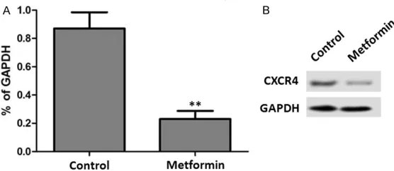
The RT-PCR result showed that metformin ex- posure resulted in a significant reduction of CXCR4 mRNA levels in MFC cells (P < 0.05) (Figure 2A). In agreement with this result, we- stern bolt analysis showed that the CXCR4 protein expression was also significantly de- creased after metformin treatment (P < 0.05) (Figure 2B).
Metformin inhibits the tumor growth and pro-longs the survival time
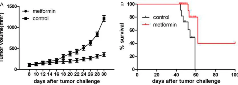
To further examine the effect of metformin on gastric tumor growth in vivo, MFC cells were injected subcutaneously to the back of inbre- eding mice. After inoculation for 7 days, the experimental group (n = 18) was injected i.v. with 10 mg/kg metformin, while the control group (n = 18) received an injection of sa- line. The tumor volume was significantly decre- ased after metformin exposure, as compared to the control group. In contrast, the survival time was significantly increased in the metfor-min-treated group, compared with the control group (P < 0.05) (Figure 3A and 3B).
Discussion
In this study, we demonstrated that metformin reduced the gastric cancer cell proliferation and tumor growth by inhibition of CXCR4 gene and protein expression.
[image:3.612.93.371.72.193.2]suppressive effects on cancer growth: (1) acti-vation of LKB1/AMPK pathway; (2) induction of cell cycle arrest and/or apoptosis; (3) inhibi-tion of protein synthesis; (4) reducinhibi-tion in circu-lating insulin levels; (5) inhibition of the un- folded protein response (UPR); (6) activation of the immune system; and (7) eradication of cancer stem cells [9]. Earlier studies showed that phenformin, a biguanide that was only briefly used in humans secondary to its in- creased propensity to cause lactic acidosis, could result in better inhibition of tumor cell growth, when added to conventional chemo-therapeutic agents [10-12]. Epithelial tumor cells exploit several mechanisms including che-mokine systems that normally regulate leuko-cyte trafficking and homing. The distinct pat-tern of chemokine receptor expression by tu- mor cells plays a critical role in tumor biology. In particular, CXCR4 expression is associated with more aggressive behaviors of various tu- mor cells [13-15]. CXCR4 is widely expressed on hematopoietic cells including CD34+ HSC, T-lymphocytes, B-lymphocytes, monocytes and macrophages, neutrophils and eosinophils as well as by brain, lung, colon, heart, kidney, and liver, and endothelial and epithelial cells, mi- croglia, astrocytes and neuronal cells, and pro-genitor cells including endothelial and smoo- th muscle progenitors. Functional CXCR4 is ex- pressed on embryonic pluripotent stem cells and several types of tissue-committed stem cells, for example, neural tissue, skeletal mus-cles, heart, liver, endothelium, and renal tu- bular- and retina pigment-epithelium [16]. The expression of CXCR4 on malignant epithelial cells and on cells from several hematopoietic malignancies implies that the CXCL12/CXCR4 Figure 2. Metformin inhibits CXCR4 expression. A. The effect of metformin on
CXCR4 mRNA expression. CXCR4 mRNA levels were determined by real-time quantitative PCR. B. The effect of metformin on CXCR4 protein expression. CXCR4 protein levels were determined by western blot. Control, saline.
pathway may influence cancer biology and play a pivotal role in directing the metastasis of CXCR4+ tumor cells to organs that expre- ss CXCL12 (e.g., lymph nodes, lungs, liver, or bones). Several CXCR4+ cancers metastasize to the bones and lymph nodes in a CXCL12-dependent manner, in which the bone marrow provides a protective environment for tumor cells [17]. Therefore, we investigated the ex- pression and function of CXCR4 in gastric can-cer cells, and attempted to correlate clini- copathological factors of gastric cancer with CXCR4 expression.
We found that MFC cells contained CXCR4 tran-scripts and cytosolic CXCR4 protein, but did not express membrane CXCR4. CXCR4 plays an important role in the spread and progression of sarcomas, neuroblastomas, and melanomas, and cancers of the breast, ovary, bladder and cervix [18, 19]. Genetic lesions and aberrant signaling networks within cancer cells have been the main focus in cancer biology over the past decades. As a consequence of this onco-gene- and tumor suppressor-centric view, most current cancer therapies target the tumor cells, which frequently acquire resistance owing to their inherent genomic instability. Signals from the tumor microenvironment may make pivotal contributions to the progression of hemato- poietic and epithelial malignancies, thus incre- asing emphasis is being placed on targeting the tumor cell microenvironment. The CXCR4-CXCL12 axis plays a central role in the spread and progression of many different types of tumors [20, 21]. Therefore, the CXCR4-CXCL12 axis may be involved in the role of metformin in
the treatment of gastric cancer. However, more evidence should be explored.
In the present study, we showed that metformin inhibits the gastric cancer cell proliferation and tumor growth, and prolongs the survival time by downregulating CXCR4 expression.
Acknowledgements
This study was supported by 2013 Army Medical Science and Technology Projects (No. CLZ13JB03), 2013 Natural Science Foundation of Gansu Province (No. 1308RJYA045), and 2014 National Medical Special Fund Project (No. L2014073).
Disclosure of conflict of interest
None.
Address correspondence to: Dr. Weiqiang Wu, De- partment of Colorectal Surgery, Lanzhou General Hospital of Lanzhou Command, PLA, 333 Nanbin Road, Qilihe District, Lanzhou 730050, China. Tel: +86-15117202099; E-mail: weiqiangewu@126.com
References
[1] Smith JK, McPhee JT, Hill JS, Whalen GF, Sullivan ME, Litwin DE, Anderson FA and Tseng JF. National outcomes after gastric resection for neoplasm. Arch Surg 2007; 142: 387-393. [2] Akhavan-Niaki H and Samadani AA. Molecular
Insight in Gastric Cancer Induction: An Overview of Cancer Stemness Genes. Cell Biochem Biophys 2014; 68: 463-473.
[image:4.612.99.516.73.222.2][3] Lordick F and Siewert JR. Recent advances in multimodal treatment for gastric cancer: a re-view. Gastric Cancer 2005; 8: 78-85.
[4] Oh DY and Bang YJ. Adjuvant and neoadjuvant therapy for gastric cancer. Curr Treat Options Oncol 2013; 14: 311-320.
[5] Wiernsperger NF and Bailey CJ. The antihyper-glycaemic effect of metformin: therapeutic and cellular mechanisms. Drugs 1999; 58 Suppl 1: 31-39.
[6] Muller A, Homey B, Soto H, Ge N, Catron D, Buchanan ME, McClanahan T, Murphy E, Yuan W, Wagner SN, Barrera JL, Mohar A, Verastegui E and Zlotnik A. Involvement of chemokine receptors in breast cancer me- tastasis. Nature 2001; 410: 50-56.
[7] Effect of intensive blood-glucose control with metformin on complications in overweight pa-tients with type 2 diabetes (UKPDS 34). UK Prospective Diabetes Study (UKPDS) Group. Lancet 1998; 352: 854-865.
[8] Bost F, Sahra IB, Le Marchand-Brustel Y and Tanti JF. Metformin and cancer therapy. Curr Opin Oncol 2012; 24: 103-108.
[9] Kourelis TV and Siegel RD. Metformin and can-cer: new applications for an old drug. Med Oncol 2012; 29: 1314-1327.
[10] Shikhman AR, Lebedev K and Dil’man VM. Inhibiting effect of phenformin (phenethyl big-uanide) on the growth of Ehrlich carcinoma. Vopr Onkol 1981; 27: 67-69.
[11] Cohen MH and Strauss B. Enhancement of the antitumor effect of 1, 3-bis(2-chloroethyl)-l-ni-trosourea (BCNU) by phenylethylbiguanide (phenformin). Oncology 1976; 33: 257-259. [12] Dilman VM, Berstein L, Ostroumova MN, Fe-
dorov SN, Poroshina TE, Tsyrlina EV, Buslaeva VP, Semiglazov VF, Seleznev IK, Bobrov YuF, Vasilyeva IA, Kondratjev VB, Nemirovsky VS and Nikiforov YF. Metabolic immunodepres-sion and metabolic immunotherapy: an at-tempt of improvement in immunologic re-sponse in breast cancer patients by correction of metabolic disturbances. Oncology 1982; 39: 9-13.
[13] Burger JA and Kipps TJ. CXCR4: a key receptor in the crosstalk between tumor cells and their microenvironment. Blood 2006; 107: 1761-1767.
[14] Kucia M, Jankowski K, Reca R, Wysoczynski M, Bandura L, Allendorf DJ, Zhang J, Ratajczak J and Ratajczak MZ. CXCR4-SDF-1 signalling, lo-comotion, chemotaxis and adhesion. J Mol Histol 2004; 35: 233-245.
[15] Lee HJ, Kim SW, Kim HY, Li S, Yun HJ, Song KS, Kim S and Jo DY. Chemokine receptor CXCR4 expression, function, and clinical implications in gastric cancer. Int J Oncol 2009; 34: 473-480.
[16] Ratajczak MZ, Zuba-Surma E, Kucia M, Reca R, Wojakowski W and Ratajczak J. The plieo-tropic effects of the SDF-1-CXCR4 axis in or-ganogenesis, regeneration and tumorigenesis. Leukemia 2006; 20: 1915-1924.
[17] Meads MB, Hazlehurst LA and Dalton WS. The bone marrow microenvironment as a tu-mor sanctuary and contributor to drug resis-tance. Clin Cancer Res 2008; 14: 2519-2526. [18] Furusato B, Mohamed A, Uhlén M and Rhim
JS. CXCR4 and cancer. Pathol Int 2010; 60: 497-505.
[19] Cabioglu N, Sahin A, Doucet M, Yavuz E, Igci A, O Yildirim E, Aktas E, Bilgic S, Kiran B, Deniz G and Price JE. Chemokine receptor CXCR4 ex-pression in breast cancer as a potential predic-tive marker of isolated tumor cells in bone marrow. Clin Exp Metastasis 2005; 22: 39-46. [20] Domanska UM, Kruizinga RC, Nagengast WB,
Timmer-Bosscha H, Huls G, de Vries EG and Walenkamp AM. A review on CXCR4/CXCL12 axis in oncology: no place to hide. Eur J Cancer 2013; 49: 219-230.