Original Article
Molecular subtyping of breast cancer patients with long
time follow up and its prognostic value on survival: a
single center analysis
Nuket Ozkavruk Eliyatkin1, Safiye Aktas2, Baha Zengel3, Hakan Postaci4, Adam Uslu3, Ayse Yagci4
1Department of Pathology, Faculty of Medicine, Adnan Menderes University, Aydin, Turkey; 2Institute of Oncology,
Dokuz Eylul University, Basic Oncology, Izmir, Turkey; Departments of 3General Surgery, 4Pathology, Turkish
Ministry of Health-Izmir Bozyaka Research and Training Hospital, Izmir, Turkey
Received January 24, 2016; Accepted May 4, 2016; Epub June 15, 2016; Published June 30, 2016
Abstract: The importance of molecular subtyping in breast cancer is an unresolved issue. In this study we aimed to evaluate the significance of molecular subtyping, and the correlation between the disease-free, and overall survival in breast cancer based on molecular subtypes. A total of 536 patients with the diagnosis of breast cancer between the years 1980 and 2014 were included in the study. Tumors were divided into five molecular subtypes accord-ing to their expression profiles as follows: Luminal A: (n=220; 41%); Luminal B: (n=72; 13.4%); Luminal B-like: (n=97, 18.1%); HER2: (n=44;8.2%); and Triple-negative (n=103;19.2%). We found significant differences between molecular subtypes, and histological subtype of the tumor (P=0.004) in terms of local recurrence (P=0.043), and metastasis (P=0.006). A statistically significant difference was found between the number of metastases, and mo-lecular subgroups. (P=0.037). Among all momo-lecular subtypes, local recurrences (11.4%), and metastasis (38.6%) were most frequently seen in the HER2 subtype, while the least number of metastases (15.3%) were detected in the Luminal A subtype. A statistically significant difference was found between Luminal A, and HER2 subgroups as for incidence of metastatic lesions (P=0.007). However in the Luminal A subgroup metastases developed in the long term (at the end of 50 months after onset of the disease). Overall, and disease-free survival curves in the Luminal A subgroup indicated risk of mortality in the long run. Based on molecular subtyping the worst, and the most favour-able survival rates were observed in the HER2, and Luminal A subgroups, respectively. Impact: In this study which encompassed multiple number of breast cancer patients encountered within 30 years, HER2 tumors had the worst survival rates Interestingly, Luminal A subgroup which displayed a very favourable prognosis during the early stage of the follow-up period, demonstrated a bad prognosis in the long term.
Keywords: Breast cancer, molecular subtype, prognosis, survival
Introduction
Breast cancer is one of the most prevalently seen malignant tumors among women in the world. Efficient use of routine screening meth-ods, and imaging modalities, increase in the opportunities of early diagnosis, common, and appropriate use of treatment protocols, discov-ery of target treatment facilities have decreased mortality rates. Still an increase in the inci-dence of breast cancer is seen.
Breast cancer is known to demonstrate varia-tions in many ways including its clinical pre- sentation, biological behaviour, and treatment response. Traditional histological classification
fails to meet this multifaceted heterogeneity. Therefore nowadays, breast cancer researches have changed from histological to molecular classification. In the year 2000, a Californian group of researchers suggested molecular clas-sification system based on gene expression pattern in breast cancer which defined four subgroups [1]. In more recent studies per-formed by different breast cancer study groups, these defined groups were confirmed, and received global acceptance.
the year 2013, criteria used to define these five different subgroups were defined clearly. Based on these criteria the subgroups were classified as follows: 1- Luminal A (ER positive, HER2 neg-ative, lower Ki67 proliferation index, and higher PR positivity), 2- Luminal B (HER2 negative): ER positive, HER2 negative, and higher Ki67 pro- liferation index or lower PR, 3- Luminal B-like (HER2 positive): ER positive, HER2 positive (overexpressed or amplified), Ki67, and PR at any state, 4- HER2 positive: HER2 overex-pressed or amplified, ER and PR negative, 5- Triple-negative: ER, PR, and HER2 negative. These molecular groups correlate with bio-chemical biomarkers. In new studies, differ-ences in response to treatment, and course of the disease have been demonstrated.
In this study we aimed to investigate whether molecular subgrouping in breast cancer is superior over conventional histopathological evaluation. We also intended to evaluate the correlation between predefined molecular sub-groups of breast cancer as for event-free sur-vival (EFS), and overall sursur-vival (OS). A cohort containing 536 breast cancer patient groups with a survival time of 30 years was re-classi-fied according to molecular subtypes. In this subtyping, widely used biomarkers as oestro-gen receptor (ER), progesterone receptor (PgR), HER2, and Ki67 were utilized.
Material and method
The patients treated, and followed-up with the diagnosis of breast cancer between 1980, and 2014 years in Izmir-Bozyaka Training and Re- search Hospital were included in the study. The patients were treated with modifed radical mastectomy, lumpectomy, breast-sparing sur-gery, and one of the surgical interventions rec-ommended within the years where cases of cancer were detected. Later on clinicopatho-logical evaluation was made, then in line with the decisions of the council, chemotherapy, radiotherapy, and the appropriate treatment selected among target treatment programs valid during the years of diagnosis were applied. Electronic database related to the the time period extending from 1980 up to September 2014 contained information about a total of 768 patients. The patients whose hormone receptors (n=54), and cerbB2 protein (n=66) expressions, and Ki67 proliferation indices
(n=112) were not evaluated or recorded were excluded from the study leaving 536 patients as study participants.
Histopathological, immunohistochemical evaluations
Histopathological, and immunohistopathologi-cal diagnoses were based on the assessments of pathologists working in the pathology labora-tory, and a senior pathologist was consulted for each pathology slide. Clinical, and histopatho-logical data were retrieved from archival files. Local recurrence was accepted as any localized lesion in the breast tissue, mastectomy scar, ipsilateral axillary lymph nodes. Distant metas-tasis was defined as any localized lesion other than breast tissue, mastectomy scar, ipsilateral axillary and/or supraclavicular lymph nodes. Cases with bilateral breast cancers were included in the study. A time interval of 3 months was accepted as a criterion used to make a differentiation between metachronous, and synchronous bilateral breast cancers. ER, and PgR positivity were determined using immunohistochemical. assessments, and all data were retrieved, and recorded from elec-tronic media screening. Staining of more than 1% of tumor cells was accepted as ER or PgR positivity. Ki-67 proliferation index was also determined based on the results of immunohis-tochemical evaluation. Nuclear staining detect-ed in more than 15% of tumor cells was accept-ed as a criterion of positivity. HER2-receptor state was determined based on immunohisto-chemically evaluated archival data. IHC 2+ tumor specimens were also evaluated using florescence in-situ hybridization (FISH) method with signal amplification. Apart from IHC-negative (score 0, and 1) or positive (score 3) cases, HER2-receptor state of each case was determined based on the FISH results.
Statistical analysis
Continuous data were expressed as means ± standard deviations (SD) and categorical data as frequencies, counts and percentages. One-Way ANOVA test was used to evaluate
[image:3.612.93.520.86.613.2]differ-ences between molecular subtypes. P values were considered significant if less than 0.05. Event free survival (EFS) was considered as the time interval between the time of the diag-nosis, and detection of local recurrence and/or metastasis. Overall survival (OS) was estimated
Table 1. Descriptive statics for the 536 breast cancer cases
Luminal A Luminal B Luminal B-like HER2 Triple negative Total Number (%) 220 (41) 72 (13.4) 97 (18.1) 44 (8.2) 103 (19.2) 536 Mean age (SD) 55 (13.4) 51.9 (13.4) 52.8 (12.2) 51.9 (10.7) 49.9 (12.8) 53.3 (13) Menopausal status
Premenopausal 72 (32.9) 34 (47.2) 38 (39.6) 20 (45.5) 57 (55.3) 221 (41.2) Postmenopausal 145 (66.2) 38 (52.8) 57 (59.4) 23 (52.3) 45 (43.7) 308 (57.7) Laterality
Unilaterality 203 (92.3) 69 (95.8) 88 (90.7) 41 (93.2) 99 (96.1) 500 (93.3)
Bilaterality 17 (7.7) 3 (4.2) 41 (93.2) 3 (6.8) 4 (39.0) 36 (6.7) Tumor type
IDC 157 (72) 58 (80.6) 75 (77.3) 39 (88.6) 61 (59.8) 390 (73.2)
ILC 30 (13.8) 8 (11.1) 7 (7.2) 1 (2.3) 15 (14.7) 61 (11.4)
IDC+ILC 14 (6.4) 3 (4.2) 9 (9.3) 2 (4.5) 8 (7.8) 36 (6.8)
Mucinous 11 (5.0) 1 (1.4) 3 (3.1) - 1 (1.0) 16 (3)
Medullary - - 1 (1.0) - 9 (8.8) 10 (1.9)
Tubular 3 (1.4) - - - 1 (1) 4 (0.7)
Cribriform 1 (0.5) 1 (1.4) - - - 2 (0.4)
Papillary 1 (1.4) - - 1 (1) 2 (0.4)
Other 2 (0.9) - 2 (2.1) 2 (4.5) 6 (5.9) 12 (2.3)
Tumor sizea n (%)
T1 67 (30.5) 15 (20.8) 20 (20.6) 9 (20.5) 29 (28.2) 140 (26.2)
T2 103 (46.8) 37 (51.4) 58 (59.8) 21 (47.7) 52 (50.5) 271 (50.7)
T3 20 (9.1) 10 (13.9) 6 (6.2) 7 (15.9) 8 (7.8) 51 (9.5)
T4 19 (8.6) 5 (6.9) 6 (6.2) 3 (6.8) 5 (4.9) 38 (7.1) LN statusa
N0 86 (39.3) 23 (31.9) 33 (34.0) 14 (31.8) 48 (46.6) 204 (38.1)
N1 56 (26.6) 15 (20.8) 25 (25.8) 9 (20.5) 24 (23.3) 129 (24.1)
N2 41 (18.7 23 (31.9) 20 (20.6) 9 (20.5) 12 (11.7) 105 (19.6) N3 26 (4.6) 11 (15.3 16 (16.5) 11 (2.5) 15 (14.6) 79 (14.8) Stageb
I 46 (21.0) 5 (6.9) 12 (12.4) 3 (6.8) 20 (19.4) 86 (16.1)
II 88 (40.2) 26 (36.1) 41 (42.3) 18 (40.9) 45 (43.7) 218 (40.7) III 69 (31.5) 35 (48.6) 39 (40.2) 21 (47.7) 28 (27.2) 192 (35.9) IV 8 (3.7) 2 (2.8) 1 (1.0) - 1 (1.0) 12 (2.2) Unknown 8 (3.7) 4 (5.6) 4 (4.1) 2 (4.5) 8 (3.7) 27 (5.0) Local nux 5 (2.3) 1 (1.4) 5 (5.2) 5(11.4) 4 (3.9) 20 (3.7) Metastasis 33 (15.3) 13 (18.3) 25 (25.8) 17 (38.6) 20 (19.6) 108 (20.1) Metastasis type
Single 27 (12.5) 8 (11.3) 20 (20.8) 9 (20.5) 16 (15.5) 80 (14.9)
Multipl 15 (6.9) 7 (9.9) 9 (9.4) 8 (18.2) 7 (6.9) 46 (8.6)
from the date of cancer diagnosis to the date of death from any cause. The impact of subtypes on EFS and OS was assessed by means of Kaplan-Meier test. SPSS 18.0 sofware was used for statistical analysis.
Results
Distinctive clinicopathological features
A total of 536 cases (male, n=5, 0.5%; and female, n=531, 99.5%) with invasive breast carcinoma were included in the study. Two hun-dred and twenty-one patients (41.4%) were in their premenopausal period. Mean age of the patients was 52.3 years (23-84 years). Involvement of the right (n=236, 44%), left (n=264; 49.3%), and both (n=36; 6.7%) breasts was detected in respective number of patients. Most widely encountered morphological cate-gory was invasive ductal carcinoma (n=390; 73.2%) Other types, included lobular carcino-ma, mixed carcinocarcino-ma, cribriform carcinocarcino-ma, papillary carcinoma and mucinous carcinoma. Mean tumor size was 3.2 cm (range 0.1-14 cm). In 204 (38.1%) patients lymph node metasta-sis was not found. In most of the patients (n=184; 34.3%) more than 3 metastatic lymph nodes were detected. The patients were fol-lowed up for an average period of 81 months (0.4-401.3 mos).
Distribution of molecular subtypes
The distribution of subtypes was as follows: Luminal A (n=220; 41%); Luminal B (n=72 cases; 13.4%), Luminal B-like (n=97; 18.1%); HER2 (n=44; 8.2%); Triple negative (n=103; 19.2%). All clinicopathological characteristics
0.004), local recurrences (P=0.043), and meta- stases (P=0.006). Since, priorly we wanted to evaluate the correlation between molecular subtyping, and prognosis in breast cancer, the number of metastases (single or multiple) was also assessed. A statistically significant differ-ence was detected between molecular sub-groups regarding the number of metastases (P=0.037).
[image:4.612.92.394.86.242.2]Student-T test for independent variables was performed to determine which subtypes were responsible for this difference. Comparisons between molecular subgroups are seen in
Table 2.
Among all molecular subtypes local recurrence was seen most frequently (11.4%) in HER2 sub-group. Local recurrences were seen in decreas-ing order of frequency as follows: Luminal B-like (5.2%), Triple-negative (3.9%), Luminal A (2.3%), and Luminal B 1.4%. Similarly, metastases were most frequently (38.6%) seen in the HER2 subgroup followed by Luminal B-like (25.8%), Triple-negative (19.6%), Luminal B (18.3%), and Luminal A (15.3%).
Mean occurrence of metastases in all molecu-lar subgroups after diagnosis of breast cancer varied widely (HER2, 25.6±20.41 mos; Luminal B-like, 35.0±26.7 mos; Triple-negative, 46.5± 30.0 mos; Luminal B, 47.8±29.7 mos; Luminal A, 50.3±41.6 mos). Frequency of metatases, and shorter interval from the time of diagnosis up to the development of metastases correlat-ed between subgroups. A statistically signifi-cant correlation was detected between groups as for the time elapsed from the diagnosis of the disease up the occurrence of metastases
Table 2. Comparisons between molecular subgroups
Comparison of Subtype Tumor Type Local nux Metastasis Metastasis TypeSingle/Multipl Luminal A x Luminal B 1.216 0.048 0.069 0.114
Luminal A x Luminal B-like 0.014 0.012 0.014
Luminal A x HER2 1.412 0.029 0.108 0.103
Luminal A x Triple negative 0.265 0.023 0.045 0.107
Luminal B x Luminal B-like 0.679 0.020 0.055 0.116
Luminal B x HER2 1.080 0.016 0.039 0.009
Luminal B x Triple negative 0.466 0.026 0.108 0.203
Luminal B-like x HER2 0.906 0.030 0.035 0.080
Luminal B-like x Triple negative 0.578 0.071 0.179 0.276
HER2 x Triple negative 0.271 0.160 0.343 0.508
of the patients according to molecular subtypes are given separately in Table 1.
as evaluated using one-way ANOVA test (P= 0.081). Statistically significant differences (if any) between groups were investigated using independent Student’s T test. A statistically significant difference was detected between Luminal A, and HER2 subgroups as for the time to the occurrence of metastases (P=0.007). We encountered local recurrences, and meta-static lesions after a long term in patients with
tumor is included in histopathological evalua-tion [6].
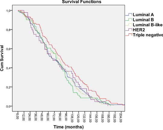
[image:5.612.91.366.75.294.2]In the present study, we priorly evaluated fre-quency of molecular subtypes in a cohort con-sisting of numerous, and widely distributed breast cancer patients with longer follow-up who were treated in a single center in Turkey. We based our assessments on widely used, and precisely defined immunohistochemical Figure 1. Kaplan-Meier survival curve for overall survival.
Figure 2. Kaplan-Meier survival for event free survival.
Luminal A subtype breast cancer (at the end of 75, and 50 months after onset of the disease, re- spectively). Kaplan-Meier sur- vival analysis was performed between subgroups, and log-rank test did not reveal any sta-tistically significant differences between subgroups (P=0.159) (Figures 1 and 2).
Discussion
[image:5.612.91.362.338.555.2]evaluation criteria related to ER, PgR, HER2, and Ki67. The most prevalently seen breast cancer type was Luminal A subtype (41%). Various reports coming from many regions of the world have indicated Luminal A subtype as the most prevalently seen type [7-11]. Discrimination between Luminal A and Luminal B tumor subtypes is made based on the cut-off point of Ki67 proliferation index which is a complex, and problematic method. In this study we determined a cut-off value of 15% for Ki67 proliferation index in line with currently valid recommendations of St Gallen International Expert Consensus [3]. However, since Ki-67 is a continuous variable, increasing its cut-off value to 20% was discussed critically in 13th St Gallen Consensus Conference. At present cut-off value for Kİ67 has not been standardized yet. Therefore relatively limited number of stud-ies have been compared based on Ki67 prolif-eration indices so far [12].
As reported in many studies, in our study Luminal B was the second most prevalently seen subtype [13, 14]. In many studies the inci- 14]. In many studies the inci-14]. In many studies the inci-dence of triple negative-basal like subtype has been reported to vary between 15, and 20 per-cent [11, 15-18]. We also obtained similar inci- 15-18]. We also obtained similar inci-15-18]. We also obtained similar inci-dence rate (19.2%). However in some other studies very high incidence rates have been reported [19, 20]. Diverse incidence rates have been also reported for HER2 subtype. Our inci-dence rates for HER2 subtype were also in con-sistent with the outcomes of the studies per-formed separately by Cherbal et al. and Zheng et al. [11, 20]. In various studies very high or very low incidence rates have been reported [15, 21]. Widely different range of values detected for HER2, and triple negative sub-types may be attributed to different genetic backgrounds, geographic factors, and etiologi-cal heterogeneity.
In this study in addition to classifying breast cancer patients into 4 molecular subgroups, we also evaluated correlations between histopath-ological subtype, TNM stage, local recurrence and metastasis between these subgroups. Since we detected correlations between molec-ular subgroups as for local recurrence, and metastasis, after this part of the study we analyzed the difference between disease-free, and overall survival rates. As can be expected, Luminal A has the most favourable
greater number of patients, this erroneous grouping, and the number of inadequately treated patients did not effect statistical evalu-ations adversely.
In summary, our results represent numerous breast cancer patients during a long-term observation period. Most of the studies on sur-vival rates have evaluated a period of 5-10 years, we performed molecular subtyping in a patient group which we followed up for more than 20 years. We detected many findings com-pliant with the literature. Luminal A subgroup which displayed a good prognosis in the early stages of the follow-up period, while as an inter-esting observation in the long-term it had a bad prognosis. Future studies should take long-term follow-up data covering more than 10 years, and different population analysis criteria as race, and geographic etiologies into con- sideration when investigating the correlation between molecular subtyping, and survival rates.
Disclosure of conflict of interest
None.
Address correspondence to: Dr. Nuket Eliyatkin, Department of Pathology, Faculty of Medicine, Adnan Menderes University, Aydin, Turkey. Tel: +90 506 4173659; E-mail: drnuket2003@yahoo.com
References
[1] Perou CM, Sorlie T, Eisen MB, van de Rijn M, Jeffrey SS, Rees CA, Pollack JR, Ross DT, Johnsen H, Akslen LA, Fluge O, Pergamen- schikov A, Williams C, Zhu SX, Lonning PE, Borresen-Dale AL, Brown PO, Botstein D. Molecular portraits of human breast tumours. Nature 2000; 406: 747-752.
[2] Kreienberg R, Albert US, Follmann M, Kopp IB, Kühn T, Wöckel A. Interdisciplinary GoR level III Guidelines for the Diagnosis, Therapy and Follow-up Care of Breast Cancer: Short ver- sion - AWMF Registry No.: 032-045OL AWMF-Register-Nummer: 032-045OL - Kurzversion 3.0, Juli 2012. Geburtshilfe Frauenheilkd 2013; 73: 556-583.
[3] Untch M, Gerber B, Harbeck N, Jackisch C, Marschner N, Möbus V, von Minckwitz G, Loibl S, Beckmann MW, Blohmer JU, Costa SD, Decker T, Diel I, Dimpfl T, Eiermann W, Fehm T, Friese K, Jänicke F, Janni W, Jonat W, Kiechle M, Köhler U, Lück HJ, Maass N, Possinger K, Rody A, Scharl A, Schneeweiss A, Thomssen C,
Wallwiener D, Welt A. 13th st. Gallen interna-tional breast cancer conference 2013: primary therapy of early breast cancer evidence, con-troversies, consensus-opinion of a german team of experts (zurich 2013). Breast Care (Basel) 2013; 8: 221-229.
[4] Colombo PE, Milanezi F, Weigelt B, Reis-Filho JS. Microarrays in the 2010s: the contribution of microarray-based gene expression profiling to breast cancer classification, prognostication and prediction. Breast Cancer Res 2011; 13: 212.
[5] Sorlie T, Perou CM, Tibshirani R, Aas T, Geisler S, Johnsen H, Hastie T, Eisen MB, van de Rijn M, Jeffrey SS, Thorsen T, Quist H, Matese JC, Brown PO, Botstein D, Lonning PE, Borresen-Dale AL. Gene expression patterns of breast carcinomas distinguish tumor subclasses with clinical implications. Proc Natl Acad Sci U S A 2011; 98: 10869-10874.
[6] Weigelt B, Baehner FL, Reis-Filho JS. The con-tribution of gene expression profiling to breast cancer classification, prognostication and pre-diction: a retrospective of the last decade. J Pathol 2010; 220: 263-280.
[7] Sotiriou C, Pusztai L. Gene-expression signa-tures in breast cancer. New Engl J Med 2009; 360: 790-800.
[8] Engstrom MJ, Opdahl S, Hagen AI, Romundstad PR, Akslen LA, Haugen OA, Vatten LJ, Bofin AM. Molecular subtypes, histopathological grade and survival in a historic cohort of breast can-cer patients. Breast Cancan-cer Res Treat 2013; 140: 463-473.
[9] Salhia B, Tapia C, Ishak EA, Gaber S, Berghuis B, Hussain KB, DuQuette RA, Resau J, Carpten J. Molecular subtype analysis determines the association of advanced breast cancer in Egypt with favorable biology. BMC Womens Health 2011; 11: 44-52.
[10] Zhu X, Ying J, Wang F, Wang J, Yang H. Estrogen receptor, progesterone receptor, and human epidermal growth factor receptor 2 status in invasive breast cancer: a 3,198 cases study at National Cancer Center, China. Breast Cancer Res Trea 2014; 147: 551-555.
[11] Cherbal F, Gaceb H, Mehemmai C, Saiah I, Bakour R, Rouis AO, Boualga K, Benbrahim W, Mahfouf H. Distribution of molecular breast cancer subtypes among Algerian women and correlation with clinical and tumor characteris-tics: A population-based study. Breast Dis 2015; 35: 95-102.
[13] Elkablawy MA, Albasri AM, Hussainy AS, Nouh MM, Alhujaily A. Molecular Profiling of Breast Carcinoma in Almadinah, KSA: Immunopheno- typing and Clinicopathological Correlation. Asian Pac J Cancer Prev 2015; 16: 7819-7824. [14] Inwald EC, Koller M, Klinkhammer-Schalke
M, Zeman F, Hofstädter F, Gerstenhauer M, Brockhoff G, Ortmann O. 4-IHC classification of breast cancer subtypes in a large cohort of a clinical cancer registry: use in clinical routine for therapeutic decisions and its effect on sur-vival. Breast Cancer Res Treat 2015; 153: 647-658.
[15] Bhargava R, Striebel J, Beriwal S, Flickinger JC, Onisko A, Ahrendt G, Dabbs DJ. Prevalence, morphologic features and proliferation indices of breast carcinoma molecular classes using immunohistochemical surrogate markers. Int J Clin Exp Pathol 2009; 2: 444-455.
[16] Caldarella A, Buzzoni C, Crocetti E, Bianchi S, Vezzosi V, Apicella P, Biancalani M, Giannini A, Urso C, Zolfanelli F, Paci E. Invasive breast can-cer: a significant correlation between histologi-cal types and molecular subgroups. J Cancer Res Clin Oncol 2013; 139: 617-23.
[17] Shomaf M, Masad J, Najjar S, Faydi D. Distribution of breast cancer subtypes among Jordanian women and correlation with histo-pathological grade: molecular subclassifica-tion study. JRSM Short Rep 2013; 4: 1-6. [18] Elesawy BH, Abd El hafez A, Shawky Ael-A,
Arafa M. Immunohistochemistry-based subtyp-ing of breast carcinoma in Egyptian women: a clinicopathologic study on 125 patients. Ann Diagn Pathol 2014; 18: 21-26.
[19] El-Hawary AK, Abbas AS, Elsayed AA, Zalata KR. Molecular subtypes of breast carcinoma in Egyptian women: clinicopathological features. Pathol Res Pract 2012; 208: 382-386. [20] Zheng S, Song QK, Ren Y, Feng WL, Kong YN,
Huang R, Xu F, Li J, Zhang BN, Fan JH, He JJ, Qiao YL. The characteristics of breast cancer subtypes: Implications for treatment guide-lines and individualized treatment strategies in China. Appl Immunohistochem Mol Morphol 2014; 22: 383-389.
[21] Akbar M, Akbar K, Naveed D. Frequency and correlation of molecular subtypes of breast cancer with clinicopathological features. J Ayub Med Coll Abbottabad 2014; 26: 290-293.
[22] Haque R, Ahmed SA, Inzhakova G, Shi J, Avila C, Polikoff J, Bernstein L, Enger SM, Press MF. Impact of Breast Cancer Subtypes and Treatment on Survival: An Analysis Spanning Two Decades. Cancer Epidemiol Biomarkers Prev 2012; 21: 1848-55.
[23] Blows FM, Driver KE, Schmidt MK, Broeks A, van Leeuwen FE, Wesseling J, Cheang MC, Gelmon K, Nielsen TO, Blomqvist C, Heikkilä P, Heikkinen T, Nevanlinna H, Akslen LA, Bégin LR, Foulkes WD, Couch FJ, Wang X, Cafourek V, Olson JE, Baglietto L, Giles GG, Severi G, McLean CA, Southey MC, Rakha E, Green AR, Ellis IO, Sherman ME, Lissowska J, Anderson WF, Cox A, Cross SS, Reed MW, Provenzano E, Dawson SJ, Dunning AM, Humphreys M, Easton DF, García-Closas M, Caldas C, Pharoah PD, Huntsman D. Subtyping of breast cancer by immunohistochemistry to investigate a rela-tionship between subtype and short and long term survival: a collaborative analysis of data for 10,159 cases from 12 studies. PLoS Med 2010; 7: e1000279.
[24] Chang HR, Glaspy J, Allison MA, Kass FC, Elashoff R, Chung DU, Gornbein J. Differential response of triple-negative breast cancer to a docetaxel and carboplatin-based neoadjuvant treatment. Cancer 2010; 116: 4227-4237. [25] Yerushalmi R, Hayes MM, Gelmon KA, Chia S,