Original Article
Changes in postoperative cognitive function during
off-pump coronary artery bypass graft
surgery: dose response of propofol
Wenqian Zhai, Jiapeng Liu, Yifei Si, Jiange Han
Department of Anesthesiology, Chest Hospital of Tianjin, Tianjin 300051, China
Received November 23, 2015; Accepted April 14, 2016; Epub June 15, 2016; Published June 30, 2016
Abstract: To determine if concentration of propofol is related to the incidence of postoperative cognitive dysfunc-tion (POCD) during off-pump coronary artery bypass grafting (OPCABG) surgery in geriatric patients. A total of 218 patients scheduled for elective OPCABG surgery were enrolled into three groups based on propofol dose, including TCI plasma concentration of 0.5-1.0 µg/ml (Group P1), 1.1-1.5 µg/ml (Group P2) and 1.6-2.0 µg/ml (Group P3). Neuropsychological testing was performed before surgery and at day 1, day 3, day 7, 3 months, and 6 months after surgery. S100-β and neuron-specific enolase (NSE) were measured at the startand end of surgery and at 6, 12, and 24 hours after surgery. As compared to groups P1 and P2, MMSE scores in group P3 decreased significantly at day 1 and day 3 postoperatively (P<0.05). S100-β protein and NSE were significantly differentin group P3 as compared to groups P1 and P2 (P<0.05). POCD incidence may correlate with concentration of propofol in patients undergoing OPCABG surgery.
Keywords: Propofol, postoperative cognitive dysfunction, off-pump coronary artery bypass grafting
Introduction
POCD is a well-recognized complication of CAB [1, 2]. The CABG surgery (off-pump or on-pump) impacts cognitive function for 3-6 months [3],with no major difference in POCD occurrence between OPCABG group (21% at 3 months and 30.8% at 12 months) and CPB group (29% at 3 months and 33.6% at 12 months) [4]. Total intravenous anesthesia (TIVA) is more suitable than inhalation anesthesia for senile patients [5]. The incidence of early POCD in patients under propofol anesthesia is lower than inhalation anesthesia [6]. Although many studies have investigated POCD after CABG surgery, the dose of the anesthetic is rarely mentioned. We investigate whether the dose of propofol is associated with POCD incidence during OPCABG surgery in geriatric patients. Materials and methods
The duration of this prospective, randomized clinical study was eight months, from February to December 2013. Ethical approval was pro-vided by the Clinical Research Ethics Commi-
ttee (Approved ID: 2012KY-001-01). Informed written consent was obtained from all patients. The retrospective registration work was com-pleted on www.chictr.org.cn (ChiCTR-OOC-15- 005978).
Patients
The study was performed on 218 patients, between 60 and 75 years of age, ASA III, preop-erative Euroscore 4-6 points, who were referred for elective OPCABG surgery. Patients were excluded if they met any of the following crite-ria: (1) emergency surgery; (2) preoperative cerebrovascular disease; (3) history of mental illness, long-term use of sedatives and antide-pressants; (4) history of pneumothorax; (5) liver and kidney dysfunction; (6) secondary surgery; (7) ejection fraction <40%; (8) level of educa-tion <7 years; (9) serious visual, auditory and language barriers.
Grouping
Anesthesia
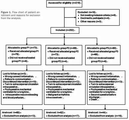
[image:2.612.92.524.74.477.2]All preoperative cardiac medica-tions except anticoagulants were administered until the morning of surgery. Diabetic patients on oral anti-diabetic agents were preoper-atively switched to insulin therapy. All patients received a standard pre-operative treatment: 0.1 mg/ kg morphine and 0.3 mg sco- polamine by in tramuscular injec-tion, 30 minutes before surgery. In the operating room, the patients were monitored with 5-lead elec-trocardiogram, oxygen saturation and electroencephalogram (BIS). A peripheral line was established Figure 1. Flow chart of patient
en-rollment and reasons for exclusion from the analysis.
Table 1. Baseline characteristics
Variables
All patients (n=156)
P value Group P1
(n=60) Group P2 (n=52) Group P3 (n=44) Age (yr) 69±4.7 67±3.7 65±4.5 0.362 Education (yr) 10.5±3.2 10.6±3.5 11.0±3.5 0.462 Female gender 21 (35%) 16 (31%) 14 (32%) 0.563 Height (cm) 170±8.6 169±7.5 171±8.2 0.517 Weight (kg) 73±5.2 73±5.9 74±6.3 0.498 Hypertension 50 (83%) 45 (86%) 38 (86%) 0.221 Diabetes mellitus 35 (58%) 28 (53%) 22 (50%) 0.139 Old myocardial infarction 14 (23%) 10 (19%) 9 (20%) 0.235 Preoperative EF (%) 54±4.6 54±5.2 55±3.9 0.735 ASA classification II (II~III) II (II~III) II (II~III) 0.869 Preoperative Euroscore 2.0±0.1 2.0±0.2 2.0±0.1 0.935
P1, n=73), 1.1-1.5 µg/ml (Group P2, n=69) and
[image:2.612.91.353.510.693.2]or right radial or brachial artery catheterization. Then, anesthesia was induced with etomidate 0.15-0.3 mg/kg, sufentanil 1.0-1.5 µg/kg, and rocuronium 0.6 mg/kg. Oralintubation was per-formed. The indices of ventilation were tidal vol-ume 6-8 ml/kg; respiratory rate 12-15 breaths/ min, and end-tidal carbon dioxide (PETCO2) 30-40 mmHg. Central venous catheter was placed through the right internal jugular vein or subclavian vein, and a single cavity venous catheter was placed retrograde to the jugular vein bulb through the right internal jugular vein. A pulmonary artery catheter was inserted, when necessary. Anesthesia was maintained with propofol and BIS targeted between 40 and 60. During surgery, sufentanil (0.8-1.2 μg/mL) and cisatracurium (0.2-0.3 mg/kg/h) were con-tinuously pumped. Intra-operative monitoring
and 24 hours after surgery to measure S100-β protein and NSE.
MMSE
All patients underwent neuropsychological test-ing ustest-ing MMSE scale before surgery and at day 1, day 3, day 7, 3 months, and 6 months after surgery.
Statistical analysis
[image:3.612.91.370.84.190.2]Data were presented as mean ± standard error of mean. The D’Agostino normality test was applied before performing the ANOVA test. Differences among the three groups at each time point were determined by one-way ANOVA followed by Newman-Keuls post hoc test. Differences across the six time points in each Table 2. Intraoperative vital signs
Variables
All patients (n=156)
P value Group P1
(n=60) Group P2 (n=52) Group P3 (n=44)
Heart rate (bpm) 69±4.2 67±6.1 73±5.4 0.458 MAP (mmHg) 73±6.6 70±4.5 68±6.1 0.452 SpO2 (%) 99±1.2 99±0.8 99±1.1 0.884 Bis 48±2.5 46±1.9 43±4.2 0.377 ETCO2 (mmHg) 31±2.2 33±4.4 32±5.1 0.548
Table 3. Clinic record
Variables
All patients (n=156)
P
value Group P1
(n=60) Group P2 (n=52) Group P3 (n=44) Number of graft 2 (2~3) 3 (2~4) 3 (2~4) 0.946 Anesthesia duration (hour) 3.5±0.5 4.1±0.7 4.0±0.6 0.645 ICU stay (day) 2.2±0.2 2.0±0.1 2.3±0.2 0.424 Hospital stay (day) 8.3±1.2 7.2±1.3 7.9±1.2 0.197
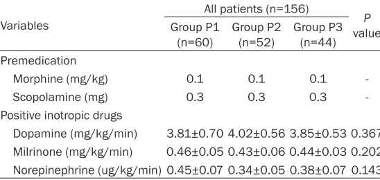
Table 4. Complication and drugs
Variables
All patients (n=156)
P
value Group P1
(n=60) Group P2 (n=52) Group P3 (n=44) Premedication
Morphine (mg/kg) 0.1 0.1 0.1 Scopolamine (mg) 0.3 0.3 0.3 -Positive inotropic drugs
Dopamine (mg/kg/min) 3.81±0.70 4.02±0.56 3.85±0.53 0.367 Milrinone (mg/kg/min) 0.46±0.05 0.43±0.06 0.44±0.03 0.202 Norepinephrine (ug/kg/min) 0.45±0.07 0.34±0.05 0.38±0.07 0.143
included electrocardiogram, ar- terial pressure, pulse oxygen saturation, end-tidal expiratory carbon dioxide, central venous pressure, nasopharyngeal tem-perature, and urine output. Na- sopharyngeal temperature was maintained above 35°C, and systolic blood pressure above 60 mmHg.
OPCABG surgery was performed by the same group of surgeons. Sternotomy was performed at the midline and the number of bypass grafts was 2-4. Intravenous heparin (1.0 mg/ kg) was administered and the activated clotting time (ACT) was controlled between 250-350 s and then lowered to 90-120 s after protamine neu-tralization. At the end of surgery, the patients were transferred to the Intensive Care Unit (ICU) without extubation. Subsequen- tly, follow-up personnel record-ed decannulation time, time back to the general ward, and discharge time.
Blood samples
[image:3.612.91.365.352.482.2]group were assessed by repeated measures one-way ANOVA and Tukey’smulti-comparison test. P<0.05 was considered to be statistically significant.
Results
During the studyperiod, 218 patients were screened, of which 202 matched the criteria. Among the eligible patients, 46 were excluded due to the following reasons: (1) intraoperative finding of malignant arrhythmia; (2) intraopera-tiveswitched to extracorporeal circulation; (3) intraoperative massive bleeding and blood per-fusion; (4) second surgery within short term after operation; (5) lengthy postoperative mechanical ventilation support; (6) lost to fol-low-up; and (7) death. Finally, 156 patients were enrolled in the study (Figure 1).
Analysis of general condition
There were no statistical differencesin epide-miological features, history, ASA classification, and preoperative Euroscore (Table 1). There
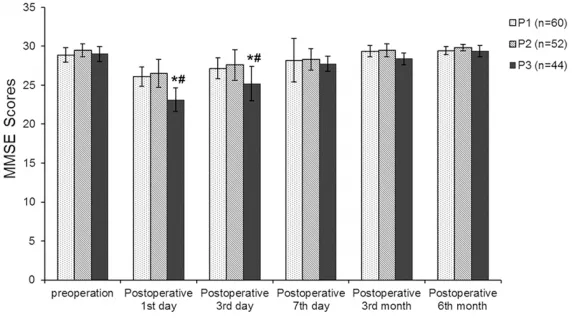
[image:4.612.91.377.195.354.2]There was no significant difference in the pre-operative MMSE scores among the three groups. As compared to groups P1 and P2, MMSE scores of group P3 were lower and sig-nificantly differenton day 1 and day 3 postop-eration (P<0.05) by one-way ANOVA followed by Newman-Keuls post hoc test. The MMSE results of day 1 and day 3 were statistically different from other time points in group P3 (P<0.05) by repeated measures one-way ANOVA and Tukey’s multi-comparison test. MMSE scores of patients in groups P1 and P2 were in the normal range during the entire post-operative follow-up period (P>0.05) (Table 5; Figure 2).
S100-β protein levels
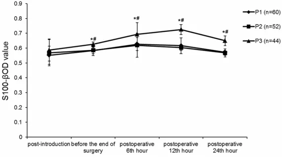
We investigated the differences in S100-β between the three groups after surgery by one-way ANOVA. The average OD value of S100-β in group P3 significantly increased from the end of surgery to 24 hours postoperation (P<0.05), and were higher atthe end of surgery, and 6, 12, and 24 hours post operation in group P3 Table 5. MMSE scores
Group Preoperation Postoperative 1st day Postoperative 3rd day Postoperative 7th day Postoperative 3rd month Postoperative 6 th
month P1 (n=60) 28.88±0.93 26.12±1.25 27.14±1.35 28.20±2.78 29.38±0.71 29.43±0.53 P2 (n=52) 29.50±0.84 26.50±1.79 27.60±1.95 28.33±1.37 29.50±0.84 29.83±0.41 P3 (n=44) 29.00±0.99 23.13±1.51*,# 25.2±2.21*,# 27.75±0.98 28.38±0.74 29.38±0.75
Results are expressed as mean ± SEM of the whole groups. *P<0.05, ANOVA one-way, post hoc Newman-Keuls test. #P<0.05,
ANOVA one-way and Tukey’s, multi-comparison test.
Figure 2. Differences in MMSE scores among the three groups before and af-ter off-pump coronary araf-tery bypass graft surgery. Error bars represent stan-dard error. *P<0.05, ANOVA one-way post hoc Newman-Keuls test. #P<0.05, ANOVA one-way and Tukey’s multi-comparison test. P1= propofol TCI 0.5-1.0 µg/ml; P2= propofol TCI 1.1-1.5 µg/ml; P3= propofol TCI 1.6-2.0 µg/ml.
were nosignificant difference-sin the heart rate, MAP, SpO2, BIS value, and PETCO2 amo- ng the three groups (Table 2), and the number of grafts, anesthesia duration, ICU and hospital stay among the groups (Table 3). No differ-ences were found between the groups in premedication and positive inotropic drugs support (Table 4). If cardiac adverse events (sudden car-diac death, congestive heart failure, angina, myocardial in- farction, arrhythmia) and mor-tality existed, the patient was excluded.
as compared to groups P1 and P2 (P<0.05). The values of group P1 were slightly higher than group P2 postoperation (P>0.05) (Table 6; Figure 3).
NSE levels
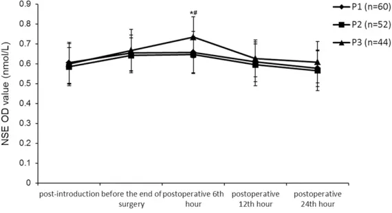
The OD values of NSE increased perioperatively and were statistically different between group P3 and the other two groups at 12 hours post-surgery (P<0.05) (Table 7; Figure 4).
Discussion
Age, as an independent factor, affects the men-tal state after anesthesia. This study selects geriatric patients (60-75 years) with coronary heart disease. A study proposes that decreas-ing age is a risk factor for early POCD [7], and the increasing age is a dominant potential risk factor for POCD, irrespective of gender [8]. Older patients are the most affected with POCD after surgery, based on risk factors and high-risk groups.
There is increasing evidence that long-term or permanent neuronal and neurological changes could occur following administration of
anes-nal jugular vein, and can accurately reflect the relationship between cerebral oxygen supply and oxygen consumption. Therefore, we collect internal jugular venous blood to measure the biomarkers. Blood S100-β protein is a sensitive biomarker of blood-brain barrier damage [12]. POCD indeed correlates with the concentra-tions of peripheral inflammatory markers, par-ticularly IL-6 and S-100β protein [13]. Therefore, S100-β protein content is closely related to the severity of brain damage, and can predict adverse neurological outcome. NSE may be an useful biomarker to identify patients with cogni-tive performance impairment [14].
Whether a causal relationship existed between propofol dose and occurrence of POCD remains unknown. Although some anesthetics have shown neuroprotective effects in preclinical studies (cell culture systems or animal models of focal or global cerebral ischemia), the evi-dence in human studies has been inconsistent and controversial [15-18]. Non-sedative doses of propofol does not produce dyskinesia in rats [19]. We analyze why different doses of propo-fol anesthesia produce different levels of post-Table 6. S100-β protein OD value
Group Post-introduction Before the end of surgery Postoperative 6hour th Postoperative 12hour th Postoperative 24th hour
P1 (n=60) 0.552±0.062 0.584±0.022# 0.627±0.043# 0.616±0.053# 0.573±0.022
P2 (n=52) 0.569±0.089 0.586±0.036# 0.619±0.080# 0.603±0.017# 0.568±0.027
P3 (n=44) 0.587±0.075 0.626±0.016#,* 0.693±0.078#,* 0.725±0.033#,* 0.650±0.033*
Results are expressed as mean ± SEM of the whole groups. *P<0.05, ANOVA one-way post hoc Newman-Keuls test. #P<0.05,
[image:5.612.90.376.195.356.2]ANOVA one-way and Tukey’s multi-comparison test.
Figure 3. The plasma OD values of S100-β protein.Error bars represent stan-dard error. *P<0.05, ANOVA one-way post hoc Newman-Keuls test. #P<0.05, ANOVA one-way and Tukey’s multi-comparison test. P1= propofol TCI 0.5-1.0 µg/ml; P2= propofol TCI 1.1-1.5 µg/ml; P3= propofol TCI 1.6-2.0 µg/ml.
thetics [9]. High-dose fentanyl is not associated with a differ-ence in the inciddiffer-ence of POCD at 3 or 12 months after sur-gery. However, low-dose fen-tanyl is associated with a greater incidence of POCD, one week after surgery [10]. Sufentanil is stable for gener-al anesthesia and has the least adverse effect on hemo-dynamics [11]. In our study, we use high-dose sufentanil-as the analgesic, and findno significant difference amon-gthe three groups.
inter-operative cognitive function in this study, and the reasons maybe associated with the following:
The BIS values of the high-dose propofol group are slightly lower than the low-dose propofol groups during anesthesia. A report confirms the influence of extremely low BIS value on delirium by multivariate analysis [20]. Further- more, high dose of propofol strengthens the function of GABAA receptors, inhibits synaptic prolonged enhanced expression of hippocam-pal cells and affects cognitive function. Addi- tionally, an inflammatory response may be involved in the occurrence of POCD [21, 22]. Reasonable combination of sedative and anal-gesic drugs not only guarantees reduction of cerebral metabolism and blood flow but also hemodynamicstability to reduce the stress response.
The major limitation of the present study is the sample size of each group. Despite the power analysis to calculate the sample size, the effec-tive group size was much smaller due to the inability of patients to perform cognitive tests
ence in the treatment cannot be avoided due to technical reasons.
POCD incidence may correspond with the con-centration of propofol during OPCABG surgery. Disclosure of conflict of interest
None.
Address correspondence to: Jiange Han, Depart- ment of Anesthesiology, Chest Hospital of Tianjin, Tianjin 300051, China. Tel: +86 13820622082; Fax: +86-022-88185156; E-mail: hanjiange@hotmail. com
References
[1] Liu YH, Wang DX, Li LH, Wu XM, Shan GJ, Su Y, Li J, Yu QJ, Shi CX, Huang YN and Sun W. The effects of cardiopulmonary bypass on the number of cerebral microemboli and the inci-dence of cognitive dysfunction after coronary artery bypass graft surgery. Anesth Analg 2009; 109: 1013-1022.
[image:6.612.89.524.85.150.2][2] Newman MF, Kirchner JL, Phillips-Bute B, Gaver V, Grocott H, Jones RH, Mark DB, Reves
Table 7. NSEOD value
Group Post-introduction Before the end of surgery Postoperative 6hour th Postoperative 12hour th Postoperative 24hour th P1 (n=60) 0.605±0.103 0.654±0.090 0.657±0.107# 0.609±0.100 0.578±0.092
P2 (n=52) 0.585±0.096 0.642±0.088 0.646±0.092# 0.595±0.105 0.565±0.101
P3 (n=44) 0.599±0.102 0.668±0.106 0.734±0.103*,# 0.627±0.093 0.608±0.104
Results are expressed as mean ± SEM of the whole groups. *P<0.05, ANOVA one-way post hoc Newman-Keuls test. #P<0.05,
ANOVA one-way and Tukey’s multi-comparison test.
Figure 4. The concentrations of NSE. Error bars represent standard error. *P<0.05, ANOVA one-way post hoc Newman-Keuls test. #P<0.05, ANOVA one-way and Tukey’s multi-comparison test. P1= propofol TCI 0.5-1.0 µg/ml; P2= propofol TCI 1.1-1.5 µg/ml; P3= propofol TCI 1.6-2.0 µg/ml.
and several missing values that had to be amended. On the other hand, the differenc-es in the number of dropouts in the study groups have to be interpreted as results of the intervention.
Another limitation is the ab- sence ofa standardizedtest to excludeddelirium as a global cerebral deficit. The CAM-ICU has been a suitable tool to detect hyper-and hypo-active delirium [23].
[image:6.612.91.376.195.350.2]interfer-JG and Blumenthal JA. Longitudinal assess-ment of neurocognitive function after coro-nary-artery bypass surgery. N Engl J Med 2001; 344: 395-402.
[3] van Dijk D, Moons KG, Nathoe HM, van Aarnhem EH, Borst C, Keizer AM, Kalkman CJ and Hijman R. Cognitive outcomes five years after not undergoing coronary artery bypass graft surgery. Ann Thorac Surg 2008; 85: 60-64.
[4] Van Dijk D, Jansen EW, Hijman R, Nierich AP, Diephuis JC, Moons KG, Lahpor JR, Borst C, Keizer AM, Nathoe HM, Grobbee DE, De Jaegere PP and Kalkman CJ. Cognitive out-come after off-pump and on-pump coronary artery bypass graft surgery: a randomized trial. JAMA 2002; 287: 1405-1412.
[5] Cai Y, Hu H, Liu P, Feng G, Dong W, Yu B, Zhu Y, Song J and Zhao M. Association between the apolipoprotein E4 and postoperative cognitive dysfunction in elderly patients undergoing in-travenous anesthesia and inhalation anesthe-sia. Anesthesiology 2012; 116: 84-93. [6] Xu D, Yang W and Zhao G. Effect of propofol
and inhalation anesthesia on postoperative cognitive dysfunction in the elderly: a meta-analysis. Nan Fang Yi Ke Da Xue Xue Bao 2012; 32: 1623-1627.
[7] Moller JT, Cluitmans P, Rasmussen LS, Houx P, Rasmussen H, Canet J, Rabbitt P, Jolles J, Larsen K, Hanning CD, Langeron O, Johnson T, Lauven PM, Kristensen PA, Biedler A, van Beem H, Fraidakis O, Silverstein JH, Beneken JE and Gravenstein JS. Long-term postopera-tive cognipostopera-tive dysfunction in the elderly ISPOCD1 study. ISPOCD investigators. Interna- tional Study of Post-Operative Cognitive Dys- function. Lancet 1998; 351: 857-861. [8] Kotekar N, Kuruvilla CS and Murthy V.
Post-operative cognitive dysfunction in the elderly: A prospective clinical study. Indian J Anaesth 2014; 58: 263-268.
[9] Hanning CD. Postoperative cognitive dysfunc-tion. Br J Anaesth 2005; 95: 82-87.
[10] Silbert BS, Scott DA, Evered LA, Lewis MS, Kalpokas M, Maruff P, Myles PS and Jamrozik K. A comparison of the effect of high- and low-dose fentanyl on the incidence of postopera-tive cognipostopera-tive dysfunction after coronary artery bypass surgery in the elderly. Anesthesiology 2006; 104: 1137-1145.
[11] Sebel PS and Bovil JG. Cardiovascular effects of sufentanil anesthesia. Anesth Analg 1982; 61: 115-119.
[12] Bailey DM, Evans KA, McEneny J, Young IS, Hullin DA, James PE, Ogoh S, Ainslie PN, Lucchesi C, Rockenbauer A, Culcasi M and
Pietri S. Exercise-induced oxidative-nitrosative stress is associated with impaired dynamic ce-rebral autoregulation and blood-brain barrier leakage. Exp Physiol 2011; 96: 1196-1207. [13] Peng L, Xu L and Ouyang W. Role of peripheral
inflammatory markers in postoperative cogni-tive dysfunction (POCD): a meta-analysis. PLoS One 2013; 8: e79624.
[14] Baranyi A and Rothenhausler HB. The impact of S100b and persistent high levels of neuron-specific enolase on cognitive performance in elderly patients after cardiopulmonary bypass. Brain Inj 2013; 27: 417-424.
[15] Roach GW, Newman MF, Murkin JM, Martzke J, Ruskin A, Li J, Guo A, Wisniewski A and Mangano DT. Ineffectiveness of burst suppres-sion therapy in mitigating perioperative cere-brovascular dysfunction. Multicenter Study of Perioperative Ischemia (McSPI) Research Group. Anesthesiology 1999; 90: 1255-1264. [16] Kanbak M, Saricaoglu F, Avci A, Ocal T, Koray
Z and Aypar U. Propofol offers no advantage over isoflurane anesthesia for cerebral protec-tion during cardiopulmonary bypass: a prelimi-nary study of S-100beta protein levels. Can J Anaesth 2004; 51: 712-717.
[17] Zaidan JR, Klochany A, Martin WM, Ziegler JS, Harless DM and Andrews RB. Effect of thiopen-tal on neurologic outcome following coronary artery bypass grafting. Anesthesiology 1991; 74: 406-411.
[18] Michenfelder JD. A valid demonstration of bar-biturate-induced brain protection in man--at last. Anesthesiology 1986; 64: 140-142. [19] Pain L, Angst MJ, LeGourrier L and Oberling P.
Effect of a nonsedative dose of propofol on memory for aversively loaded information in rats. Anesthesiology 2002; 97: 447-453. [20] Radtke FM, Franck M, Lendner J, Kruger S,
Wernecke KD and Spies CD. Monitoring depth of anaesthesia in a randomized trial decreas-es the rate of postoperative delirium but not postoperative cognitive dysfunction. Br J Anaesth 2013; 110 Suppl 1: i98-105.
[21] Hajjar LA, Vincent JL, Galas FR, Nakamura RE, Silva CM, Santos MH, Fukushima J, Kalil Filho R, Sierra DB, Lopes NH, Mauad T, Roquim AC, Sundin MR, Leao WC, Almeida JP, Pomerantzeff PM, Dallan LO, Jatene FB, Stolf NA and Auler JO Jr. Transfusion requirements after cardiac surgery: the TRACS randomized controlled tri-al. JAMA 2010; 304: 1559-1567.
restric-tive transfusion in high-risk patients after hip surgery. N Engl J Med 2011; 365: 2453-2462. [23] Klugkist M, Sedemund-Adib B, Schmidtke C,
Schmucker P, Sievers HH and Huppe M.