Original Article
Research of the effects of metformin on the
proliferation of non-invasive bladder cancer cells in vitro
Quan Yuan1*, Zhong Wang1*, Xiaolin Xu1, Feng Liu1, Lintao Liu1, Jianjun Yu1,2
1Department of Urinary Surgery, Shanghai Fengxian District Central Hospital, Shanghai, China; 2Department of
Urinary Surgery, Affiliated Sixth People’s Hospital of Shanghai Jiao Tong University, Shanghai, China. *Equal
con-tributors and co-first authors.
Received January 22, 2017; Accepted February 24, 2017; Epub May 15, 2017; Published May 30, 2017
Abstract: Objective: To study the effects and mechanisms of metformin on the proliferation of 253J non-invasive bladder cancer cell line. Methods: 253J cells were treated in vitro with metformin at different concentrations (5 mmoL/L, 10 mmoL/L and 20 mmoL/L). The effects of metformin on the proliferation of 253J cells, cell cycle, cell apoptosis and the expression of cell proliferation related proteins were respectively tested by CCK-8 method, flow cytometry, Annexin V-FITC/PI dual fluorescent and the Western blot method. Results: Metformin could apparently restrain the proliferation of 253J cells (P=0.004). Besides, with the increase of metformin concentration and the extension of treatment time, the survival rate of 253J cells decreased gradually. At the same time, metformin could promote 253J cell apoptosis (P=0.003) and make the cell stagnate in G0/G1 phase (P=0.002), and its func-tion gradually increased with the increase of metformin concentrafunc-tion. The results of Western blot indicated that the expression levels of AMPK and p38 and their phosphorylated proteins in the experimental group were signifi-cantly higher than those in the control group, while the expression of Cyclin D1 protein was signifisignifi-cantly decreased (P=0.001). Conclusion: Metformin can restrain the proliferation and promote the apoptosis of 253J non-invasive bladder cancer cell line, and the mechanisms may be related to the regulation of Cyclin D1 expression by AMPK signaling pathway.
Keywords: Metformin, non-invasive bladder cancer, AMPK signaling pathway
Introduction
In recent years, the incidence of bladder tumor increased year by year [1]. In clinical treatment, the pathological type of the majority of patients with bladder cancer was non-muscle invasive. Besides, it had a high recurrence rate and a low 5-year survival rate after chemotherapeu- tic medicine lavage and transurethral resection of bladder tumor (TURBT) [2, 3]. Obviously, the prognosis of the bladder cancer is not optimis-tic. Hence, finding a safe and effective drug against bladder cancer has been the focus of scholars.
Metformin is a first-line oral biguanides hypo-glycemic drug, which was widely used in treat-ments of patients with type 2 diabetes mellitus [4]. Recent studies have shown that metformin can not only control blood glucose levels and the fatality rate of patients with diabetes, but
Research of the effects of metformin
Materials and methods
Experimental materials
Cell lines and cell culture: Human 253J non-invasive bladder cancer cell line was purchas- ed from the American type culture collection (ATCC). Cells were inoculated in the RPMI-1640 culture medium containing 10% fetal bovine serum and antibiotics (100 mg/L streptomycin and 1*105 U/L penicillin), and then placed in an incubator (5% CO2, 37°C); the cells at loga-rithmic phase were then used for subsequent experiments.
Main reagents and materials: RPMI-1640 cul-ture medium, fetal bovine serum, trypsin and EDTA were all purchased from the American Gibco Company; Mouse anti-human AMPK, p38, p-AMPK, p-p38 and Cyclin D1 protein were primary antibodies and all purchased from the American Abcam Company; the sec-ondary antibodies labeled goat anti-mouse horseradish peroxidase were purchased from the American Santa Cruz Company; metformin and ECL chemiluminescence reagent were purchased from the American Sigma Com- pany; CCK-8 kit was purchased from DOJINDO Laboratories; Annexin V-FITC/PI Apoptosis kit was purchased from BD Biosciences Company (USA); enzyme linked immunosorbent assay instrument was from Thermo Company; flow cytometry was from BD Company (USA) and Gel imaging system was from Bio-Rad Company.
Experimental methods
The effects of metformin on the proliferation of 253J non-invasive bladder cancer cells were detected by CCK-8 method: The 253J cells were inoculated in the 96-well plate at a densi-ty of 2*103 cells/well, and placed in an incuba-tor (5% CO2, 37°C). They were randomly divided into a control group and other experimental groups, while the experimental groups were respectively stimulated at different concentra-tions of metformin (5 mmoL/L, 10 mmoL/L and 20 mmoL/L) and each group had 5 repeat wells. After the cells being respectively stimu-lating by metformin for 24 h, 48 h and 72 h, 10 μL CCK-8 was added into each well. After 2-hour routine culture, the 96-well plate was implanted into enzyme linked immunosorbent assay instrument to examine the absorbance value (OD value) of each well at wave length of
450 nm. An empty tube with only culture liquid and CCK-8 reagent was used as the blank control. According to the OD values of each group: the cell survival rate (%) = OD value of drug group/OD value of control group *100%. Metformin was not added in the control group while other processing methods were as same as the experimental group.
The effects of metformin on the cell cycle of 253J non-invasive bladder cancer cells were detected by flow cytometry: The 253J cells were inoculated in the 6-well plate at a den- sity of 4*104 cells/well. After being stimulated by different concentrations of metformin (5 mmoL/L, 10 mmoL/L and 20 mmoL/L) for 48 h, the cells were digested and washed with pre-cooled PBS and then collected in the flow cytometry tube. Seventy percent of ice alcohol was used to re-suspended cells and placed at -20°C for one hour. The cells were washed with PBS, then centrifuged at 2000 r/min for 5 min and re-suspended by HBSS solutioncon-taining 50 μg/mL PI. Finally, the cell cycle of 253J cells of each group was detected by the flow cytometry and analyzed by the ModFit software.
The effects of metformin on the cell apoptosis of 253J non-invasive bladder cancer cells were detected by flow cytometry: The 253J cells which were treated with different concentra-tions of metformin for 48 h, were washed with PBS and then collected in the flow cytometry tube. After double staining with 10 μL Annexin V-FITC and 5 μL PI, incubated the cells in dark environment with room temperature for 15 min. Then, the cell apoptosis of 253J cells was detected by the flow cytometry and ana-lyzed by the ModFit software.
The expression of protein involved in the cell proliferation was detected by the western blot:
4°C for overnight. Next, the secondary antibod-ies were added after washing the membrane at room temperature with TBS-T solution contain-ing 0.1% of Tween 20 and then incubated at room temperature for one hour. Finally, the membrane was washed again with TBS-T solu-tion, then the protein was reacted with the ECL chemiluminescence reagent and exposed and scanned by Gel imaging system. The β-actin was taken as the internal reference.
Statistical analysis
The data were statistically analyzed by SPSS 17.0 software and the measurement data were expressed by mean ± standard deviation. The comparison among samples of each group was carried on by one-way ANOVA, and the compari-son between two independent samples was
carried on by t-test. P<0.05 indicated that the differences were statistically significant. Results
The effects of metformin on the proliferation ability of 253J cells
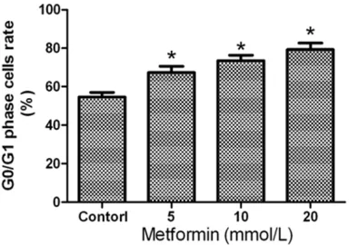
253J non-invasive bladder cancer cells were treated with different concentrations of met- formin in this study, the survival rate of 253J cells were detected by CCk-8 method. As sh- own in Figure 1A, after being treated with dif-ferent concentrations of metformin (5 mmoL/L, 10 mmoL/L and 20 mmoL/L), the cell viability of 253J cells were (71.3±8.2)%, (65.4±9.1)% and (52.3±10.1)% respectively. When com-pared with the control group, the proliferation ability of 253J cells in the experimental group was significantly inhibited (P=0.004). With the increase of metformin concentrations and ex- tension of stimulant time, the survival rate of 253J cells under the effects of the metformin gradually reduced, which indicated that met- formin significantly inhibited the proliferation of 253J non-invasive bladder cancer cells in a time and concentration dependent manner (Figure 1A and 1B).
The effects of metformin on cell cycle of 253J
cells
[image:3.612.95.520.75.233.2]After 253J non-invasive bladder cancer cells being treated for 48 h by different concentra-tions of metformin (5 mmoL/L, 10 mmoL/L and 20 mmoL/L), the changes of cell cycle were detected. The results showed that the cell ratio
Figure 1. The effects of metformin on the proliferation ability of 253J non-invasive bladder cancer cells. *P<0.05
vs. control group.
Figure 2. The effects of metformin on cell cycle of 253J cells. *Compared with the control group,
[image:3.612.93.285.291.426.2]Research of the effects of metformin
of G0/G1 phase in metformin treatment group was (67.4±3.2)%, (73.5±2.9)% and (79.3±3.4)%, respectively; compared with the control group, its cell ratio of G0/G1 phase increased signifi-cantly (P=0.002), and with the increasing con-centration of metformin, the ratio of 253J cells in G0/G1 phase increased gradually. It indicat-ed that metformin could block 253J non-inva-sive bladder cancer cells in the G0/G1 phase (Figure 2).
The effects of metformin on non-invasive blad
-der cancer cell apoptosis
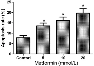
The concentrations of metformin (5 mmoL/L, 10 mmoL/L and 20 mmoL/L) were respective- ly applied to 253J non-invasive bladder cancer cells for 48 h, then Annexin V-FITC/PI was used to stain necrotic and apoptotic cells. The results showed that the apoptotic ratio were (13.5± 1.4)%, (16.1±1.7)% and (19.7±2.1)%, respec-tively. Compared with the control group, the
dif-ference was statistically significant (P=0.003). And with the increasing concentration of met-formin, its apoptotic effects on 253J cells in- creased gradually. It indicated that metformin could significantly promote the apoptosis of 253J (as shown in Figures 3 and 4).
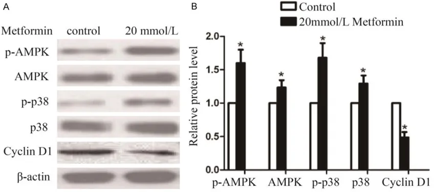
Regulation mechanisms of metformin on 253J non-invasive bladder cancer cells proliferation
Western Blot was performed to detect the pro-tein expression levels of p-AMPK, AMPK, p38, p-p38 and Cyclin D1 in the 253J cells after the treatment of metformin for 48 h. The results showed that, compared with the control group, the protein expression levels of p-AMPK, AM- PK, p38, p-p38 were strengthened significant- ly after the treatment of metformin at the con-centration of 20 mmoL/L. However, the protein expression level of Cyclin D1 was decreased significantly (P<0.05, Figure 5A and 5B).
Discussion
[image:4.612.97.518.76.188.2]Because metformin has such characteristics as less side-effect, hypoglycemic stability, lac-tic acidosis and hypoglycemia, it has been widely used in the treatment of Type 2 mellitus. Recent studies showed that metformin could significantly inhibit the proliferation of esopha-geal carcinoma, breast cancer, lung cancer and other types of tumor cells [8, 9]. Consistent with the previous studies, this study found that metformin had significant inhibitory effects on the proliferation of low grade 253J non-invasive bladder cancer cells, which also showed time and concentration dependence (Figure 1A and 1B). As for cell cycle and cell apoptosis, metfor-min could significantly promote the apoptosis of 253J cells and made the cell cycle block in
Figure 3. The effects of metformin on apoptosis of 253J non-invasive bladder cancer cells. A: Control; B: 5 mmoL/L; C: 10 mmoL/L; D: 20 mmoL/L.
Figure 4. Apoptosis ratio of 253J non-invasive blad-der cancer cells after treatment of different concen-tration of metformin. *Compared with the control
[image:4.612.92.285.244.379.2]the G0/G1 phase and these effects would be strengthened gradually with the increased con-centration of metformin. Obviously, besides the effects on decreasing blood sugar, metformin also could prevent from non-invasive bladder cancer.
Other studies showed that metformin did not only have antitumor activity, but also could significantly reduce tumor incidence rate and relevant mortality. It promoted the apoptosis of tumor cells mainly by stagnating cell cycle and activating signal transduction pathway of cell apoptosis [10, 11]. The relationship be- tween cell cycle and tumor development has always been a hot spot. In a series of cytokines regulating cell cycle, regulators for G1 phase had mutations or changes which could cause uncontrolled cell proliferation or even tumors [12]. Cyclin D1, one of the most important tein in cell cycle control, was regulated by pro-tein kinase with cell cycle dependence. Some studies suggested that when the expression of Cyclin D1 was out of control, it could lead to the abnormal proliferation of the cells and the overexpression which was connected with the occurrence and the development of the tumors [13]. For example, in the cells of pan- creatic cancer, metformin could block the cell cycle of the tumor in G0/G1 phase by lower- ing the expression of Cyclin D1 and thus inhi- bit the proliferation of the cells [14]. And other studies confirmed that Cyclin D1 proteins in low-grade and well-differentiated bladder
can-cer cells were usually overexpression and were related to the recurrence of the superficial bladder cancer [15]. In our research, the results showed that metformin could block 253J cells in G0/G1 phase to inhibit the proliferation of the cells by lowering the expression of Cyclin D1, suggesting that Cyclin D1 proteins might play an important role in the occurrence and the development of low-grade bladder tumors (Figures 2 and 5).
The mutation and abnormal expression of MAPK signaling pathway and its downstream target gene, Cyclin D1, are related with the occurrence and the development of various kinds of tumors. MAPK family includes many subfamilies such as p38, ERK and c-jun. These families form many signaling pathways, among which the p38 and AMPK are two major ways. As the energy receptor of the cells, MAPK sig-naling pathway plays an important role in regu-lating the energy metabolism, apoptosis and proliferation of the cells [16]. The deactivation of AMPK signaling pathway could lead to the abnormal proliferation of bladder cancer cells and could also inhibit the proliferation and the biosynthesis of the cells by two ways. One is inhibiting the expression of Cyclin D1 proteins through P53/p21 signal pathway to block the cells in G1 phase. Some studies confirmed that metformin could realize the antiproliferative effects of breast cancer by activating AMPK [17, 18]. Moreover, in the T24 cell lines of blad-der tumors, metformin could inhibit the
[image:5.612.93.519.75.263.2]Research of the effects of metformin
eration of the T24 cells by activating AMPK [19]. Other studies also suggested that met- formin could facilitate the apoptosis through AMPK signaling pathway [20]. For example, in the non-small cells of lung cancer, metformin could activate AMPK and then inhibit mTOR sig-naling pathway, which could thus facilitate the apoptosis of cells [21]. In addition, the activa-tion of p38 AMPK signaling pathway could str- engthen the antitumor effects of metformin. Some studies suggested that p38 AMPK block-er could inhibit the prolifblock-eration of SKOV3 ovar-ian cancer cell line. However, metformin could enhance the effects of the inhibition [22]. P38-AMPK signaling pathway played an important role in the apoptosis of cells and applying spe-cific p38 MAPK blocker could lower the induc-tion effects of metformin on the apoptosis of cell lines of lung adenocarcinoma [23]. In this study, we found that metformin could also ac- tivate AMPK and p-38 in 253J non-invasive bladder cancer cell, and these facts suggested that metformin could inhibit the cell prolifera-tion and facilitate the cell apoptosis (Figure 5). However, the occurrence and the development of the bladder tumors are multistep and com-plex processes. Besides AMPK signaling path-way and Cyclin D1 proteins, there may be other signaling pathways affecting metformin’s inhi-bition effects on the proliferation and induction function of apoptosis of non-invasive bladder cancer cell. In addition, the anti-tumor effects of metformin still needs further confirmation from the multi-center randomized controls and clinical studies with a larger sample size. As a kind of hypoglycemic agent, the blood concen-tration of the maximum dosage of metformin in human body is 10 mmoL/L, which is lower than that of this study. Whether the human body owns good tolerance of high concentration of metformin also needs to be confirmed by a larg-er numblarg-er of studies.
In conclusion, metformin could inhibit the pro-liferation of low-grade non-invasive bladder cancer cell and facilitate its apoptosis and make its cell cycle block in the G0/G1 phase. The mechanisms of action might be related with the decrease of Cyclin D1 expression and the activation of AMPK signaling pathway, sug-gesting that metformin might become the new aided drug to patients with non-invasive blad-der cancer, which might provide a new method for the clinical treatment.
Disclosure of conflict of interest
None.
Address correspondence to: Jianjun Yu, Department of Urinary Surgery, Shanghai Fengxian District Cen- tral Hospital, No.6600 Nanfeng Road, Fengxian Dis- trict, Shanghai 201499, China; Department of Urinary Surgery, Affiliated Sixth People’s Hospital of Shanghai Jiao Tong University, No.600 Yishan Road, Shanghai 200233, China. Tel: +86-021-57424577; +86-021-64369181; Fax: +86-021-57424577; +86- 021-64701361; E-mail: Yujj917@163.com
References
[1] Heidari F, Abbas Zade S, Mir Hosseini SH and Ghadian A. Metformin for the prevention of bladder cancer recurrence: is it effective? Nephrourol Mon 2016; 8: e30261.
[2] Nargund VH, Tanabalan CK and Kabir MN. Management of non-muscle-invasive (superfi-cial) bladder cancer. Semin Oncol 2012; 39: 559-572.
[3] Chen GF, Shi TP, Wang BJ, Wang XY and Zang Q. Efficacy of different resections on non-mus-cle-invasive bladder cancer and analysis of the optimal surgical method. J Biol Regul Homeost Agents 2015; 29: 465-470.
[4] Wojciechowska J, Krajewski W, Bolanowski M, Krecicki T and Zatonski T. Diabetes and can-cer: a review of current knowledge. Exp Clin Endocrinol Diabetes 2016; 124: 263-275. [5] Wu GF, Zhang XL, Luo ZG, Yan JJ, Pan SH, Ying
XR, Pan JG, Zhang GF. Metformin therapy and prostate cancer risk: a meta-analysis of obser-vational studies. Int J Clin Exp Med 2015; 8: 13089-98.
[6] Franciosi M, Lucisano G, Lapice E, Strippoli GF, Pellegrini F and Nicolucci A. Metformin therapy and risk of cancer in patients with type 2 dia-betes: systematic review. PLoS One 2013; 8: e71583.
[7] Zhang T, Guo P, Zhang Y, Xiong H, Yu X, Xu S, Wang X, He D and Jin X. The antidiabetic drug metformin inhibits the proliferation of bladder cancer cells in vitro and in vivo. Int J Mol Sci 2013; 14: 24603-24618.
[8] Pan Q, Yang GL, Yang JH, Lin SL, Liu N, Liu SS, Liu MY, Zhang LH, Huang YR, Shen RL, Liu Q, Gao JX and Bo JJ. Metformin can block precan-cerous progression to invasive tumors of blad-der through inhibiting STAT3-mediated signal-ing pathways. J Exp Clin Cancer Res 2015; 34: 77.
squamous cell carcinoma. BMC Cancer 2012; 12: 517.
[10] Guo LS, Li HX, Li CY, Zhang SY, Chen J, Wang QL, Gao JM, Liang JQ, Gao MT and Wu YJ. Vitamin D3 enhances antitumor activity of metformin in human bladder carcinoma SW-780 cells. Pharmazie 2015; 70: 123-128. [11] Park SH, Lee DH, Kim JL, Kim BR, Na YJ, Jo MJ,
Jeong YA, Lee SY, Lee SI, Lee YY and Oh SC. Metformin enhances TRAIL-induced apoptosis by Mcl-1 degradation via Mule in colorectal cancer cells. Oncotarget 2016; 7: 59503-59518.
[12] Manning AL and Dyson NJ. pRB, a tumor sup-pressor with a stabilizing presence. Trends Cell Biol 2011; 21: 433-441.
[13] Reis-Filho JS, Savage K, Lambros MB, James M, Steele D, Jones RL and Dowsett M. Cyclin D1 protein overexpression and CCND1 amplifi-cation in breast carcinomas: an immunohisto-chemical and chromogenic in situ hybridisa-tion analysis. Mod Pathol 2006; 19: 999-1009.
[14] Liu Q, Yuan W, Tong D, Liu G, Lan W, Zhang D, Xiao H, Zhang Y, Huang Z, Yang J, Zhang J and Jiang J. Metformin represses bladder cancer progression by inhibiting stem cell repopula-tion via COX2/PGE2/STAT3 axis. Oncotarget 2016; 7: 28235-28246.
[15] Garcia-España A, Salazar E, Sun TT, Wu XR and Pellicer A. Differential expression of cell cycle regulators in phenotypic variants of transgeni-cally induced bladder tumors: implications for tumor behavior. Cancer Res 2005; 65: 1150-1157.
[16] Acosta AM and Kadkol SS. Mitogen-activated protein kinase signaling pathway in cutaneous melanoma: an updated review. Arch Pathol Lab Med 2016; 140: 1290-1296.
[17] Cai H, Zhang Y, Han TK, Everett RS and Thak- ker DR. Cation-selective transporters are criti-cal to the AMPK-mediated antiproliferative ef-fects of metformin in human breast cancer cells. Int J Cancer 2016; 138: 2281-2292. [18] Peng M, Huang Y, Tao T, Peng CY, Su Q, Xu
W, Darko KO, Tao X, Yang X. Metformin and gefitinib cooperate to inhibit bladder cancer growth via both AMPK and EGFR pathways joining at Akt and Erk. Sci Rep 2016; 6: 28611. [19] Zhu J, Zheng Y, Zhang H and Sun H. Target-
ing cancer cell metabolism: the combination of metformin and 2-Deoxyglucose regulates apoptosis in ovarian cancer cells via p38 MAPK/JNK signaling pathway. Am J Transl Res 2016; 8: 4812-4821.
[20] Queiroz EA, Puukila S, Eichler R, Sampaio SC, Forsyth HL, Lees SJ, Barbosa AM, Dekker RF, Fortes ZB and Khaper N. Metformin induces apoptosis and cell cycle arrest mediated by oxidative stress, AMPK and FOXO3a in MCF- 7 breast cancer cells. PLoS One 2014; 9: e98207.
[21] Sayyid RK and Fleshner NE. Potential role for metformin in urologic oncology. Investig Clin Urol 2016; 57: 157-164.
[22] Xie Y, Peng Z, Shi M, Ji M, Guo H and Shi H. Metformin combined with p38 MAPK inhibitor improves cisplatin sensitivity in cisplatinresis-tant ovarian cancer. Mol Med Rep 2014; 10: 2346-2350.