Original Article
Expression of CD151/Tspan24 and
integrin alpha 3 complex in aid of prognostication
of HER2-negative high-grade ductal carcinoma in situ
Hanna M Romanska1, Piotr Potemski2, Renata Kusinska1, Janusz Kopczynski3, Rafal Sadej4, Radzislaw
Kordek1
1Department of Pathology Medical University of Łόdź, Poland; 2Department of Chemotherapy,Medical University of Łόdź and M. Kopernik Memorial Hospital, Poland; 3Holycross Cancer Centre, Kielce, Poland; 4Department of Molecular Enzymology, Intercollegiate Faculty of Biotechnology, Medical University of Gdańsk, Poland
Received June 1, 2015; Accepted July 21, 2015; Epub August 1, 2015; Published August 15, 2015
Abstract: The pro-tumorigenic and pro-metastatic functions of the tetraspanin protein CD151 (Tspan24) are thought to be dependent on its ability to form complexes with laminin-binding integrin receptors (i.e. alpha6beta1, alpha-3beta1, alpha6beta4). We have previously reported that in invasive ductal carcinoma (IDC), CD151/alpha3beta1 complex was of prognostic value in patients with HER2-negative tumors. Extrapolating these findings to the pre-invasive setting, we aimed to make an assessment of a potential relationship between expression of the CD151/ alpha3beta1 complex in DCIS and Van Nuys prognostic index (VNPI) in high-grade ductal carcinoma in situ (DCIS) in relationto the HER2 status. Protein distributions were analyzed in 49 samples of pure DCIS using immunohis-tochemistry. For each case immunoreactivity was assessed in at least 5 ducts (325 ducts in total) and an average score was taken for statistical analyses. When analyzed in the whole cohort, there was no statistical association between the VNPI and any of the proteins scored either separately or in combination. When stratified according to the HER2 status, in the HER2-negative subgroup, CD151 assessed in combination with alpha3beta1 was signifi -cantly correlated with VNPI (P = 0.044), while neither protein analyzed individually showed any significant link with the prognostic index. Expression of the CD151/alpha3beta1 complex in HER2-negative DCIS might reflect tumor behavior relevant to the patient outcome and thus might aid prognostication of the disease.
Keywords: CD151/tspan24, integrin alpha3, DCIS, HER2-negative, invasive progression
Introduction
Traditionally considered as the non-obligate precursor of invasive breast cancer (BCa), DCIS is characterized by proliferation of neoplastic cells within the duct lumen. However, increas-ing evidence indicates that, contrary to the gen-erally accepted paradigm of a stepwise evolu-tion of BCa, the molecular program conferring invasive growth is already switched on at the BCa pre-invasive stages, i.e., at the inception of the DCIS→IDC transition [1, 2]. This implicates that application of the knowledge of IDC biology to the DCIS setting is likely to unravel several aspects of DCIS pathophysiology relevant to the clinic. In practical terms, molecular markers identifying subsets of cells driving disease
progression might be shared by IDC and DCIS [3].
coordinate integrin-dependent signalling net-works [11]. Our finding that in IDC expression of CD151 in combination with integrin alpha3be-ta1 represents a more stringent indicator of poor survival than CD151 alone seems to reflect this interaction [12].
Results of our recent study indicate that CD151 may play an important role also in the develop-ment of pre-invasive lesions in the mammary gland. CD151 was found elevated in human DCIS and was shown to correlate with a higher tumor grade [13]. There was no association with disease pattern and the frequency of posi-tivity for CD151 was similar in the pure cases to those with established invasion [13]. In a model of DCIS based on the HB2 non-tumorigenic mammary epithelial cell line, it was demon-strated that CD151 promoted proliferation of cells both in vivo (mouse xenografts) and in a 3-D in vitro set-ups. Although under experimen-tal conditions proliferative activity of CD151 appeared independent of its association integ-rin alpha3beta1, the presence of fully function-al CD151 was not sufficient to function-allow prolifera-tion of α3β1-negative cells in 3D ECM [13]. Interdependence between CD151 and α3β1 in the intricate environment of developing human DCIS and its impact on disease prognosis has not been studied.
In clinical practice, morphological examination of DCIS samples using conventional histology and classification according to the Van Nuys scoring system remains the ‘Gold Standard’ for estimation of the anticipated biological
behav-ior of the tumor and, consequently, the guide-line for treatment options [14]. However, the biology and natural history of DCIS are still poorly understood. Numerous attempts to iden-tify biological markers of a risk of invasive pro-gression have not produced conclusive results and a value of HER2 as a sole prognostic indi-cator in DCIS remains controversial [15-19]. We have shown previously that an impact of CD151 on IDC patient’s survival was inversely corre-lated with the level of HER2 expression [12]. The aim of the study was therefore to assess a potential clinical significance of the CD151/ α3β1 complex in DCIS by evaluating its relation-ship with the Van Nuys prognostic index (VNPI) in relation to the HER2 status. As high-grade DCIS is considered to progress more rapidly to invasive disease [20], the study focused exclu-sively on grade 3 DCIS lesions.
Material and methods
Patient selection and samples
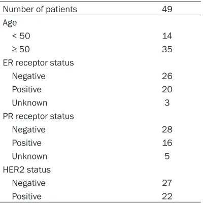
[image:2.612.90.290.83.283.2]Specimens of high-grade pure DCIS were obtained from 49 patients treated at the Oncology Department of Copernicus Memorial Hospital in Łódź, Poland and the Holycross Cancer Center in Kielce, Poland, between 2011 and 2015. The characteristics of the popula-tion relevant to the study are summarized in the Table 1. The use of the samples was approved by the Local Research Ethics Committee (# RNN/284/13/KE).
Immunohistochemistry
The initial pathological diagnosis was con-firmed on haematoxylin/eosin-stained sec-tions. ER/PR/HER2 status was determined by routine histological assessment. Serial 5 µm paraffin sections of formalin-fixed blocks we- re processed for immunohistochemistry for CD151 (monoclonal mouse anti-human; 1:100; Novocastra, UK) and integrin alpha3beta1 (INTA3) (polyclonal goat anti-human; 1:200, Santa Cruz, UK) using protocols described pre-viously [12]. As a negativecontrol for the immu-nostaining, primary antibodies were replaced by non-immune sera.
Scoring of immunostaining for CD151 was based on the Guidelines for Scoring recom-mended by the manufacturer of the Hercep- TestTM (Dako, Denmark) and modified as
fol-lows: i) 0/negative-no reactivity or only partially Table 1. Patient characteristics
Number of patients 49
Age
< 50 14
≥ 50 35
ER receptor status
Negative 26
Positive 20
Unknown 3
PR receptor status
Negative 28
Positive 16
Unknown 5
HER2 status
Negative 27
membranous reactivity in ≤ 10% of tumor cells; ii) 1+/negative-faint membranous or partially membranous in ≥ 10% of tumor cells; iii) 2+/ positive-weak to moderate complete membra-nous in ≥ 10% of tumor cells; iv) 3+/positive-strong complete membranous in ≥ 30% of the tumor cells. Taking into account well recognized heterogeneity of DCIS lesions within an individ-ual case, for each specimen, immunoreactivity was assessed in at least five the largest ducts (325 ducts in total) and an average score was taken for statistical analyses. Scoring of immu-noreactivity for INTA3 was carried out as fol-lows: i) 0/negative-no reactivity, ii) 1+/positive-weak to moderate membranous and/ or cyto-plasmic staining in ≤ 10% of tumor cells; iii) 2+/ positive-moderate membranous and/or cyto-plasmic staining in ≥ 10% of tumor cells; iv) 3+/ positive-strong membranous and/or cytoplas-Table 2. Association between CD151 and/or INTA3 expression and tumor phenotypic charac-teristics
Feature
P value INTA3
(high: n = 33) (high: n = 25)CD151 CD151/INTA3 (high: n = 16) 0.610
CD151 0.610
HER2 (-) 0.468 0.064 0.468
ER (-) 0.065 0,041 0.334
PR 0.421 0.075 0.317
[image:4.612.90.303.108.226.2]ER/PR 0.065 0.041 0.334
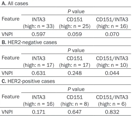
Table 3. Association between CD151 and/or INTA3 expression and VNPI
A. All cases
Feature
P value INTA3
(high: n = 33) (high: n = 25)CD151 CD151/INTA3 (high: n = 16)
VNPI 0.597 0.059 0.070
B. HER2-negative cases
Feature
P value INTA3
(high: n = 17) (high: n = 17)CD151 CD151/INTA3 (high: n = 10)
VNPI 0.631 0.248 0.044
C. HER2-positive cases
Feature
P value INTA3
(high: n = 16) (high: n = 8)CD151 CD151/INTA3 (high: n = 6)
VNPI 0.171 0.647 0.832
mic staining in ≥ 30% of the tumor cells. Immunohistochemical staining was evaluated and scored independently by two observers (HR, RK*). The agreement on staining intensity was > 90%. Where there was disagreement, intensity was determined by consensus. As epithelial cells of the normal gland displayed strong immunoreactivty for CD151 and much weaker for ITNA3, final scores were dichoto-mized into: a) ‘negative’ and b) ‘positive’ for CD151/0-2; INTA3/0 and CD151/3 and INTA3/1-3, respectively.
Statistical analysis
The data were assessed by unpaired t test and chi-square or Fisher exact test using the StatsDirect software (StatsDirect Ltd, Altrincham, UK). Two-sided P value < 0.05 was considered as significant.
All cases were reviewed and stained for ErbB2/ HER2 using HercepTestTM (Dako). Immunohis-
tochemical staining was recorded using a semiquantitative scoring system recommend-ed by the manufacturer.
Follow-up data were available only in 6 cases precluding a DFS analysis.
Results
Expression of CD151 and INTA3 in normal
mammary gland
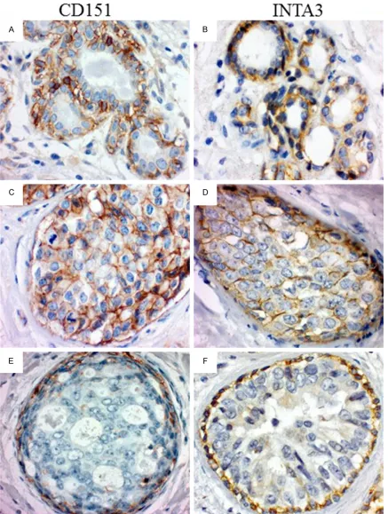
Both CD151 and INTA3 showed moderate to strong, predominantly, membranous immuno-reactivity, confined to the basal and lateral sur-faces of the basal layer cells, with no or very weak staining in luminal epithelial cells (Figure 1A, 1B).
Expression of CD151 and INTA3 in DCIS
[image:4.612.90.305.276.466.2]present in all cells of the lesion (Figure 1D) and restricted to the cells at the tumor-stroma inter-face (Figure 1F), were observed in 17 and 16 cases, respectively.
No apparent histological differences between DCIS specimens positive and negative for CD151 and/or INTA3 were noticed.
Expression of CD151 was inversely associated with ER (P = 0.041) and hormone receptor sta-tus (ER and/or PR) (P = 0.041) and there was a trend towards statistical significance of an inverse relationship with HER2 (P = 0.064). Level of INTA3 expression did not correlate with any of the tumor phenotypic characteristics (Table 2). Expression of neither CD151 nor INTA3 when assessed alone or in combination, correlated with the VNPI.
As reported previously in IDC, CD151/integrin alpha3 was of prognostic value only in pa- tients with HER2-negative tumors [12]. Assu- ming that the traits of cell invasiveness are maintained throughout the BCa development, we looked at the relationship between expres-sion of CD151/INTA3 and an equivalent of prognostic indicator in DCIS, the VNPI, in the subpopulation of HER2-negatvie patients. In- deed, levels of CD151/INTA3 expression corre-lated with VNPI in HER2-negative patients (P = 0.044), whereas there was no significant rela-tionship with either CD151 (P = 0.248) or INTA3 (P = 0.631) when assessed individually (Table 3). Respective values in HER2-positive cases were for: i) CD151/INTA3 P = 0.834, ii) CD151, P= 0.647 and iii) INTA3 P = 0.171.
Discussion
Following our report implicating the tetraspanin CD151 in the development of mammary carci-noma in situ [13], here we have undertaken an evaluation of a relationship between CD151 in complex with its principal molecular partner, the integrin alpha3beta1 and a prognostic indi-cator in high grade DCIS. Due to low availability of pure high grade DCIS, the number of speci-mens was relatively low. However, results of our statistical analyses clearly demonstrate that in a subgroup of HER2-negative tumors, CD151 and integrin apha3 assessed in combination, were significantly correlated with VNPI, while neither protein analyzed individually showed any significant link with the prognostic index.
To date, HER2/ErbB2 is one of the most exten-sively studied biological prognostic factors in IDC, but available data on its importance in DCIS is scarce and contradicting [15-19]. While there have been several reports on lack of sig-nificant association between HER2 and the risk of recurrence after a DCIS [16, 18, 19], a recent study by Zhou et al. demonstrated that a high level of HER2 overexpression was highly predic-tive of disease relapse [21]. Furthermore, HER2 status determined the type of recurrence. HER2+DCISs were more likely to recur as new in situ lesions, while HER2-tumors were related to recurrences being invasive. This is consis-tent with the observation by Lu et al., that although HER2 robustly promotes growth fac-tor-independent cell proliferation, it is unable to induce basement membrane breakdown and subsequent invasive growth [22]. Activation of additional mechanisms acting in cooperation with HER2-mediated signaling are required to induce invasive potential in HER2-positive cells, conferring a high risk of progression to IDC [22].
the intrinsic changes associated with genetic mutations, other events, and in particular, interaction with the stroma may play a critical role in the progression to invasion [25]. Like its invasive counterpart, DCIS is characterized by a high degree of both inter-and intra-tumor het-erogeneity in terms of conventional histological grades, prognostic biomarkers and patterns of growth [26]. It is likely that in certain biological contexts, this evolution is due to the competi-tion for dominance among multiple cell sub-clones co-existing within an individual tumor [26]. The selective pressure of dominant HER2-negative cells might contribute to the develop-ment of IDC and provide an explanation for the low incidence of HER2-positive invasive tumors (20-25%), as compared to a high frequency of HER2 expression seen in DCIS (50-60%) [20]. In light of previous reports and current findings, it is tempting to speculate that the presence of CD151 on the membrane of HER2-negative cell might be a common trait to pre-invasive and malignant breast cancer cells, that makes them more responsive to the stimuli released by the environment, thus conferring their invasive phenotype.
The Van Nuys prognostic index (VNPI), described as a numerical representation of measurable prognostic factors (tumor size, margin width, nuclear grade, the presence or absence of comedonecrosis and, recently added age), was introduced in 1996 as an aid to be used in con-junction with clinical experience in the treat-ment decision-making process [14]. A multivari-ate regression analysis shown that, unlike its individual components, the VNPI treated as whole is a good prognostic indicator of a risk of local recurrence [27, 28]. VNPI has been based on recurrence data from large series of DCIS patients and shown a good correlation with dis-ease outcome but a biological meaning of this empirically developed indicator in terms of a relationship to molecular mechanisms underly-ing DCIS progression remains poorly under-stood. Results of our study demonstrating a significant correlation between VNPI and CD151 seem to shed some light on pathophysi-ology of a subtype of DCIS tumors and suggest that CD151 expression might be a trait, com-mon to IDC and DICS, identifying a subpopula-tion of DCIS HER2-negative cells likely to drive an invasive progression.
In summary, our results suggest that acting in complex with the integrin alpha3beta1 CD151 might play a role in the pathophysiology of DCIS and contribute to the process of invasive pro-gression of HER2-negative lesions. Further analysis of a large cohort of DCIS patients with a long follow-up as well as mechanistic studies using experimental models are required to assess the emerging possibility for identifica-tion of DCIS lesions with invasive characteris-tics, potentially to be guiding clinical evaluation and management.
Acknowledgements
This work was supported by the National Science Centre (grant No. UMO-2013/09/B/ NZ4/02512 to HR).
Disclosure of conflict of interest
None.
Address correspondence to: Dr. Hanna M Romans- ka, Department of Pathology Medical University of Łόdź, 92-213 Łódź, Poland. Tel: +48-42 272 5605; Fax: +48-42 272 5604; E-mail: hanna.romanska@ gmail.com
References
[1] Ma XJ, Salunga R, Tuggle JT, Gaudet J, Enright E, McQuary P, Payette T, Pistone M, Stecker K, Zhang BM, Zhou YX, Varnholt H, Smith B, Gadd M, Chatfield E, Kessler J, Baer TM, Erlander MG and Sgroi DC. Gene expression profiles of human breast cancer progression. Proc Natl Acad Sci U S A 2003; 100: 5974-5979. [2] Husemann Y, Geigl JB, Schubert F, Musiani P,
Meyer M, Burghart E, Forni G, Eils R, Fehm T, Riethmuller G and Klein CA. Systemic spread is an early step in breast cancer. Cancer Cell 2008; 13: 58-68.
[3] Clark SE, Warwick J, Carpenter R, Bowen RL, Duffy SW and Jones JL. Molecular subtyping of DCIS: heterogeneity of breast cancer reflected in pre-invasive disease. Br J Cancer 2011; 104: 120-127.
[4] Boucheix C and Rubinstein E. Tetraspanins. Cell Mol Life Sci 2001; 58: 1189-1205. [5] Hemler ME. Tetraspanin functions and
associ-ated microdomains. Nat Rev Mol Cell Biol 2005; 6: 801-811.
[7] Romanska HM and Berditchevski F. Tetras- panins in human epithelial malignancies. J Pathol 2011; 223: 4-14.
[8] Yang XH, Richardson AL, Torres-Arzayus MI, Zhou P, Sharma C, Kazarov AR, Andzelm MM, Strominger JL, Brown M and Hemler ME. CD151 accelerates breast cancer by regulat-ing alpha 6 integrin function, signalregulat-ing, and molecular organization. Cancer Res 2008; 68: 3204-3213.
[9] Sadej R, Romanska H, Baldwin G, Gkirtzimanaki K, Novitskaya V, Filer AD, Krcova Z, Kusinska R, Ehrmann J, Buckley CD, Kordek R, Potemski P, Eliopoulos AG, Lalani E and Berditchevski F. CD151 regulates tumorigenesis by modulating the communication between tumor cells and endothelium. Mol Cancer Res 2009; 7: 787-798.
[10] Kwon MJ, Park S, Choi JY, Oh E, Kim YJ, Park YH, Cho EY, Kwon MJ, Nam SJ, Im YH, Shin YK and Choi YL. Clinical significance of CD151 overexpression in subtypes of invasive breast cancer. Br J Cancer 2012; 106: 923-930. [11] Sadej R, Grudowska A, Turczyk L, Kordek R and
Romanska HM. CD151 in cancer progression and metastasis: a complex scenario. Lab Invest 2014; 94: 41-51.
[12] Novitskaya V, Romanska H, Kordek R, Potemski P, Kusinska R, Parsons M, Odintsova E and Berditchevski F. Integrin alpha3beta1-CD151 complex regulates dimerization of ErbB2 via RhoA. Oncogene 2014; 33: 2779-2789. [13] Novitskaya V, Romanska H, Dawoud M, Jones
JL and Berditchevski F. Tetraspanin CD151 regulates growth of mammary epithelial cells in three-dimensional extracellular matrix: im-plication for mammary ductal carcinoma in situ. Cancer Res 2010; 70: 4698-4708. [14] Silverstein MJ, Lagios MD, Craig PH, Waisman
JR, Lewinsky BS, Colburn WJ and Poller DN. A prognostic index for ductal carcinoma in situ of the breast. Cancer 1996; 77: 2267-2274. [15] Provenzano E, Hopper JL, Giles GG, Marr G,
Venter DJ and Armes JE. Biological markers that predict clinical recurrence in ductal carci-noma in situ of the breast. Eur J Cancer 2003; 39: 622-630.
[16] Ringberg A, Anagnostaki L, Anderson H, Idvall I and Ferno M. Cell biological factors in ductal carcinoma in situ (DCIS) of the breast-relation-ship to ipsilateral local recurrence and his- topathological characteristics. Eur J Cancer 2001; 37: 1514-1522.
[17] Roses RE, Paulson EC, Sharma A, Schueller JE, Nisenbaum H, Weinstein S, Fox KR, Zhang PJ and Czerniecki BJ. HER-2/neu overexpression as a predictor for the transition from in situ to invasive breast cancer. Cancer Epidemiol Bio- markers Prev 2009; 18; 1386-1389.
[18] Latta EK, Tjan S, Parkes RK and O’Malley FP. The role of HER2/neu overexpression/amplifi -cation in the progression of ductal carcinoma in situ to invasive carcinoma of the breast. Mod Pathol 2002; 15: 1318-1325.
[19] Park K, Han S, Kim HJ, Kim J and Shin E. HER2 status in pure ductal carcinoma in situ and in the intraductal and invasive components of in-vasive ductal carcinoma determined by fluo -rescence in situ hybridization and immunohis-tochemistry. Histopathology 2006; 48: 702-707.
[20] Allegra CJ, Aberle DR, Ganschow P, Hahn SM, Lee CN, Millon-Underwood S, Pike MC, Reed SD, Saftlas AF, Scarvalone SA, Schwartz AM, Slomski C, Yothers G and Zon R. National Institutes of Health State-of-the-Science Con- ference statement: Diagnosis and Manage- ment of Ductal Carcinoma In Situ September 22-24, 2009. J Natl Cancer Inst 2010; 102: 161-169.
[21] Zhou W, Johansson C, Jirstrom K, Ringberg A, Blomqvist C, Amini RM, Fjallskog ML and Warnberg F. A Comparison of Tumor Biology in Primary Ductal Carcinoma In Situ Recurring as Invasive Carcinoma versus a New In Situ. Int J Breast Cancer 2013; 2013: 582134.
[22] Lu J, Guo H, Treekitkarnmongkol W, Li P, Zhang J, Shi B, Ling C, Zhou X, Chen T, Chiao PJ, Feng X, Seewaldt VL, Muller WJ, Sahin A, Hung MC and Yu D. 14-3-3zeta Cooperates with ErbB2 to promote ductal carcinoma in situ progres-sion to invasive breast cancer by inducing epi-thelial-mesenchymal transition. Cancer Cell 2009; 16: 195-207.
[23] Sadej R, Romanska H, Kavanagh D, Baldwin G, Takahashi T, Kalia N and Berditchevski F. Tetraspanin CD151 regulates transforming growth factor beta signaling: implication in tu-mor metastasis. Cancer Res 2010; 70: 6059-6070.
[24] Deng X, Li Q, Hoff J, Novak M, Yang H, Jin H, Erfani SF, Sharma C, Zhou P, Rabinovitz I, Sonnenberg A, Yi Y, Zhou P, Stipp CS, Kaetzel DM, Hemler ME and Yang XH. Integrin-associated CD151 drives ErbB2-evoked mam-mary tumor onset and metastasis. Neoplasia 2012; 14: 678-689.
[26] Allred DC, Wu Y, Mao S, Nagtegaal ID, Lee S, Perou CM, Mohsin SK, O’Connell P, Tsimelzon A and Medina D. Ductal carcinoma in situ and the emergence of diversity during breast can-cer evolution. Clin Cancan-cer Res 2008; 14: 370-378.
[27] Gilleard O, Goodman A, Cooper M, Davies M and Dunn J. The significance of the Van Nuys prognostic index in the management of ductal carcinoma in situ. World J Surg Oncol 2008; 6: 61.