Original Article
Knockdown of CUL4B inhibits proliferation and
promotes apoptosis of colorectal cancer cells through
suppressing the Wnt/β-catenin signaling pathway
Baoji Song1*, Hongjie Zhan2*, Quan Bian1, Jiarui Li3
1Department of General Surgery, Tianjin Hospital, Tianjin 300211, China; 2Department of Gastric Cancer, Tianjin
Cancer Hospital, Key Laboratory of Cancer Prevention and Treatment of Tianjin City, Tianjin Medical University, Tianjin 300060, China; 3Department of Emergency Medicine, Tianjin Hospital, Tianjin 300211, China. *Equal
con-tributors.
Received March 23, 2015; Accepted May 22, 2015; Epub September 1, 2015; Published September 15, 2015 Abstract: Colorectal cancer is one of the leading causes of cancer related deaths worldwide. Cullin 4B (CUL4B) is over-expressed in diverse cancer types. However, the function and precise molecular mechanism of CUL4B in colorectal cancer remains largely unknown. Therefore, in this study, we examined the expression of CUL4B in colorectal cancer cell lines and its effects on cellular proliferation and apoptosis, and the underlying mechanism
was also explored. Our results showed that CUL4B was significantly overexpressed in colorectal cancer cell lines.
Silencing CUL4B obviously inhibited proliferation and tumorigenicity of colorectal cancer cells both in vitro and in vivo, and it also promoted the apoptosis of colorectal cancer cells. Moreover, knockdown of CUL4B inhibited the
expression of β-catenin, cyclin D1 and c-Myc in colorectal cancer cells. Taken together, these results showed that
knockdown of CUL4B inhibit proliferation and promotes apoptosis of colorectal cancer cells through suppressing the
Wnt/β-catenin signaling pathway. Therefore, CUL4B may represent a novel therapeutic target for colorectal cancer
treatment.
Keywords:Cullin 4B (CUL4B), colorectal cancer, proliferation, apoptosis, Wnt/β-catenin pathway
Introduction
Colorectal cancer is the third leading cause of cancer associated death in the United States of America [1] and the second most prevalent cancer in China. Although the diagnosis and treatment of colorectal cancer have been
improved in the past years, the efficacy of sur -gery and chemotherapy remains unsatisfacto-ry. This is largely attributed to a lack of com-plete understanding of the exact cause and mechanisms for this malignancy. Therefore, further understanding of the molecular mecha-nisms of cancer progression and the develop-ment of new therapeutic tools based on these mechanisms are required.
Cullin-RING ligases (CRLs) complexes comprise the largest known class of ubiquitin ligases [2]. CRLs regulate diverse cellular processes, including cell cycle progression, transcription, signal transduction and development [3]. CRLs
are multisubunit complexes composed of a cul-lin, RING protein and substrate-recognition sub-unit, which was linked by an adaptor. Cullin 4B (CUL4B) is a component of the Cullin 4B-Ring E3 ligase complex (CRL4B) that functions in proteolysis [4]. It has been reported that loss-of-function mutation in the X-linked CUL4B
molecular mechanism of CUL4B in colorectal cancer, however, remains largely unknown. Therefore, in this study, we examined the expression of CUL4B in colorectal cancer cell lines and its effects on cellular proliferation and apoptosis, and the underlying mechanism was explored.
Materials and methods
Cell culture
Healthy human colon mucosa cell line (NCM460) and colorectal cancer cell lines (SW480, HT-29 and HCT116) were purchased from American Type Culture Collection (ATCC, Manassas, VA), and all the cell lines were
main-tained in Dulbecco’s modified Eagle’s medium (DMEM; Sigma, St. Louis, MO, USA) supple -mented with 10% fetal bovine serum (FBS; Sigma, St. Louis, MO, USA), streptomycin (100 mg/ml), and penicillin (100 mg/ml). All cell lines were cultured at 37°C under 5% CO2.
RT-PCR
Total RNA was extracted from colorectal cancer tissues and cells using Trizol reagent (Abcam, Cambridge, UK) according to the
manufacturer’s instructions. cDNA was synthe -sized from the extracted RNA (4 µg) using
the EasyScript First-Strand cDNA Synthesis
SuperMix kit (Invitrogen, Carlsbad, CA, USA).
PCR amplification was performed using the following primers: CUL4B, 5’-CCTGGAGTTTGT-AGGGTTTGAT-3’ (sense) and 5’-GAGACGGTG-GTAGAAGATTTGG-3’ (antisense); and β-actin, 5’-TTAGTTGCGTTACACCCTTTC-3’ (sense) and 5’-ACCTTCACCGTTCCAGTTT-3’ (antisense). The
PCR conditions included an initial denatu- ration step of 94°C for 2 min, followed by 35 cycles of 94°C for 30 s, 56°C for 30 s, and
72°C for 2 min, and a final elongation step of
72°C for 10 min.
Western blot
Total protein was extracted from colorectal can-cer tissues and cells, then washed with ice-cold PBS and lysed with RIPA Cell Lysis Buffer (Santa Cruz Biotechnology, Santa Cruz, CA, USA). The protein concentrations were determined by the
BCA method. The samples (30 μg protein/lane) were separated on 10% SDS-PAGE and trans
-ferred onto polyvinylidene fluoride (PVDF) mem -branes (Millipore, Boston, MA, USA). After
blocking in TBS buffer (50 mmol/L NaCl, 10 mmol/L Tris, pH 7.4) containing 5% nonfat milk, the blots were incubated with primary
antibodies (CUL4B, β-catenin, anti-cyclin D1, anti-c-Myc or β-actin) (Santa Cruz
Biotechnology, Santa Cruz, CA, USA) at 4°C overnight. Membranes were then washed and incubated with horseradish peroxidase-conjugated secondary antibodies. Expression was visualized by using ECL Western blotting detection reagent (Thermo Fisher Scientific,
RockFord, IL, USA).
RNA interference-mediated knockdown of CUL4B and cell transfection
CUL4B siRNA were constructed by Gimma
company in Shanghai, as follow: 5’-CAAUCUC-CUUGUUUCAGAATT-3’; siRNA con vector (the
random sequence as control that was not
related to CUL4B mRNA) 5’-UUCUCCGAAC-GUGUCACGUTT-3’. For in vitro transfection, 5 × 10⁴ cells were seeded in each cell of a 24-well micro-plate, grown for 24 h to reach 30%-50%
confluence, and then incubated with a mixture
of siRNA and Lipofectamine 2000 reagent
(Invitrogen, Carlsbad, CA, USA) in 100 μl serum-free DMEM, according to the manufacturer’s instructions. The transfection efficiency was
examined by Western blot.
Cell proliferation assay
Cell proliferation was determined by 3- (4.5-methylthiozol-2yl)-2.5-diphenyltetrazolium bromide (MTT) assay. Cells (3 × 10³ cells/well) were seeded into 96-well plates and cultured for 24, 48, 72, and 96 h. MTT (10 ml) was added into each well, and cells were cultured for an additional 4 h. The culture media was
removed and 200 ml DMSO (Sigma, St. Louis,
MO, USA) was added to each well. The absor-bance at 570 nm was measured with a
micro-plate reader (Takara Biotechnology, Dalian,
China).
Cell apoptosis assay
The cell apoptotic ratio was measured by Annexin V-FITC and PI staining followed by
fol-lowed by fixation with 70% ice-cold ethanol and
stored at -20°C overnight. The pellets were washed with cold PBS and stained with propid-ium iodide and Annexin V. Apoptotic cells were
analyzed by FACSCalibur to define as those
positive cells for Annexin V with or without PI staining.
Tumorigenicity assay
Male BALB/c nude mice were obtained (Shanghai Slac Laboratory Animal Co. Ltd,
China) and bred under specific pathogen-free
conditions. CUL4B knockdown cells (1 × 10⁶) and their control cells were injected
subcutane-ously into the opposite flanks of the same
mouse. The resulting tumors were measured once a week, and tumor volumes (mm3) were calculated using the following formula: volume = width2 × length × 0.5 [12]. Tumors were
har-vested 35 days after injection. Data were pre -sented as tumor volumes and tumor weight
(mean ± SD). Animal experiment was carried
out with the approval of the ethics committee of Tianjin Medical University in accordance with the Guide for the Care and Use of Laboratory Animals.
Statistical analysis
All results are reported as means ± SD. Statistical analysis involved Student’s t-test for
the comparison of two groups or one-way ANOVA for multiple comparisons. P < 0.05 was
considered to be significant.
Results
CUL4B is overexpressed in colorectal cancer cell lines
To study the expression of CUL4B in colorectal cancer, we used real-time PCR and Western blotting to determine the mRNA and protein expression of CUL4B in colorectal cancer cell lines. As shown in Figure 1A, CUL4B mRNA expression was obviously increased in colorec-tal cancer cell lines compared to the human colon mucosa cell line (NCM460). Consistent with the results of RT-PCR, Western blot analy-sis showed that CUL4B expression was higher in three colorectal cancer cell lines than the human colon mucosa cell line (Figure 1B).
Knockdown of CUL4B inhibits the proliferation of colorectal cancer cells
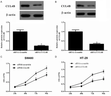
To investigate the role of CUL4B in the prolifera-tion of colorectal cancer cells, we knocked down the expression of CUL4B in SW480 and HT-29 cells using siRNA. Western blot results
showed that the target sequence significantly
decreased the expression of endogenous CUL4B in SW480 (Figure 2A) and HT-29 (Figure 2B) cells compared to the siRNA-scramble group. Then, we used the MTT assay to investi-gate the role of CUL4B in the proliferation of colorectal cancer cells. It was found that knock-down of the expression of CUL4B in SW480
cells significantly inhibited the proliferation of
[image:3.612.92.286.71.405.2]the cancer cells (Figure 2C). Similar results were also observed in HT-29 cells (Figure 2D). Figure 1.CUL4B is overexpressed in colorectal
can-cer cell lines. A. CUL4B mRNA expression was deter-mined in several colorectal cancer cell lines and the human colon mucosa cell line. B. Western blot analy-sis was performed to examine CUL4B expression in colorectal cancer cell lines and the human colon
mu-cosa cell line. β-actin was used as a loading control.
Knockdown of CUL4B promotes the apoptosis of colorectal cancer cells
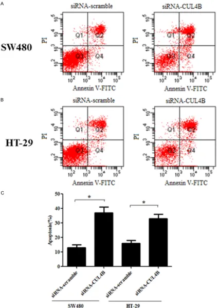
To further investigate the effect of CUL4B on colorectal cancer cell apoptosis, SW480 and HT-29 cells were treated with siRNA-CUL4B and
stained by flow cytometry-based Annexin
V-FITC/PI double staining. As shown in Figure 3A and 3C, after treatment with siRNA-CUL4B for 24 h, the apoptosis rate of SW480 cells was 37.1%, respectively, in comparison with the control group (13.2%). Also in HT-29 cells, the proportion of apoptotic cells was higher than that of the control group (33.4% vs. 16.1%) (Figure 3B and 3C).
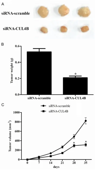
Knockdown of CUL4B inhibited the tumorige-nicity of HT-29 cells in vivo
The role of endogenous CUL4B in the tumor-igeicity of HT-29 cells was examined using a xenograft mouse model. The control cells and cells whose expression of CUL4B was
knock-down were injected into the flanks of
[image:4.612.93.525.73.448.2]4-week-old nude mice. The developed tumors were har-vested 35 days after injection. It was found that knockdown of the expression of CUL4B retard-ed the tumorigenicity of HT-29 in vivo (Figure 4A), as shown by the tumor weight and tumor volume (Figure 4B and 4C). Taken together, these results suggested that down-regulation
Figure 2. Knockdown of CUL4B expression inhibits cell proliferation in vitro. CUL4B -knockdown SW480 (A) and
Figure 3. Knockdown of CUL4B expression promotes cell apoptosis in vitro. SW480 and HT-29 cell apoptosis was
detected through PI staining and the Annexin V method after 48 h of CUL4B-siRNA transfection, followed by flow
of CUL4B attenuated the tumorigenicity of the colorectal cancer cells in vivo.
Knockdown of CUL4B promotes the apoptosis of colorectal cancer cells through Wnt/β-catenin signaling pathway
The Wnt/β-catenin signaling pathway is fre -quently upregulated in a variety of cancers, and
there is some evidence showing that
Wnt/β-catenin signaling pathway is involved in the pro-liferation and apoptosis of cancer. To further investigate a potential mechanism that could be involved in the growth and migration inhibi-tion of colorectal cancer cells, we investigated
the effect of CUL4B on the expression of
β-catenin, cyclin D1 and c-Myc. As shown
in Figure 5, knockdown of the expression of CUL4B markedly downregulated the
expres-sion of β-catenin, cyclin D1 and c-Myc in HT-29
cells. Discussion
The key findings of this study are that CUL4B was significantly upregulated in colorectal can -cer cell lines. Silencing CUL4B inhibited prolif-eration and tumorigenicity of colorectal cancer cells both in vitro and in vivo, and it promoted the apoptosis of colorectal cancer cells. Furthermore, knockdown of CUL4B inhibited
the expression of β-catenin, cyclin D1 and
c-Myc in colorectal cancer cells.
Although CUL4B has been shown to be overex-pressed in several types of cancer and has been associated with cancer progression and development, its role in human colorectal can-cer is still unclear. Herein, we found that CUL4B
was significantly upregulated in colorectal can -cer cell lines, suggesting that CUL4B may func-tion as an oncogenic protein in the develop-ment and progression of human colorectal cancer.
Several reports have provided evidence that CUL4B is involved in the survival and prolifera-tion of malignant phenotypes of cancer cells [7, 9]. For example, knockdown of CUL4B reduced the proliferation of hepatocellular carcinoma cells in vitro and inhibited tumor growth in vivo
[10]. In agreement with these reports, we found that knockdown of CUL4B inhibited prolifera-tion and tumorigenicity of colorectal cancer cells both in vitro and in vivo [10]. Collectively, these results obtained from both in vitro and in vivo experiments strongly suggest that CUL4B overexpression plays important roles in pro-moting the tumorigenicity and progression of human colorectal cancer.
CUL4A is also a member of the cullin family of proteins that composes the multifunctional ubiquitin ligase E3 complex [13]. CUL4A is pro-posed as oncogenic based on its ability to ubiq-uitinate and degrade tumor suppressors, such
as p21, p27, DDB2 and p53 [14, 15]. Like
CUL4A, CUL4B also ubiquitinates histone H2A, H3, and H4, facilitating the recruitment of
[image:6.612.91.287.71.420.2]repair proteins to the damaged DNA [16].
Figure 4. Knockdown of the expression of CUL4B
Recently, it has been shown that silencing CUL4A could promote apoptosis in human lung cancer cell lines [17]. CUL4A downregulation also induced apoptosis in human prostate can-cer cells [18]. Consistent with other reports, our results showed that knockdown of CUL4B obvi-ously promote the apoptosis of colorectal can-cer cells.
Wnt/β-catenin signaling pathway is considered
to play a critical role in promoting tumor pro-gression, dysregulation of cell cycle and apop-tosis [19-21]. Its aberrant activation is a key event in the pathogenesis and progression of
human colorectal cancers [9, 22]. β-catenin is
a main downstream effector of the canonical Wnt signaling pathway and accumulation of
β-catenin is a hallmark of Wnt signaling activa
-tion [23]. In addi-tion, cyclin D1 and c-Myc are downstream targets of Wnt/β-catenin signaling
[24]. It has been reported that emodin signifi
-cantly decreased the expression of β-catenin
and that of its various downstream targets
(cyclin D1, c-Myc, snail, vimentin, MMP-2 and
MMP-9) in colorectal cancer cells [25]. Kaur et al showed that silibinin obviously decreased
β-catenin-dependent T-cell factor-4 (TCF-4)
transcriptional activity and protein expression
of β-catenin target genes such as c-Myc and cyclin D1 in SW480 cells [26]. Consistent with
the above data, in this study, we found that knockdown of the expression of CUL4B
mark-edly downregulated the expression of β-catenin, cyclin D1 and c-Myc in HT-29 cells. Therefore,
these results suggest that CUL4B regulates cell
growth through the Wnt/β-catenin signaling
pathway.
In conclusion, we demonstrated that knock-down of CUL4B inhibits proliferation and pro-Figure 5. Knockdown of CUL4B promotes the apoptosis of colorectal cancer cells through Wnt/β-catenin signaling pathway. (A) The protein levels of β-catenin, cyclin D1 and c-Myc were determined by western blot analysis, β-actin was used as a loading control. (B) β-catenin, (C) Cyclin D1 and (D) c-Myc levels were quantified by Image J software
[image:7.612.96.523.73.412.2]motes apoptosis of colorectal cancer cells
through suppressing the Wnt/β-catenin signal -ing pathway. Therefore, CUL4B may represent a novel therapeutic target for colorectal cancer treatment.
Disclosure of conflict of interest
None.
Address correspondence to: Dr. Quan Bian,
De-partment of General Surgery, Tianjin Hospital, 406
Jiefangnan Road, Hexi District, Tianjin 300211,
China. Tel: +86-22-28255361; E-mail: quan_bian@
163.com; Dr. Jiarui Li, Department of Emergency
Medicine, Tianjin Hospital, 406 Jiefangnan Road,
Hexi District, Tianjin 300211, China. Tel:
+86-22-28255361; E-mail: jiarui_litj@163.com
References
[1] Siegel R, Naishadham D, Jemal A. Cancer sta -tistics, 2013. CA Cancer J Clin 2013; 63: 11-30.
[2] Petroski MD, Deshaies RJ. Function and regu -lation of cullin–RING ubiquitin ligases. Nat Rev Mol Cell Bio 2005; 6: 9-20.
[3] Bosu DR, Kipreos ET. Cullin-RING ubiquitin li -gases: global regulation and activation cycles.
Cell Div 2008; 3: 7.
[4] Jackson S, Xiong Y. CRL4s: the CUL4-RING E3 ubiquitin ligases. Trends Biochem Sci 2009; 34: 562-570.
[5] Tarpey PS, Raymond FL, O’Meara S, Edkins S, Teague J, Butler A, Dicks E, Stevens C, Tofts C,
Avis T. Mutations in CUL4B, which encodes a ubiquitin E3 ligase subunit, cause an X-linked mental retardation syndrome associated with aggressive outbursts, seizures, relative macro-cephaly, central obesity, hypogonadism, pes cavus, and tremor. Am J Hum Genet 2007; 80: 345-352.
[6] Zou Y, Liu Q, Chen B, Zhang X, Guo C, Zhou H, Li J, Gao G, Guo Y, Yan C. Mutation in CUL4B, which encodes a member of cullin-RING ubiq-uitin ligase complex, causes X-linked mental retardation. Am J Hum Genet 2007; 80: 561-566.
[7] Hu H, Yang Y, Ji Q, Zhao W, Jiang B, Liu R, Yuan J, Liu Q, Li X, Zou Y. CRL4B catalyzes H2AK119 monoubiquitination and coordi-nates with PRC2 to promote tumorigenesis. Cancer Cell 2012; 22: 781-795.
[8] Chen Z, Shen BL, Fu QG, Wang F, Tang YX, Hou CL, Chen L. CUL4B promotes proliferation and inhibits apoptosis of human osteosarco-ma cells. Oncol Rep 2014; 32: 2047-2053. [9] Yang Y, Liu R, Qiu R, Zheng Y, Huang W, Hu H,
Ji Q, He H, Shang Y, Gong Y. CRL4B promotes
tumorigenesis by coordinating with SUV39H1/
HP1/DNMT3A in DNA methylation-based epi -genetic silencing. Oncogene 2015; 34: 104-118.
[10] Yuan J, Han B, Hu H, Qian Y, Liu Z, Wei Z, Liang X, Jiang B, Shao C, Gong Y. CUL4B activates
Wnt/β-catenin signalling in hepatocellular
carcinoma by repressing Wnt antagonists. J Pathol 2015; 235: 784-795.
[11] Jiang T, Tang HM, Wu ZH, Chen J, Lu S, Zhou
CZ, Yan Dw, Peng ZH. Cullin 4B is a novel
prognostic marker that correlates with colon cancer progression and pathogenesis. Med Oncol 2013; 30: 1-8.
[12] Wu Z, Cui F, Yu F, Peng X, Jiang T, Chen D, Lu S,
Tang H, Peng Z. Up-regulation of CHAF1A, a poor prognostic factor, facilitates cell prolifera-tion of colon cancer. Biochem Bioph Res Co 2014; 449: 208-215.
[13] Lee J, Zhou P. Pathogenic role of the CRL4 ubiquitin ligase in human disease. Front Oncol 2012; 2: 21.
[14] Li B, Jia N, Kapur R, Chun KT. Cul4A targets p27 for degradation and regulates prolifera-tion, cell cycle exit, and differentiation during erythropoiesis. Blood 2006; 107: 4291-4299. [15] Nag A, Bagchi S, Raychaudhuri P. Cul4A
physically associates with MDM2 and partici -pates in the proteolysis of p53. Cancer Res 2004; 64: 8152-8155.
[16] Guerrero-Santoro J, Kapetanaki MG, Hsieh
CL, Gorbachinsky I, Levine AS, Rapić-Otrin V. The cullin 4B-based UV-damaged DNA-binding
protein ligase binds to UV-damaged chromatin and ubiquitinates histone H2A. Cancer Res 2008; 68: 5014-5022.
[17] Wang Y, Zhang P, Liu Z, Wang Q, Wen M, Wang Y, Yuan H, Mao JH, Wei G. CUL4A overexpres-sion enhances lung tumor growth and sensitizes lung cancer cells to Erlotinib via transcriptional regulation of EGFR. Mol Cancer 2014; 13: 252.
[18] Ren S, Xu C, Cui Z, Yu Y, Xu W, Wang F, Lu J, Wei M, Lu X, Gao X. Oncogenic CUL4A determines the response to thalidomide treatment in prostate cancer. J Mol Med 2012; 90: 1121-1132.
[19] Hoang BH, Kubo T, Healey JH, Yang R, Nathan SS, Kolb EA, Mazza B, Meyers PA, Gorlick R.
Dickkopf 3 inhibits invasion and motility of
Saos-2 osteosarcoma cells by modulating the
Wnt-β-catenin pathway. Cancer Res 2004; 64:
2734-2739.
[20] Zhang Y, Morris JP, Yan W, Schofield HK, Gurney A, Simeone DM, Millar SE, Hoey T,
Hebrok M, di Magliano MP. Canonical wnt signaling is required for pancreatic carcinogen-esis. Cancer Res 2013; 73: 4909-4922. [21] Shan BE, Wang MX, Li RQ. Quercetin inhibit
associa-tion with inhibiassocia-tion of cyclin D1 and survivin expression through Wnt/β-catenin signaling
pathway. Cancer Invest 2009; 27: 604-612. [22] Ying J, Li H, Yu J, Ng KM, Poon FF, Wong SC,
Chan AT, Sung JJ, Tao Q. WNT5A exhibits tu-mor-suppressive activity through antagonizing
the Wnt/β-catenin signaling, and is frequently
methylated in colorectal cancer. Clin Cancer Res 2008; 14: 55-61.
[23] Cheng XX, Wang ZC, Chen XY, Sun Y, Kong QY, Liu J, Li H. Correlation of Wnt-2 expression and
β-catenin intracellular accumulation in
Chinese gastric cancers: relevance with tu-mour dissemination. Cancer Lett 2005; 223: 339-347.
[24] Tetsu O, McCormick F. β-Catenin regulates ex
-pression of cyclin D1 in colon carcinoma cells.
Nature 1999; 398: 422-426.
[25] Pooja T, Karunagaran D. Emodin suppresses
Wnt signaling in human colorectal cancer cells SW480 and SW620. Eur J Pharmacol 2014; 742: 55-64.
[26] Kaur M, Velmurugan B, Tyagi A, Agarwal C, Singh RP, Agarwal R. Silibinin suppresses growth of human colorectal carcinoma SW480 cells in culture and xenograft through
down-regulation of β-catenin-dependent signaling.