Original Article
Angiotensin type 1 receptor A1166C gene
polymorphism is associated with endothelial
dysfunction and in-stent restenosis after
percutaneous coronary intervention
Yu Li, Fang Chen, Xiaoling Zhang, Yuechun Gao, Changyan Wu, Haiyan Li, Yuchen Zhang
Department of Cardiology, Beijing Anzhen Hospital, Capital Medical University, Chaoyang District 100029, Beijing, China
Received April 14, 2015; Accepted May 28, 2015; Epub June 1, 2015; Published June 15, 2015
Abstract: Background and purpose: Percutaneous coronary intervention (PCI) has been commonly used in the treat-ment of ischemic cardiovascular diseases, but the postprocedural in-stent restenosis (ISR) associated with altered endothelial functions has limited the clinical application of it; preventive medication with aspirin and statins has underlying adverse effects despite lowered risk of ISR. The purpose of this study was to investigate the role of angio-tensin type 1 receptor (AT1R) A1166C gene polymorphisms in the development of endothelial dysfunction and ISR after PCI. Methods: A total of 483 ST-segment elevation myocardial infarction (STEMI) patients undergoing PCI were prospectively genotyped using polymerase chain reaction (PCR) and restriction fragment length polymorphism as-say. The demographic, clinical, laboratory and angiographic parameters were recorded peri-procedurally and the pa-tients were followed within 3 years. The flow-mediated dilation (FMD) was used to reflect the short-term changes in endothelial functions among different genotypes. The significance of AT1R gene polymorphisms in the development of ISR was analyzed using univariable and multivariable models. Results: Amongst 483 patients, the distribution of the AT1R genotypes (AA, AC and CC) was associated with the levels of blood biomarkers of oxidative stress and deteriorated FMD after PCI (P<0.05). In univariable and multivariable logistic regression analysis, it was shown that AT1R CC genotype is strongly associated with the development of restenosis within 3 years after PCI (OR=3.736;
P<0.001; calibrated OR=4.104; P<0.001). Conclusion: The CC AT1R genotype was associated with deteriorated endothelial functions in the target vessels of PCI and intermediate to long-term ISR. Our findings contribute to the foundation of genome-based prevention for high risk groups of cardiovascular diseases and pretreatment for the patients undergoing PCI.
Keywords: Angiotensin type 1 receptor, polymorphism, percutaneous coronary intervention, in-stent restenosis
Introduction
Percutaneous coronary intervention (PCI) is commonly used in the treatment of ischemic cardiovascular diseases, including unstable angina pectoris, ST-segment elevation myocar-dial infarction (STEMI) and non-STEMI, and chronic coronary heart disease (CHD). PCI is effective in quickly increasing the blood supply to myocardial tissues and lowering the risk of major adverse cardiac events (MACEs), espe-cially in those patients that are not anatomi-cally suitable for surgical therapies. However, PCI has been associated with a series of vascu-lar complications, which are usually considered
to result from altered endothelial functions, ever since it was clinically applied [1-3]. The patients undergoing PCI are very likely to suffer postprocedural endothelial dysfunction in tar-get vessels, exposed to high risk of in-stent ste-nosis (ISR), myocardial infarction and even death. In order to modulate the endothelial functions in target vessels and lower the risk of postprocedural cardiovascular events, statins have been clinically used and considered to sig-nificantly lower the risk of thrombosis and inflammatory injuries [4-9].
statins in the patients undergoing PCI, the com-plex pharmacological mechanisms of statins have never been entirely clarified and some underlying influence on the cardiovascular sys -tem or other organs and sys-tems are usually unpredictable. As the worldwide application of PCI, the cases of PCI-related hemorrhage have also accumulated; these postprocedural hem-orrhagic complications are mainly associated with the preoperative use of anticoagulants such as warfarin and statins except for some iatrogenic injuries [10-13]. When hemorrhagic complications occur, it is a dilemma between the withdrawal of anticoagulants and the pre-vention of thrombosis. In addition, the clinical application of statins does also have its limita-tions because many of the MACEs do not have subjective premonitory symptoms, thus inter-mediate to long-term pretreatment is often impossible. Obviously, genome-based thera-pies, which are more targeted and controlled, can overcome the shortcoming of statins and other traditional preventive medications and provide long-term protection from endothelial dysfunction following PCI, but the genotypes involved in the onset and progression of endo-thelial dysfunction and ISR are still unclear. Therefore, the purpose of this prospective 3-year observational study was to investigate the significance of angiotensin type 1 receptor (AT1R) A1166C gene Polymorphism in the development of endothelial dysfunction among STEMI patients undergoing PCI.
Methods and materials
Study cohort
A total of 483 STEMI patients were treated with PCI and followed for 3 years. The patients were at the ages between 65 and 79 years, including 223 males and 260 females. On the basis of genotyping results, the patients were divided into AT1R AA (n=216), AC (n=155) and CC groups (n=112). The inclusion criteria were: 1) STEMI patients undergoing successful PCI; 2) complete clinical records; 3) signing informed consent; 4) consistent results from AT1R geno-typing twice. The exclusion criteria were: 1) past history of cardiovascular diseases treated with invasive modalities; 2) medication with tra-ditional Chinese medicine; 3) life-threatening comorbidities such as malignant tumors; 4) serious hepatic or renal dysfunction; 4) chronic inflammatory diseases or systemic autoim
-mune diseases; 5) coagulation defects; 6) loss to follow-up. During follow-ups, 307 patients were diagnosed as ISR based on the coronary angiographic results.
PCI procedure
Each patient was preprocedurally administered with aspirin, 200 mg/d, and clopidogrel, 300 mg. The bare metal stents were deployed according to the results of quantitative coro-nary angiography. A successful PCI procedure was defined as a residual stenosis less than 20% and TIMI flow grade III. Each patient was postprocedurally administered with aspirin, 200 mg/d, along with clopidogrel, 75 mg/d, for 4 weeks, and then asked to take aspirin, 100 mg/d, for a lifetime unless otherwise advised during follow-ups. A definite diagnosis of ISR was defined as a restenosis no less than 50% within a distance of 5 mm to the stented site, which was detected using coronary angiogra- phy.
Genotyping and detection of blood biomarkers
The AT1R genotyping of STEMI patients was performed using polymerase chain reaction (PCR) restriction fragment-length polymor-phism assay [14]. All of the samples were geno-typed twice; the samples delivering inconsis-tent results were discarded and the corre- sponding cases were excluded from the study cohort. For each patient, fasting peripheral venous blood was drawn before and one day after the procedure, and immediately tested for the levels of Ang II, vWF, hs-CRP and IL-6. The attending physicians were blinded from these results so the management was not affected for any of the patients.
Statistical analysis
between AT1R genotypes and ISR, univariate and multivariate Logistic regression analyses were performed with odds ratio (OR) and the 95% confidence interval (95% CI) were used to reflect relative risk. P<0.05 was considered statistically significant.
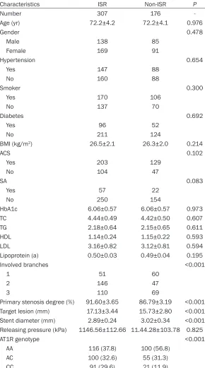
[image:3.612.93.383.97.618.2]cases were not significantly different in prepro -cedural FMD (P=0.257), but ISR cases showed a more significant decrease in FMD than non-ISR cases when the PCI was completed (P<0.001). The blood biomarkers of inflamma -tory reactions, vWF, hs-CRP and IL-6, increased Table 1. Comparison of demographic and clinical data: ISR versus
non-ISR cases
Characteristics ISR Non-ISR P
Number 307 176
-Age (yr) 72.2±4.2 72.2±4.1 0.976
Gender 0.478
Male 138 85
Female 169 91
Hypertension 0.654
Yes 147 88
No 160 88
Smoker 0.300
Yes 170 106
No 137 70
Diabetes 0.692
Yes 96 52
No 211 124
BMI (kg/m2) 26.5±2.1 26.3±2.0 0.214
ACS 0.102
Yes 203 129
No 104 47
SA 0.083
Yes 57 22
No 250 154
HbA1c 6.06±0.57 6.06±0.57 0.973
TC 4.44±0.49 4.42±0.50 0.607
TG 2.18±0.64 2.15±0.65 0.611
HDL 1.14±0.24 1.15±0.22 0.593
LDL 3.16±0.82 3.12±0.81 0.594
Lipoprotein (a) 0.50±0.03 0.49±0.04 0.195
Involved branches <0.001
1 51 60
2 146 47
3 110 69
Primary stenosis degree (%) 91.60±3.65 86.79±3.19 <0.001 Target lesion (mm) 17.13±3.44 15.73±2.80 <0.001 Stent diameter (mm) 2.89±0.24 3.02±0.34 <0.001 Releasing pressure (kPa) 1146.56±112.66 11.44.28±103.78 0.825
AT1R genotype <0.001
AA 116 (37.8) 100 (56.8)
AC 100 (32.6) 55 (31.3)
CC 91 (29.6) 21 (11.9)
Results
Demographic and clinical data
Sex, age and body mass index (BMI) distribution was similar between ISR and non-ISR groups (P>0.05). The per-centages of hypertension, smoker, diabetes, acute coro-nary syndrome (ACS) and stable angina (SA) were also similar between the two groups (P>0.05). Hemoglobin A1c (HbA1c), total cholesterol (TC), triglycerides (TG), high-density lipoprotein (HDL), low-density lipoprotein (LDL) and Lipoprotein (a) were also evenly distributed between the two groups (P>0.05). Involved branches, primary stenosis degree, target lesion and stent diameter were dif-ferent between the two gro- ups (P<0.01), which reflected the significance of primary lesion severity and stenting degree in the prognosis of PCI, except that 2 involved branches had a higher rate of postoperative ISR. Most im- portantly, ISR and non-ISR groups have significantly dif -ferent compositions of AT1R genotypes (P<0.001); the percentage of 3-year ISR was respectively 53.7%, 64.5% and 81.3% in AT1R AA, AC and CC cases. See Table 1 for details.
after PCI procedure, as suggested that stenting contributed to the activation of inflammatory factors; ISR and non-ISR cases were not signifi -cantly different in levels of blood biomarkers of inflammation (P>0.05), but ISR cases showed a more significant increase after PCI in compari -son with the non-ISR cases (P<0.001). Interestingly, ISR and non-ISR cases were sig-nificantly different in pre- and postprocedural level of Ang II (P<0.01). See Table 2 for details. To investigate the association between
endo-was no longer a significant factor (P=0.094), but AT1R CC genotype was still significant (P<0.001). See Table 4 for details.
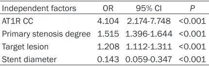
[image:4.612.90.378.93.307.2]All the demographic and clinical parameters were included into the final multivariable regression model with forward method (likeli-hood ratio) and the Hosmer-Leme-show test gave a p-value of 0.834. The independent risk factors to suffer postprocedural ISR were AT1R CC [OR=4.104 (2.174-7.748); P<0.001], primary stenosis degree [OR=1.515 (1.396-1.644); Table 2. Comparison of pre- and postprocedural FMD and blood
biomarkers in different genotypes
ISR non-ISR P
FMD (%)
Before 6.43±1.13 6.56±1.24 0.257
After 4.32±1.48 5.52±1.34 <0.001 vWF (IU/dL)
Before 156.10±50.30 150.65±50.40 0.253 After 202.46±55.88 174.32±50.86 <0.001 Hs-CRP (mg/L)
Before 2.16±0.97 2.25±0.93 0.345
After 3.74±1.26 2.89±0.96 <0.001 IL-6 (pg/dL)
Before 2.76±0.14 2.75±0.14 0.658
After 3.22±0.25 3.12±0.21 <0.001 Ang II (pmol/L)
Before 15.36±0.86 13.12±0.75 <0.001 After 16.82±1.14 14.30±0.94 <0.001
Table 3. Comparison of Blood biomarkers among different AT1R genotypes
AA AC CC P
FMD (%)
Before 6.66±1.14 6.32±1.19 6.35±1.18 0.008 After 5.12±1.38 4.50±1.60 4.43±1.62 <0.001 vWF (IU/dL)
Before 155.15±50.43 149.33±52.50 158.72±46.95 0.297 After 189.69±56.12 189.11±59.51 201.33±48.56 0.141 hs-CRP (mg/L)
Before 2.18±0.96 2.15±0.99 2.27±0.91 0.603 After 3.32±1.23 3.40±1.25 3.71±1.17 0.023 IL-6 (pg/mL)
Before 2.75±0.15 2.75±0.14 2.78±0.14 0.249 After 2.95±0.32 3.25±0.33 3.55±0.31 <0.001 Ang II (pmol/L)
Before 14.36±1.43 14.55±1.38 14.87±1.14 0.006 After 15.69±1.71 15.86±1.62 16.37±1.33 0.001
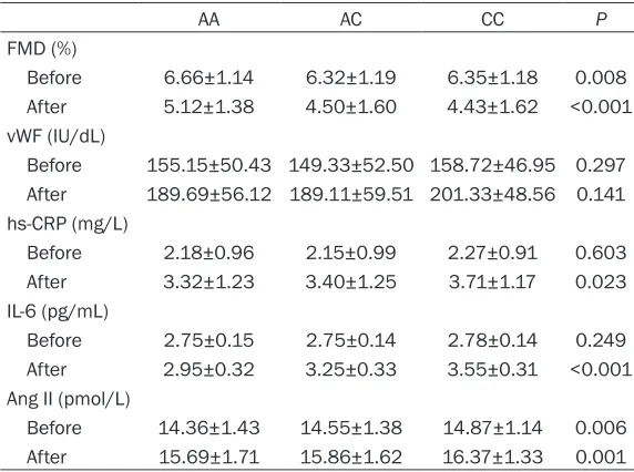
FMD, vWF, hs-CRP, IL-6 and Ang II were performed among three AT1R genotypes (Table 3). Before PCI, the three AT1R genotypes were significantly different in FMD and Ang II (P<0.01), with the AT1R CC genotype showing the lowest FMD and highest level of Ang II, but no significant difference was found in levels of blood biomarkers of inflammation among the three genotypes (P>0.05). However, after PCI, the three AT1R genotypes were significantly different in all the parameters above (P<0.01), with the AT1R CC genotype showing the lowest FMD, and highest level of Ang II and the blood biomarkers of inflam-mation.
[image:4.612.90.376.350.563.2]1.311); P<0.001] and stent diameter [OR= 0.143 (0.059-0.347); P<0.001] (Table 5). Discussion
For the patients with serious ischemic cardio-vascular diseases such as STEMI, conservative treatment is not actually a good option. The progression of ischemic myocardial injuries is usually accelerated as the abnormal anatomic structure remains untreated, leading to life-threatening MACEs. In comparison with surgi-cal therapies, PCI is less invasive and most of the patients can be peri-procedurally compli-ant. Although surgical treatment is still reserved for those patients with complex and severe car-diovascular lesions, PCI has been increasingly used in the treatment of different ischemic car-diovascular diseases. However, one of the most prominent limitations of PCI is the high risk of ISR and subsequent MACEs associated with altered endothelial functions. However, a recent large-scale multi-center study has shown that a considerable number of patients undergoing PCI without pretreatment of statins did not suffer higher risk of in-hospital
compli-cations and mortality [15]; it suggested that not all of the patients undergoing serious endothe-lial dysfunction after PCI and that those patients suffering post-procedural ISR might be attributed to some gene-level mechanisms. The concrete mechanisms of post-procedural endothelial dysfunction amongst the patients undergoing PCI have not been entirely clarified, although past reports have identified a number of risk factors for ISR and MACEs. A study enrolling 687 acute coronary syndrome (ACS) or stable angina pectoris patients undergoing successful PCI showed that high low-density lipoprotein cholesterol (LDL-C)/high-density lipoprotein cholesterol (HDL-C) ratio predicts a high risk of MACEs after PCI [16]. A recent study examining 238 patients who suffered recurrent cardiac ischemia after PCI also suggested that LDL-C/HDL-C ratio contributes to the onset of newly developed coronary artery diseases induced by restenosis [17]. The prognostic sig-nificance of C-reactive protein (CRP) has also been mentioned in many reports regarding the patients undergoing PCI. According to a large-scale study including 1800 stable or unstable angina patients, the difference between base-line and peaked CRP values during treatment, rather than the baseline values, was closely associated with the risk of in-stent restenosis amongst the patients undergoing PCI, which suggested that restenosis is mainly attributed to the inflammatory reactions induced by stent -ing [18]. A long-term follow-up performed amongst 850 angina pectoris patients under-going PCI suggested that CRP levels reflect the inflammatory reactions triggered by stenting and predict the risk of restenosis [19]. Similarly, a retrospective study examined 513 patients undergoing PCI and coronary angiography showed that preprocedural CRP levels were associated with in-stent restenosis and a dia-betic milieu, LDL-C, non-HDL-C and total choles -terol (TC) levels also play roles in the progres-sion of coronary leprogres-sions [20]. Some past reports have also pointed that obstructive sleep apnea (OSA) leads to a hypoxic milieu which contrib-utes to the risk of post-PCI restenosis [21, 22]. Regular physical exercise has been considered protective for those patients undergoing PCI from restenosis since it promotes lipid metabo-lism and increases aerobic capacity [23, 24]. Many reports have provided direct implications in the significance of altered glucolipid metabo -Table 4. Logistic regression for 3-year risk of
post-procedural ISR among elderly STEMI patients Independent factors OR 95% CI P
Univariable model
AT1R AA 1.000 -
AT1R AC 1.567 1.026-2.395 0.038 AT1R CC 3.736 2.167-6.440 <0.001 Calibrated model 1
AT1R AA 1.000 -
AT1R AC 1.567 1.027-2.401 0.037 AT1R CC 3.764 2.181-6.496 <0.001 Calibrated model 2
AT1R AA 1.000 -
[image:5.612.91.306.94.267.2]AT1R AC 1.651 0.918-2.970 0.094 AT1R CC 5.352 2.643-10.839 <0.001
Table 5. Multivariable logistic regression for 3-year risk of postprocedural ISR among STEMI patients
Independent factors OR 95% CI P
[image:5.612.91.306.324.392.2]lism in the development of post-PCI restenosis [25-28]. Thus, it is very likely that oxidative stress and related inflammatory reactions play an essential role in the onset and progression of endothelial functional disorders.
Existing literature has suggested that the endo-thelial functional disorders after PCI are associ-ated with oxidative stress. AT1R A1166C poly-morphisms play a key role in the oxidative stress during the progression of myocardial degeneration. A large-scale study including 3416 patients undergoing successful PCI has suggested that AT1R 1166CC genotype is sig-nificantly associated with intermediate-term post-PCI restenosis [29]; our study made fur-ther investigation into the long-term outcomes of the patients undergoing PCI and showed similar associations between AT1R 1166CC genotype and ISR. That is to say, our results have suggested that AT1R 1166CC genotype was not only strongly associated with interme-diate-term post-PCI restenosis, but also with long-term post-PCI restenosis. In addition, our study cohort included STEMI patients only and guaranteed the homogeneity to some extent. Considering the significance of other gene poly -morphisms, which has been analyzed in some of the past reports [30, 31], gene-gene interac-tions may also play a role in the pathogenesis of post-PCI restenosis; it is a promising research direction in the future.
Our study does still have some limitations. For example, it was a single-center observational study, so the sample size is limited and the par-ticipants were at a relatively high age (≥65 y). However, our study has suggested AT1R gene polymorphisms as a long-term prognostic fac-tor for ISR among the patients undergoing PCI for the first time and primarily provided some evidence on the significance of AT1R gene poly -morphisms in the future genome-based thera-pies, as a supplement to the existing research. Our study may also have some implications in the future investigation into other types of gene polymorphisms associated with post-procedur-al ISR among the patients undergoing PCI. Conclusion
Our study has delivered the first report on the significance of AT1R CC genotype in the deterio -ration of endothelial functions and the develop-ment of ISR in the long term, contributing to the
foundation of genome-based prevention for high risk groups of cardiovascular diseases and pretreatment for the patients undergoing PCI. In the future, other types of gene polymor-phisms should be investigated to clarify the gene-gene interactions in the development of ISR among the patients undergoing PCI and a genome-based therapy will be made possible. Disclosure of conflict of interest
None.
Address correspondence to: Dr. Yuchen Zhang, Department of Cardiology, Beijing Anzhen Hospital, Capital Medical University, Beijing, China. E-mail: [email protected]
References
[1] Monnink SH, Tio RA, Veeger NJ, Amoroso G, van Boven AJ and van Gilst WH. Exercise-in-duced ischemia after successful percutane-ous coronary intervention is related to distal coronary endothelial dysfunction. J Investig Med 2003; 51: 221-226.
[2] Thanyasiri P, Kathir K, Celermajer DS and Ad -ams MR. Endothelial dysfunction and resteno-sis following percutaneous coronary interven-tion. Int J Cardiol 2007; 119: 362-367. [3] Izzo P, Macchi A, De Gennaro L, Gaglione A, Di
Biase M and Brunetti ND. Recurrent angina af -ter coronary angioplasty: mechanisms, diag-nostic and therapeutic options. Eur Heart J Acute Cardiovasc Care 2012; 1: 158-169. [4] Briguori C, Colombo A, Airoldi F, Violante A,
Fo-caccio A, Balestrieri P, Paolo Elia P, Golia B, Lepore S, Riviezzo G, Scarpato P, Librera M, Bonizzoni E and Ricciardelli B. Statin adminis-tration before percutaneous coronary interven-tion: impact on periprocedural myocardial in-farction. Eur Heart J 2004; 25: 1822-1828. [5] Chang SM, Yazbek N and Lakkis NM. Use of
statins prior to percutaneous coronary inter-vention reduces myonecrosis and improves clinical outcome. Catheter Cardiovasc Interv 2004; 62: 193-197.
[6] Patti G, Pasceri V, Colonna G, Miglionico M, Fischetti D, Sardella G, Montinaro A and Di Sciascio G. Atorvastatin pretreatment im-proves outcomes in patients with acute coro-nary syndromes undergoing early percutane-ous coronary intervention: results of the ARMYDA-ACS randomized trial. J Am Coll Car -diol 2007; 49: 1272-1278.
may prevent restenosis after successful coro-nary intervention, independent of lipid-lower-ing effect and CRP level. Fukuoka Igaku Zasshi 2007; 98: 260-269.
[8] Di Sciascio G, Patti G, Pasceri V, Gaspardone A, Colonna G and Montinaro A. Efficacy of atorv -astatin reload in patients on chronic statin therapy undergoing percutaneous coronary in-tervention: results of the ARMYDA-RECAPTURE (Atorvastatin for Reduction of Myocardial Dam -age During Angioplasty) Randomized Trial. J Am Coll Cardiol 2009; 54: 558-565.
[9] Ko DT, Wijeysundera HC, Yun L, Austin PC, Can -tor WJ and Tu JV. Effectiveness of preproce-dural statin therapy on clinical outcomes for patients with stable coronary artery disease after percutaneous coronary interventions. Circ Cardiovasc Qual Outcomes 2011; 4: 459-466.
[10] Edris A, Patel PM, Krishnam MS and Sengupta PP. Intramyocardial hemorrhage after percuta-neous coronary intervention. Echocardiogra-phy 2012; 29: E50-51.
[11] Zia MI, Ghugre NR, Connelly KA, Joshi SB, Strauss BH, Cohen EA, Wright GA and Dick AJ. Thrombus aspiration during primary percuta-neous coronary intervention is associated with reduced myocardial edema, hemorrhage, mi-crovascular obstruction and left ventricular re-modeling. J Cardiovasc Magn Reson 2012; 14: 19.
[12] Maluenda G, Mitulescu L, Ben-Dor I, A Gaglia M Jr, Weissman G, Torguson R, F Satler L, Pich-ard AD, BernPich-ardo NL, Waksman R. Retroperito -neal hemorrhage after percutaneous coronary intervention in the current practice era: clinical outcomes and prognostic value of abdominal/ pelvic computed tomography. Catheter Cardio-vasc Interv 2012; 80: 29-36.
[13] Grines CL and Schreiber T. The hemorrhage of information to reduce bleeding complications after percutaneous coronary intervention. J In-terv Cardiol 2013; 26: 639-640.
[14] Hingorani AD and Brown MJ. A simple molecu -lar assay for the C1166 variant of the angioten-sin II type 1 receptor gene. Biochem Biophys Res Commun 1995; 213: 725-729.
[15] Kenaan M, Seth M, Aronow HD, Naoum J, Wun -derly D, Mitchiner J, Moscucci M and Gurm HS; Blue Cross Blue Shield of Michigan Cardiovas-cular Consortium (BMC2). Preprocedural statin use in patients undergoing percutane-ous coronary intervention. Am Heart J 2014; 168: 110-116, e113.
[16] Matsumoto I, Miyake Y, Mizukawa M and Taka-gi Y. Impact of low-density lipoprotein choles-terol/high-density lipoprotein cholesterol ratio on long-term outcome in patients undergoing percutaneous coronary intervention. Circ J 2011; 75: 905-910.
[17] Endo A, Yoshida Y, Kageshima K, Sato H, Suga T, Nasu H, Takahashi N and Tanabe K. Contrib-utors to newly developed coronary artery dis-ease in patients with a previous history of per-cutaneous coronary intervention beyond the early phase of restenosis. Intern Med 2014; 53: 819-828.
[18] Dibra A, Mehilli J, Braun S, Hadamitzky M, Baum H, Dirschinger J, Schuhlen H, Schomig A and Kastrati A. Inflammatory response after intervention assessed by serial C-reactive pro-tein measurements correlates with restenosis in patients treated with coronary stenting. Am Heart J 2005; 150: 344-350.
[19] Saleh N and Tornvall P. Serum C-reactive pro-tein response to percutaneous coronary inter-vention in patients with unstable or stable an-gina pectoris is associated with the risk of clinical restenosis. Atherosclerosis 2007; 195: 374-378.
[20] Xu HY, Qiao SB, Zhang JF, Dong QT and Li JJ. Different impacts of C-reactive protein and lip -id profile on coronary lesions following a percu -taneous coronary intervention. Coron Artery Dis 2012; 23: 181-187.
[21] Yumino D, Tsurumi Y, Takagi A, Suzuki K and Kasanuki H. Impact of obstructive sleep apnea on clinical and angiographic outcomes follow-ing percutaneous coronary intervention in pa-tients with acute coronary syndrome. Am J Car-diol 2007; 99: 26-30.
[22] Steiner S, Schueller PO, Hennersdorf MG, Beh-rendt D and Strauer BE. Impact of obstructive sleep apnea on the occurrence of restenosis after elective percutaneous coronary interven-tion in ischemic heart disease. Respir Res 2008; 9: 50.
[23] Munk PS, Staal EM, Butt N, Isaksen K and Larsen AI. High-intensity interval training may reduce in-stent restenosis following percutane-ous coronary intervention with stent implanta-tion A randomized controlled trial evaluating the relationship to endothelial function and inflammation. Am Heart J 2009; 158: 734-741.
[24] Lee HY, Kim JH, Kim BO, Byun YS, Cho S, Goh CW, Ahn H, Rhee KJ and Kim C. Regular exer-cise training reduces coronary restenosis after percutaneous coronary intervention in pa-tients with acute myocardial infarction. Int J Cardiol 2013; 167: 2617-2622.
[25] Novo G, Maniglia D, Corrado E, Muratori I, Sutera F, Evola S, Ferrara F, Hoffmann E and Novo S. Peripheral atherosclerosis is associat-ed with the occurrence of restenosis after per-cutaneous coronary intervention. Coron Artery Dis 2007; 18: 627-631.
per-cutaneous coronary intervention raises the risk for restenosis in patients without Type 2 diabetes mellitus. Diabet Med 2008; 25: 228-231.
[27] Lupi A, Secco GG, Rognoni A, Rossi L, Lazzero M, Nardi F, Rolla R, Bellomo G, Bongo AS and Di Mario C. Plasma fibrinogen levels and reste -nosis after primary percutaneous coronary in-tervention. J Thromb Thrombolysis 2012; 33: 308-317.
[28] Marfella R, Sasso FC, Siniscalchi M, Paolisso P, Rizzo MR, Ferraro F, Stabile E, Sorropago G, Calabro P, Carbonara O, Cinquegrana G, Pi-scione F, Ruocco A, D’Andrea D, Rapacciuolo A, Petronella P, Bresciani A, Rubino P, Mauro C and Paolisso G. Peri-procedural tight glycemic control during early percutaneous coronary tervention is associated with a lower rate of in-stent restenosis in patients with acute ST-ele-vation myocardial infarction. J Clin Endocrinol Metab 2012; 97: 2862-2871.
[29] Wijpkema JS, van Haelst PL, Monraats PS, Bru-inenberg M, Zwinderman AH, Zijlstra F, van der Steege G, de Winter RJ, Doevendans PA, Waltenberger J, Jukema JW and Tio RA. Reste-nosis after percutaneous coronary interven-tion is associated with the angiotensin-II type-1 receptor 1166A/C polymorphism but not with polymorphisms of angiotensin-converting en-zyme, angiotensin-II receptor, angiotensinogen or heme oxygenase-1. Pharmacogenet Ge-nomics 2006; 16: 331-337.
[30] Vogiatzi K, Apostolakis S, Voudris V, Thomo-poulou S, Kochiadakis GE and Spandidos DA. Interleukin 8 gene polymorphisms and suscep-tibility to restenosis after percutaneous coro-nary intervention. J Thromb Thrombolysis 2010; 29: 134-140.