The influence of fentanyl injection followed
by infusion on the intraocular pressure, pupil
size and aqueous tear production in healthy
non-painful dogs
Petr Rauser*, Hana Nemeckova, Marketa Mrazova, Jana Burova,
Lukas Novak
Small Animal Clinic, Faculty of Veterinary Medicine, University of Veterinary and Pharmaceutical Sciences, Brno, Czech Republic
*Corresponding author: rauserp@vfu.cz
Citation: Rauser P, Nemeckova H, Mrazova M, Burova J, Novak L (2019): The influence of fentanyl injection followed by infusion on the intraocular pressure, pupil size and aqueous tear production in healthy non-painful dogs. Veterinarni Medicina 64, 448–455.
Abstract: The goal of the presented research was to assess the influence of continuously administered fentanyl on the intraocular pressure, pupil size and aqueous tear production in dogs. A prospective, randomised, double “blind” clinical study was performed. Twenty-five non-painful dogs, 13 breeds, a body weight of 10.0 ± 5.4 kg (mean ± SD) and age of 6.5 ± 3.3 years, 12 males and 13 females with no ocular abnormalities were randomly allocated into two groups receiving an intravenous injection of saline (SAL) 0.3 ml/kg followed by an infusion 2 ml/kg/h or an intravenous injection of fentanyl (FEN) 0.005 mg/kg (diluted in 0.3 ml/kg) followed by an infusion 0.005 mg/kg/h (diluted in 2 ml/kg/h). The intraocular pressure (IOP), pupil size (PS), pulse rate (PR), respiratory frequency (fR) and systolic and diastolic arterial pressures (SAP, DAP) were measured before (baseline) and at 2, 5, 10, 20 and 30 minutes after the premedication. The Schirmer Tear Test I (STT-I) was measured prior to and at 30 min after the premedication. The data were analysed by Bartlett’s, Anderson-Darling and Dunnett’s tests, the t-test and an analysis of variance (ANOVA) (P < 0.05). Relative to the baseline, in the fentanyl group, the PS was significantly decreased at all time points, the PR was significantly decreased at T30 and the fR was significantly decreased at T5, T10, T20 and T30. There were no other significant changes in the IOP, STT-I, SAP and DAP relative to the baseline. Compared to the control group, in the fentanyl group, the PS was significantly smaller at T2, T5, T10, T20 and T30, the PR was significantly lower at T2, T20 and T30 and the fR was significantly higher at T20. Within thirty minutes of a constant rate infusion of fentanyl in the healthy non-painful dogs, the intraocular pressure and aqueous tear production were not affected. However, the fentanyl significantly decreased pupil size. This fact should be considered, when planning analgesia where miosis is undesirable.
Keywords: canine; analgesia; opioids; pupil diameter; CRI
Supported by the Ministry of Education, Youth and Sports of the Czech Republic (Research Project IGA VFU No. 101/2019/FVL).
Intraocular pressure (IOP) is an important param-eter for the normal function of a healthy eye. The phys-iological value of a canine’s IOP is 10–25 mmHg (Renwick 2002). In addition to many other
mecha-nisms, IOP can also be affected by the pupil size (PS) or the drugs used (Almeida et al. 2004).
procedures were performed in accordance with the current law for animal protection and the Ethics Committee of the University. All animal owners provided consent to have their animals participate in the study. The presented study was designed as a prospective, randomised, “double-blind”, placebo-controlled clinical study.
The experimental protocol was adopted and modified from a previous study (Mrazova et al. 2018) describing the influence of a single injec-tion of medetomidine, acepromazine, fentanyl or butorphanol used for anaesthesia premedication on the IOP and pupil size in dogs.
Animals. Twenty-five client-owned healthy dogs, 13 breeds, with a body weight of 10.0 ± 5.4 kg (mean ± SD) and age of 6.5 ± 3.3 years, 12 males and 13 females were enrolled in this study. All the dogs underwent periodontal treatment under general anaesthesia. They were clinically healthy non-painful without any ocular abnormalities and des-ignated as ASA I or II according to the American Society of Anesthesiologists (ASA). The dogs were fasted for 12 h, with free access to water prior to the anaesthesia.
A complete clinical ophthalmic examination, in- cluding slit lamp biomicroscopy, applanation to-nometry, gonioscopy and a Schirmer Tear Test I (STT-I), was performed by the same experien-ced individual person blinded to the treatment groups. Only the dogs with an IOP measured at 15–25 mmHg prior to the sedation, with an STT-I more than 12 mm/min and without any ophthalmic abnormalities were enrolled.
To avoid the influence of breed, age or body mass on the IOP, only the dogs without any eye abnor-malities weighing between 5 and 20 kg and 1 to 10 years old were enrolled. The Siberian Husky and brachycephalic breeds were also excluded, be-cause they may have higher IOP (Taylor et al. 2007). Age is a factor that may affect the IOP (Gelatt and MacKay 1998). We also excluded too small and too large dogs to avoid a relative error in the fen-tanyl dosing. Likewise, dogs with an IOP outside of the ranges or any other health problems or eye pathology were excluded.
Study protocol. A randomising software (www. randomizer.org) was utilised to allocate the animals to two groups – the control (SAL, saline; n = 10) or the fentanyl (FEN, treatment; n = 15) groups. An in-travenous catheter was placed into the cephalic vein in all the dogs. Subsequently, the intraocular pres-of the IOP (Feldman 2015). The increase in the IOP
is unacceptable in patients affected by glaucoma, a penetrating eye globe injury or undergoing in-traocular surgery. Therefore, perioperative care must prevent an increase in the IOP. Especially in patients with ocular blood flow abnormalities (Jantzen 1988), it is not appropriate to use drugs (analgesics) that could increase the IOP.
A µ-opioid fentanyl is one of the most commonly used drugs of pain management in dogs (Epstein 2015). It has a fast onset and duration, as well as low incidence of cardiovascular side-effects (Grimm et al. 2005). It is administered as a single bolus or infusion. Use of fentanyl as an infusion helps maintain a stable plasma concentration with a re-duction of undesirable resulting reactions caused by the readministration of boluses (Guedes 2012). Administration of fentanyl in combination with other drugs in people during the induction of an-aesthesia can help prevent increases in the IOP (Fukuda 2015). Mrazova et al. (2018) described the important increases of the IOP five and ten minutes after a single bolus injection of fentanyl in dogs. However, the influence of fentanyl on the IOP af-ter a constant rate infusion in dogs has not been described yet.
The pupil size has, among other functions, a significant effect on the IOP. In general, mio-sis decreases and mydriamio-sis increases the IOP. Opioids in people and dogs tend to cause miosis (Jollife 2016).
Aqueous tear production is another important function maintaining ocular homeostasis especially in the eyelids, conjunctiva and cornea. Many opi-oids, including fentanyl, have been shown to reduce tear production (Biricik et al. 2004).
The goal of the presented study is to assess the in-fluence of a fentanyl injection followed by thirty minutes of a constant rate infusion (CRI) on the IOP, PS and aqueous tear production in dogs, which has not been published yet. We hypothesised that fentanyl will increase the intraocular pressure and decrease the pupil size without affecting the aque-ous tear production in dogs within thirty minutes of administration.
MATERIAL AND METHODS
the cuff width was set as 40% of the circumference of the limb. The collected data included the systolic and diastolic arterial blood pressure.
All the measurements were performed in the morning after the patients’ acclimatisation to the lighting conditions in a quiet room for 10 min after the intravenous catheterisation.
Statistical analysis. All the data were analysed using a GraphPad InStat 3.06 (GraphPad Software Inc.), KyPlot 2.0 beta 15 (Koichi Yoshioka) and MS Excel (Microsoft). The measured parameters – IOP, PS, STT-I, PR, fR, SAP and DAP measured at the same time points in both groups were compared to each other. The intraocular pressure, PS, PR, fR, SAP and DAP measured at T2, T5, T10, T20 and T30
were compared to the baseline. The Schirmer Tear Test I measured at T30 was compared to
the base-line as well. The Anderson-Darling and Bartlett’s tests were used to confirm the normal distribu-tion of the data and the homogeneity of variance, respectively. The parametric data are reported as the mean ± standard deviation (SD), the nonpara-metric data were transformed using a natural log (ln) scale and are also reported as the mean ± SD. For the multiple comparisons between the baseline and T2, T5, T10, T20 and T30 within each group,
Dunnett’s test was used. For the comparisons of the STT-I between the baseline and T30 within each
group, the dependent t-test for paired compari-sons was used. All the variables between the groups were compared at each specific time point using the t-test for the unpaired comparison. The level of significance was set at P < 0.05.
RESULTS
We have not observed any significant differences between the SAL and FEN groups with respect to the sex, body weight, age or measured parameters at the baselines (IOP, PS, STT-I, PR, fR, SAP or DAP).
In the fentanyl group, the IOP was maintained between 16 and 30 mmHg in all the dogs through-out the measurement time (Figure 1). Relative to the baseline, the PS decreased significant-ly at all the measured times (at T2 P = 0.0392,
at T5P = 0.006, at T10P = 0.006, at T20P = 0.0014,
at T30P = 0.003; Figure 2). The STT-I insignificantly
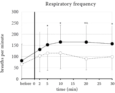
decreased in the fentanyl group over 30 minutes (Figure 3). Relative to the baseline, the PR decreased at T30 (P = 0.0044; Figure 4), the fR increased at T5
sure (IOP), pupil size (PS), Schirmer Tear Test I, pulse rate (PR), respiratory frequency (fR) and sys-tolic and diassys-tolic arterial blood pressures (SAP, DAP) were measured and recorded (baseline).
The dogs in the SAL group received a rapid injec-tion of saline (0.9% NaCl, B. Braun) 0.3 ml/kg fol-lowed by a constant rate infusion of saline 2 ml/kg/h. The dogs in the FEN group received a rapid injec-tion of fentanyl (Fentanyl Torrex, Torrex Chiesi) 0.005 mg/kg (diluted with saline in a total volu-me of 0.3 ml/kg) followed by a constant rate infu-sion of fentanyl 0.005 mg/kg/h (diluted with saline in a total volume of 2 ml/kg/h). The infusion was continuously administered by a syringe infusion pump (Perfusor Compact S, B. Braun) over a 30 min observation period.
Measurements. All the measurements were per- formed by the same person that was unaware of which drug had been administered. All the dogs were maintained in a sitting position during the measurement procedures without any manipula-tion, jugular vein or eye compression.
In all the dogs, the IOP, PS, PR, fR, SAP and DAP were measured and recorded five minutes before (baseline) and two (T2), five (T5), ten (T10), twenty
(T20) and thirty (T30) minutes after
the administra-tion of the initial bolus followed by a constant rate infusion of fentanyl or saline. The Schirmer Tear Test I measurements were taken before the admin-istration of the fentanyl or saline (baseline) and thirty (T30) minutes thereafter.
Figure 1. The intraocular pressure changes in the con-scious dogs after the rapid intravenous injection fol-lowed by the continuous infusion of fentanyl (ˉˉ) or saline (ˉˉ) (mean ± SD)
––
Intraocular pressure
time (min)
before 0 2 5 10 15 20 25 30 30
25
20
15
10
5
0
milime
tr
[image:4.595.311.516.98.266.2]es of Hg
Figure 2. The pupil size changes in the conscious dogs after the rapid intravenous injection followed by the continuous infusion of fentanyl (ˉˉ) or saline (ˉˉ) (* = significantly decreased compared to the baseline; ∨ = significantly de-creased compared to the control group) (mean ± SD)
––
Pupil size
milime
tr
es
time (min)
before 0 2 5 10 15 20 25 30 8
7 6 5 4 3 2 1 0
Figure 3. The Schirmer Tear Test I in the conscious dogs before (left column) and 30 minutes after the rapid intra-venous injection followed by the continuous infusion (right column) of fentanyl or saline (mean ± SD)
Schirmer Tear Test I
Fentanyl Saline
milime
tr
es p
er min
ut
e
22 20 18 16 14 12 10 8 6 4 2 0
18.3 17.6 19.0 18.7
Pulse rate
time (min)
before 0 2 5 10 15 20 25 30
pul
se
s p
er min
ut
e
180 160 140 120 100 80 60 40 20 0
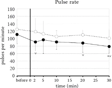
Figure 4. The pulse rate changes in the conscious dogs after the rapid intravenous injection followed by the continuous infusion of fentanyl (ˉˉ) or saline (ˉˉ) (* = significantly decreased compared to the baseline; ∨ = significantly
de-creased compared to the control group) (mean ± SD)
(P = 0.0483), T10 (P = 0.0145), T20 (P = 0.0153) and
T30 (P = 0.0319; Figure 5). There were no significant
changes in the SAP and DAP relative to the baseline (Figure 6).
In the control group, no significant changes in all the measured parameters relative to the baseline were detected.
In the fentanyl group, the PS was significant-ly smaller compared to the control group at T2
(P = 0.0329), T5 (P = 0.0053), T10 (P = 0.002), T20
(P = 0.0014) and T30 (P = 0.0012; Figure 2). In
the fentanyl group, the PR was lower compared to the control group at T2 (P = 0.0273), T20 (P = 0.0178)
and T30 (P = 0.0123; Figure 4). In the fentanyl group,
the fR was significantly higher at T20 (P = 0.0103)
compared to the control group. We have not re-corded any significant differences in the other measured parameters between the groups at any other time point.
DISCUSSION
[image:4.595.321.504.545.690.2] [image:4.595.64.289.545.698.2]effect, the fentanyl must be regularly injected re-peatedly. Re-administration of fentanyl may cause important respiratory depression (Yaksh et al. 1986), or be continuously infused with a reduction of the side effects. The pharmacokinetic properties of a continuously injected fentanyl dose in dogs were described by Sano et al. (2006). The con-tinuous intravenous administration of a fentanyl dose of 0.01 mg/kg/h caused an initial increase in the plasma concentration followed up to 30 min by its decrease. Then fentanyl concentration re-mained fairly stable (Sano et al. 2006).
The influence of a separately injected fentanyl on the IOP in conscious dogs was described in the study (Mrazova et al. 2018). No other studies investigating the effect of fentanyl administered separately on the IOP in animals or humans have been published yet. Mrazova et al. (2018) noted a marked increase of the IOP ten minutes after a single intravenous injection of fentanyl in a dose of 0.01 mg/kg. In the current study, we used a half dose of fentanyl – 0.005 mg/kg for the induction and subsequently continued with a 0.005 mg/kg/h dose. This dose is broadly recommended and used for the continuous fentanyl administration for pain management in dogs (Sinclair 2018). The fentanyl dose administered in our study is half in compari-son with the dose used in the study by Sano et al. (2006). However, the pharmacokinetics should be comparable. Although we did not measure the plas-matic concentration of fentanyl in our study, we
assume the identical situation when using a half dose of fentanyl. Important changes in the fentanyl concentration occur within 30 min after starting the continuous administration (Sano et al. 2006). After 30 min, it was supposed to be constant and its effect on the IOP, PS and cardiorespiratory variables to be without fluctuations. Therefore, we monitored the variables within 30 min of the pre-anaesthetic period only. We could no longer extend this due to the limited preoperative hospitalisa-tion. the patients did not receive any other drugs, therefore, the IOP, PS and aqueous tear production changes caused by the fentanyl only were noted.
[image:5.595.68.268.94.255.2]The IOP was maintained within 30 min of the continuous administration of the fentanyl between 16 and 30 mmHg and was without significant differ-ence compared to the baseline or the control group. By contrast, Mrazova et al. (2018) described a sig-nificant increase in the IOP five and ten minutes after the administration of the fentanyl, when using a single injection of fentanyl in a double dose. We assume that the higher IOP may be due to the high-er dose of the fentanyl used in the study by Mrazova et al. (2018). We consider a lower dose – 0.005 mg/ kg/h to be safer in ophthalmic patients even with the long-term administration relative to the IOP. It can be supposed, if the IOP changes have not occurred within 30 min of the continuous intrave-nous administration, they will not follow later on. Other authors (Stirt and Chiu 1990; Ng et al. 2000; Sator-Katzenschlager et al. 2004; Domi 2010), who
Figure 5. The respiratory frequency changes in the con-scious dogs after the rapid intravenous injection followed by the continuous infusion of fentanyl (ˉˉ) or saline (ˉˉ) (* = significantly increased compared to the base-line, ∨ = significantly increased compared to the control
group) (mean ± SD)
Respiratory frequency
time (min)
before 0 2 5 10 15 20 25 30
br
ea
ths p
er min
ut
e
300
250
200
150
100
50
[image:5.595.305.518.101.270.2]0
Figure 6. The systolic (upper two lines) and diastolic (lower two lines) arterial pressure changes in the con-scious dogs after the rapid intravenous injection fol-lowed by the continuous infusion of fentanyl (ˉˉ) or saline (ˉˉ) (mean ± SD)
Systolic and diastolic arterial pressure
time (min)
before 0 2 5 10 15 20 25 30 250
200
150
100
50
0
milime
tr
described changes to the IOP after administration of fentanyl in people, did not use fentanyl alone. Therefore, the resulting changes in the IOP were also influenced by the concomitantly administered substances.
Fentanyl significantly decreased the PS in the dogs. Blaze et al. (2009) or Zacny et al. (1992) ob-served pupil constriction in dogs after the applica-tion of other µ-opioid agonist (hydromorphone, morphine) or in people after injection of fentanyl, respectively. Mrazova et al. (2018) also describes the decrease in the PS after the intravenous injec-tion of a double dose of fentanyl. Fentanyl decreases the PS using both 0.01 mg/kg (Mrazova et al. 2018) and 0.005 mg/kg followed by 0.005 mg/kg/h within 30 min, therefore, it should be avoided when miosis is undesirable.
The influence of fentanyl on the aqueous tear production was described Biricik et al. (2004). They injected fentanyl intramuscularly in a dose of 0.01 mg/kg, after twenty minutes the STT-I de-creased significantly. In our study, we used half the dose of the fentanyl intravenously followed by a continuous infusion, after thirty minutes the STT-I decreased slightly, however insignifi-cantly. Therefore, we consider the dose 0.005 mg/kg followed by 0.005 mg/kg/h for thirty minutes safe concerning the risk of the reduction of the tear production.
Along with the eye parameters (IOP, PS and STT-I), we also monitored the vital signs (PR, fR, SAP and DAP). We wanted to assess their dependency.
Most opioids, including fentanyl, produce, be-cause of increased parasympathetic tone, a dose-de-pendent bradycardia (Laubie et al. 1974; Arndt et al. 1984; Sinclair 2018). A significant decrease in the PR was noted by Mrazova et al. (2018) ten minutes af-ter the intravenous injection fentanyl. Also, in our study, we recorded a significant decrease in the PR after the rapid injection followed by the constant rate infusion of the half dose of fentanyl compared to Mrazova et al. (2018). However, this bradycardia does not cause a fluctuation in the IOP. The risk of bradycardia must be taken into consideration after every fentanyl administration again.
Changes in the arterial pressure have a relatively low influence on the IOP. Increased systolic arte-rial blood pressure induces a transient increase in the IOP due to elevating the circulating volume in the bloodstream and, thus, an increase in the blood volume in the ciliary body arteries (Macri 1961).
Although 2, 20 and 30 min after the start of the continuous fentanyl administration, we observed a significantly lower pulse rate compared to the con-trol group, the blood pressure was not significantly different. Our results confirm Macri’s (1961) theses on the minimal impact of the cardiovascular pa-rameter changes on the IOP.
The respiratory frequency can decrease because of high or cumulative doses of fentanyl. After ad-ministration of sedative doses of fentanyl in dogs, panting is frequently observed. Hypocapnia is not necessarily present due to the dead space ventila-tion. Often hypercapnia is noticed (Sinclair 2018). Panting and a significant increase of the fR was de-tected in the presented study and in the study of Mrazova et al. (2018) as well. Panting was also report-ed by Arndt et al. (1984), but with repeatreport-edly admin-istered fentanyl, the injected dose (0.1675 mg/kg) exceeded the dose that was used in our study or the study of Mrazova et al. (2018). Arndt et al. (1984) reported an increase in the PaCO2 that may
cause a rise in the IOP (Duncalf and Weitzner 1963). We did not measure the PaCO2 in our study. Since
we have not detected an increase in the IOP, we as-sume that there was no significant rise in the PaCO2 caused by panting after the fentanyl administration.
In conclusion, within thirty minutes after a rap-id injection followed by a constant rate infusion of fentanyl in healthy dogs, the intraocular pres-sure and aqueous tear production were not affect-ed. However, the fentanyl significantly decreased the pupil size. This fact should be considered when planning analgesia where miosis is undesirable.
REFERENCES
Almeida DE, Rezende ML, Nunes N, Laus JL (2004): Evalu-ation of intraocular pressure in associEvalu-ation with cardio-vascular parameters in normocapnic dogs anesthetized with sevoflurane and desflurane. Veterinary Ophthalmol-ogy 7, 265–269.
Arndt JO, Mikat M, Parasher C (1984): Fentanyl’s analgesic, respiratory, and cardiovascular actions in relation to dose and plasma concentration in unanesthetized dogs. An-esthesiology 61, 355–361.
Biricik HS, Ceylan C, Sakar M (2004): Effects of pethidine and fentanyl on tear production in dogs. Veterinary Re-cord 155, 564–565.
Blaze C, Pine CG, Casey E, Pizzirani S (2009): The effect of intravenous hydromorphone, butorphanol, morphine, and buprenorphine on pupil size and intraocular pres-sure in normal dogs. Proceedings of 10th WCVA, AVA,
Glasgow, 134.
Cunningham AJ, Barry P (1986): Intraocular pressure-physiology and implications for anaesthetic management. Canadian Anaesthetic Society Journal 33, 195−208. Domi RQ (2010): A comparison of the effects of sufentanil
and fentanyl on intraocular pressure changes due to easy and difficult tracheal intubations. Saudi Medical Journal 31, 29−31.
Duncalf D, Weitzner SW (1963): The influence of ventila-tion and hypercapnea on intraocular pressure during anesthesia. Anesthesia & Analgesia 42, 232–237. Epstein ME (2015): Opioids. In: Gaynor JS, Muir WW III
(eds): Handbook of Veterinary Pain Management. 3rd edn.
Elsevier Mosby, St. Louis. 161–195 p.
Feldman MA (2015): Anesthesia for eye surgery. In: Miller RD (ed.): Anesthesia. 8th edn. Elsevier Saunders,
Phila-delphia. 2512–2525 p.
Fukuda K (2015): Opioid analgesics. In: Miller RD (ed.): Anesthesia. 8th edn. Elsevier Saunders, Philadelphia.
864–914 p.
Gelatt KN, MacKay EO (1998): Distribution of intraocular pressure in dogs. Veterinary Ophthalmology 1, 109–114. Grimm KA, Tranquilli WJ, Gross DR, Sisson DD, Bulmer
BJ, Benson GJ, Greene SA, Martin-Jimenez T (2005):
Cardiopulmonary effects of fentanyl in conscious dogs and dogs sedated with a continuous rate infusion of me-detomidine. American Journal of Veterinary Research 66, 1222–1226.
Guedes AGP (2012): Pain management: constant-rate infu-sion. NAVC Clinician’s Brief/March 2012, 29–33. Jantzen JP (1988): Anaesthesia and intraocular pressure.
Anaesthesia 37, 458–469.
Jollife C (2016): Ophthalmic surgery. In: Duke-Novakovski T, de Vries M, Seymour C (eds): BSAVA Manual of Canine and Feline Anaesthesia and Analgesia. 3rd edn. British Small
Animal Veterinary Association, Gloucester. 258–271 p. Laubie M, Schmitt H, Canellas J, Roquebert J, Demichel P
(1974): Centrally mediated bradycardia and hypotension induced by narcotic analgesics: dextromoramide and fen-tanyl. European Journal of Pharmacology 28, 66–75. Macri FJ (1961): Vascular pressure relationship and the
intraocular pressure. Archives of Ophthalmology 65, 133−136.
Mrazova M, Rauser P, Burova J, Georgiou G, Fichtel T (2018): Influence of medetomidine, acepromazine, fen-tanyl or butorphanol on intraocular pressure and pupil size in healthy dogs. Veterinární Medicína 63, 413–419. Ng HP, Chen FG, Yeong SM, Wong E, Chew P (2000): Effect
of remifentanil compared with fentanyl on intraocular pressure after succinylcholine and tracheal intubation. British Journal of Anaesthesiology 85, 785–787.
Renwick P (2002): Glaucoma. In: Petersen-Jones S, Crispin S (eds): BSAVA Manual of Small Animal Ophthalmology. 2nd edn. Gloucester, British Small Animal Veterinary
As-sociation. 185–203 p.
Sano T, Nishimura R, Kanazawa H, Igarashi E, Nagata Y, Mochizuki M, Sasaki N (2006): Pharmacokinetics of fen-tanyl after single intravenous injection and constant rate infusion in dogs. Veterinary Anaesthesia and Analgesia 33, 266–273.
Sator-Katzenschlager SM, Oehmke MJ, Deusch E, Dolezal S, Heinze G, Wedrich A. (2004): Effects of remifentanil and fentanyl on intraocular pressure during the mainte-nance and recovery of anaesthesia in patients undergoing non-ophthalmic surgery. European Journal of Anaesthe-siology 21, 95–100.
Sinclair M (2018): Pharmacologic and clinical application of opioid analgesics. In: Mathews KA, Sinclar M, Steele AM, Grubb T (eds): Analgesia and Anesthesia for the Ill or Injured Dog and Cat. 1st edn. Wiley Blackwell,
Hobo-ken. 119–133 p.
Taylor NR, Zele AJ, Vingrys AJ, Stanley RG (2007): Variation in intraocular pressure following application of tropi-camide in three different dog breeds. Veterinary Oph-thalmology 10, 8–11.
Yaksh TL, Noueihed RY, Durant PA (1986): Studies of the pharmacology and pathology of intrathecally
admin-istered 4-anilinopiperidine analogues and morphine in the rat and cat. Anesthesiology 64, 54–66.
Zacny JP, Lichtor JL, Zaragoza JG, de Wit H (1992): Effects of fasting on responses to intravenous fentanyl in healthy volunteers. Journal of Substance Abuse 4, 197–207.