Original Article
miR-497 inhibits the carcinogenesis of hepatocellular
carcinoma by targeting the Rictor/Akt signal pathway
Meng Zhang1*, Jianfei Wu1*, Rui Zhang1, Jihong Yang1, Quan Zhang1, Bin Liu2
Departments of 1Hepatobiliary Surgery, 2Internal Medicine-Oncology, The Affilicated Hosptial of Hebei University,
Hebei, China. *Equal contributors and co-first authors.
Received March 6, 2019; Accepted March 28, 2019; Epub June 1, 2019; Published June 15, 2019
Abstract: MicroRNAs (miRNAs) are involved in regulating various physiologic and pathologic processes of different human diseases including hepatocellular carcinoma (HCC). Our research aimed to investigate the role of miR-497 in migration, invasive ability of HepG2-GS cells and the regulating mechanism. In this study, Rictor was identified as a target gene of miR-497 by informatic software, including Microcosm Targets, miRanda, and TargetScan. MiR-497 or Rictor were silenced or overexpressed in HepG2-GS cells through transfection. The functional assay results showed that Rictor knockdown inhibited cancer cell proliferation, migration and invasion. Overexpression of Rictor inversed the effects of miR-497 on cancer cells growth inhibition. miR-497 regulated protein kinase B, PKB (Akt) signaling pathway by targeting Rictor. MiR-497 increased chemo-sensitivity of HepG2-GS through regulation of Rictor. In con-clusion, our research demonstrated that miR-497 inhibits the proliferation, invasion, metastasis, and chemotherapy resistance of hepatoma cells by targeting of Rictor/Akt signal pathway, and miR-497. Thus, Rictor has the potential to be a explored as a biomarker or therapeutic target for diagnosis and treatment of HCC.
Keywords: Hepatocellular carcinoma, miR-497, Rictor/Akt signal pathway
Introduction
Hepatocellular carcinoma (HCC), a primary ma- lignancy of the liver, is the third leading cause of cancer death worldwide [1]. The morbidity and mortality of HCC have increased in many countries over the last few decades [2]. Due to
the difficulty in early diagnosis, aggressive
nature, and resistance to chemotherapy, the 5-year survival rate of HCC is poor [3]. Re- searchers predict that the mortalities of HCC in Northern and Central Europe, North and Latin America were much higher than that in most European countries and the Americas up to 2020 [4]. Hence, it is important to investigate the molecular mechanism of HCC carcinogene-sis, to search for molecular targets and possi-ble drugs and therapeutic stategies for HCC treatment.
MicroRNAs (miRNAs), which are small and high-ly conserved noncoding RNAs, are involved in regulating physiologic and pathologic process-es including cell proliferation and death, hema-topoietic differentiation, and immunity and are
usually complementary to the 3’untranslated region (3’-UTR) of target genes [5-7]. Accu- mulating studies indicate that miRNAs contrib-ute to the development and progression of most human cancers through various mecha-nisms including the PI3K/Akt signaling pathway [8, 9]. The downregulated expression of miR-497 was found in several types of human can-cer [10, 11] and overexpression of miR-497 was able to inhibit cancer cell proliferation, migration, and invasion [12]. Some studies showed that downregulation of miR-497 was related to angiogenesis and metastasis in HCC [13]. However, the role of miR-497 in HCC carci-nogenesis and the associated mechanism remain unclear. This study aimed to investigate the role of miR-497 inmigration, invasive ability of HCC cells and the regulating mechanism.
Materials and methods
Cell culture
(Manassas, VA, USA). SK-HEP-1 and Huh-7 cells were maintained in Eagle’s Minimum Essential Medium (Gibco, Grand Island, NY, USA) (SK-
HEP-1) or Dulbecco’s Modified Eagle Medium
(DMEM) (Huh-7) containing 10% (v/v) fetal bovine serum (FBS) with penicillin and strepto-mycin, at 37°C with 5% CO2.
Cell transfection
The miR-497 mimics (miR-497), miR-497 inhib-itor (miR-497-inh), miRNA negative control and miRNA inhibitor negative control were pur-chased from Shanghai GenePharma Co., Ltd. (Shanghai, China) and transfected into SK- HEP-1 and Huh-7 cells by Lipofectamine2000 (Invitrogen, Carlsbad, CA) according to the man-ufacturer’s instructions. The sequences were referred from other research [14].
miR-497 target gene Rictor predictions, and 3’UTR luciferase reporter assay
Rictor was predicted as a targetfor miR-493 and the binding sites were analyzed by Mi- crocosm Targets, miRanda, and TargetScan. The 3’-UTR of Rictor containing miR-497 bind-ing sites was cloned downstream of luciferase gene in the pGL-3 vector (Promega, Madison, WI). SK-HEP-1 and Huh-7 cells were transfected with different miRNA and luciferase activity was measured by the dual-luciferase reporter assay system (Promega, Madison, WI).
Proliferation, migration, and invasion ability
To investigate the effects of miR-497 on the ability of proliferation, migration and invasion, transwell migration, invasion assays, and wo- und healing assay were performed. SK-HEP-1 and Huh-7 cells were transfected with Rictor
deficient plasmid (siRictor) or negative control.
The proliferation ability was evaluated by MTT assay as described previously [15]. For tran-swell assay, transfected cells were plated onto the non-coated membrane of each well in the upper chamber (24 well, diameter 6.5 mm,
pore size 8 μm) coated with matrigel extracel -lular matrix gel (ECM) (BD Bioscience, San Jose, CA, USA). Medium in thebottom chamber con-tained added hepatocyte growth factor (HGF) (Invitrogen, Carlsbad, CA, USA). The incubation times for migration and invasion was 24 h and 48 h, respectively. Cells on the upper mem-brane surface were removed and cells on the
undersurface were fixed with ethanol (v/v 95%)
and stained with crystal violet for 30 min. The number of migrated or invaded cells was count-ed under an invertcount-ed microscope (400×, six
random fields). For the wound healing assay, cells were seeded in 96-well flat-bottom micro -plate. Wounds were assembled with pipette tips and cells were observed at 0 h and 48 h by an inverted microscope.
RNA extraction and quantitative real-time (RT-PCR)
Total RNAs were extracted using from cells using Trizol reagent (Invitrogen Life Techno- logies, Carlsbad, California). To quantitate Ri- ctor expression, Equal amounts of RNA were used to synthesize cDNA by cDNA Synthesis kit (Invitrogen Life Technologies, CA, California). Quantitative real-time PCR was performed by using SYBR Green PCR Master Mix reagent kits (Promega Corporation, Madison, WI, USA). All samples were normalized to GADPH (internal control).
Protein extraction and western blot analysis
SK-HEP-1 and Huh-7 cells were harvested and lysed in cold RIPA buffer with protease and phosphatase inhibitor cocktail (Sigma-Aldrich, St. Louis, Mo. USA) and centrifuged (12,000 g for 10 min) to collect cell lysates. After
concen-trations quantification and degeneration, equal
amount of proteins were separated on Invi- trogen™ NuPAGE™ Bis-Tris gels (10%) and transferred onto PVDF membranes (Millipore, MA). After blocking, the membranes were incu-bated with primary antibodies, including anti-Rictor antibody (#9476, 1:1000, Cell Signaling
Technology Inc., Beverly, MA, USA), anti-β-actin
antibody (#8457, 1:1000, Cell Signaling Te- chnology, Inc., Beverly, MA, USA) respectively. After washing with TBST for 3 times, the mem-branes were subsequently incubated with sec-ondary antibodies conjugated with horseradish peroxidase (1:10000) for 2 hours at room tem-perature. After incubation with secondary anti-bodies (1:10000), the proteins were measured by ECL system according to the manufacturer’s instructions.
Statistical analysis
All data are presented as the means ± stan-dard deviation (SD). Data were analyzed using one-way ANOVA with Tukey’s tests using Prism 6 (San Diego, CA, USA). P < 0.05 was
Results
The levels of miR-497 and Rictor in HCC tissue and para-carcinoma tissues
The expression levels of miR-497 and Rictor in HCC tissues and corresponding para-carcino-ma tissues of HCC patients were detected by RT-PCR. As shown in Figure 1A and 1B, the level of miR-497 in HCC tissue was lower than in NT group, while Rictor was higher than in the NT group (P < 0.05). Immunohistochemical sta- ining of Rictor in HCC patients showed that the Rictor expression in HCC tissue was higher than NT group (P < 0.05) (Figure 1C). Linear re- gression was used to analyze the relationship of Rictor and miR-497, and results showed that the level of Rictor was negatively correlated with miR-497 (Figure 1D) (r = -0.7097).
miR-497 overexpression inhibited cancer cell proliferation, migration and invasion
To explore the effects of miR-497 on migratory and invasive abilities of cancer cells, we per-formed proliferation, wound healing, transwell
transwell assay results showed that the inva-sion ability of the miR-497 overexpresinva-sion group was decreased compared with the miR-NC group (Figure 2C). Conversely, the cell inva-sion ability in miR-497 group was increased compared with that of NC-inh group. Fur- thermore, the wound healing assay showed that miR-497 overexpression cells migrated more slowly compared with the miR-NC group (Figure 2D). The migration of miR-497 knock-down cells was faster than the NC-inh group. The effects of miR-497 on cell mobility were similar in SK-HEP-1 and Huh-7 cells. Taken together, these results suggest that overex-pression of miR-497 inhibits HepG2-GS cells’ proliferative and invasive ability.
Rictor is a target gene of miR-497
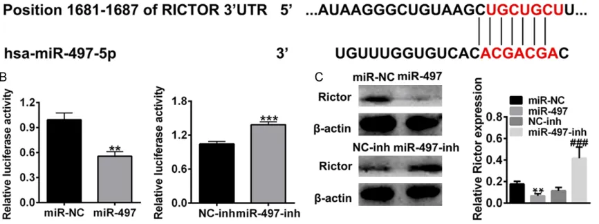
[image:3.612.92.366.69.328.2]To predict a miR-497 target, three bioinformatic databases (TargetScan, miRanda, and PicTar) were used. Rictor was selected as a putative miR-497 target due to its inhibitory effect in tumor progression and metastasis. The com-plementary sequence of miR-497 was found on the site of the 3’-UTR of Rictor mRNA (Figure
Figure 1. Level of miR-497 and Rictor in carcinoma tissue and para-carci-noma tissue of HCC patients. A. Level of miR-497 mRNA in HCC patients analyzed by RT-PCR. B. Level of Rictor mRNA in HCC patients analyzed by RT-PCR. C. Expression of Rictor in carcinoma tissue and para-carcinoma tissue of HCC patients analyzed by immunohistochemical staining. D. The linear regression of miR-497 and Rictor expression. ***P < 0.001 compared to HCC.
migration, and invasion assays upon miR-497 overexpression by transfection of miR-497 mimics (SK-HEP-1, Huh-7) or 497 knockdown with miR-497-inhibitor in human hepa-toma cells. The transfection
3A). To investigate the regulation of miR-497 on Rictor, 293T cells were transfected with Rictor luciferase reporter plasmid, with their 3’-UTR regions containing of miR-497 binding sites or corresponding mutant sites, and the 3’-UTR luciferase activity was measured. As shown in
Figure 3B, miR-497 overexpression (miR-497) reduced the transcriptional activity of Rictor compared with control (miR-NC), and miR-497 inhibitor (miR-497-inh) increased the transcrip-tional activity of Rictor compared to negative control (NC-inh). Furthermore, the protein expression of Rictor was in accordance with mRNA levels (Figure 3C). These results sug-gested that miR-497 can negatively regulate Rictor by directly targeting its 3’-UTR.
Rictor knockdown inhibited cancer cell prolif-eration, migration, and invasion
To explore the effects of Rictor on migratory and invasive abilities of HCC cancer cells, we performed proliferation, wound healing, tran-swell migration, and invasion assays in Rictor overexpression (Rictor group) human hepato-ma cells (SK-HEP-1, Huh-7) or Rictor knock-down cells (SiRictor#1 and SiRictor#2). The
transfection efficiency was confirmed by
RT-PCR. The Rictor mRNA (Figure 4A) and protein
level (Figure 4B) in the Rictor overexpression group (Rictor) were remarkably increased com-pared with the control (P < 0.001). The level of Rictor mRNA and protein were decreased in siRNA#1 and #2 group than siNC. The level of siRNA#1 was lower than #2, which indicated
that the sequence of #1 was more efficiency
than #2. MTT assay results showed that the proliferation of HCC cells was increased in Rictor overexpression cells compared with con-trol cells (Figure 4C) (P < 0.01 at 3rd day). Conversely, the proliferation of HCC cells were decreased in the Rictor knockdown group (siRictor) compared with the siNC group (P < 0.01 at 3rd day). We then investigated the role of Rictor on cell invasion. The invasion assay results showed that Rictor overexpression increased the invasive ability of the cells com-pared with controls (Figure 4D). The invasion ability of cell was decreased in Rictor knock-down cells compared with siNC group. Fur- thermore, the wound healing assay showed that Rictor overexpression cells migrated faster compared with control (Figure 4E). The migra-tion of Rictor knockdown cells was lower than the siNC group. The results were similar in SK-HEP-1 and Huh-7. Taken together, these results suggest that Rictor knockdown inhibits cell proliferative and invasive ability.
[image:5.612.93.526.155.317.2]Figure 3.Rictor is a direct target of miR-497. A. Complementary sequence of miR-497 binding sites in Rictor 3’-UTR regions. B. The luciferase activities of miR-497 mimics or miR-497-inh transfected cells at 48 h after the transfec-tion. C. Rictor expression of miR-497 or miR-497-inh transfected cells analyzed by western blotting. **P < 0.01 compared to miR-NC, ***P < 0.001 compared to miR-NC, ###P < 0.001 compared to NC-inh.
Figure 2. Effect of miR-497 overexpression or knockdown on the proliferation and motility of SK-HEP-1 and Huh-7
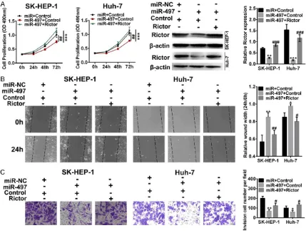
Rictor overexpression reversed the effects of miR-497 overexpression
To explore the effects of miR-497 on migratory and invasive abilities of cancer cells and to determine its regulatory effects on Rictor, SK- HEP-1 and Huh-7 cells were transfected with miR-497 agomir and/or Rictor plasmid. Pro- liferation, wound healing, transwell migration, and invasion assays were performed in differ-ent groups (miR + Control, miR-497 + control, miR-497 + Rictor). As shown in Figure 5A, MTT results showed that the proliferation of cancer cells was inhibited by miR-497 overexpression, and the effects were reversed by Rictor overex-pression. miR-497 overexpression remarkably decreased the expression of Rictor, whereas the Rictor overexpression inversed the effects of miR-497. Then the role of miR-497 on cell invasion was evaluated. The invasion assay
results showed that the miR-497 overexpres-sion inhibited the invaoverexpres-sion ability, and the effects were inversed by Rictor overexpression (Figure 5B). Furthermore, the wound healing assay showed that miR-497 overexpression inhibited the migration ability, and the inhibi-tion effects were inversed by Rictor overexpres-sion (Figure 5C). Taken together, these results suggest that miR-497 overexpression inhibits SK-HEP-1 and Huh-7 cell proliferative and inva-sive ability, while Rictor reverses the effects. miR-497 regulated Akt signaling pathway by targeting Rictor
[image:7.612.92.521.71.398.2]As our previous results demonstrated that miR-497 inhibited SK-HEP-1 and Huh-7 cell prolifer-ation, migration and invasion by targeting Ri- ctor, and other research showed that Rictor directly regulates the phosphorylation of Akt at
Figure 5. Proliferation, migration and invasion of Rictor and miR-497 overexpression cells (SK-HEP-1 and Huh-7).
Ser-473, we further explored whether the Akt signaling pathway was involved in miR-497-in-duced cell growth inhibitory effects. We ana-lyzed the phosphorylation of Aktor total Akt in miR-497 overexpression (miR-497) or miR-497-Inh transfected cells. As shown in Figure 6A, miR-497 overexpression decreased the expres-sion of Rictor and pAKT (Ser-473) but had no effects on the expression of total AKT, miR-497 inhibitor (miR-497-inh) led to the opposite effects of miR-497 expression. When cells we- re co-transfected with miR-497 and Rictor, the
level of Rictor, pAKT/AKT was significantly
in-creased compared with miR-497 overexpres-sion group (P < 0.01 vs miR-497 + control) and close to controls (miR-NC + Control).
Discussion
Hepatocellular carcinoma (HCC) is the fifth
most common cancer worldwide and displays vascular abnormalities and active metastasis [1]. Accumulating studies indicate that Mi- croRNAs (miRNAs) play important roles in tumorigenesis, metastasis and prognosis in mu- ltiple cancers including HCC [9]. Researchers found a general downregulation of miRNAs in human tumors compared with normal tissues
known. Our results indicated that the growth inhibitory effects of miR-497 were reversed by Rictor overexpression, which indirectly
con-firmed that Rictor was the target of miR-497
and related with HCC severity. Previous studies demonstrated that Rictor or mTOR directly reg-ulated the phosphorylation of Akt at Ser-473 [17]. The Akt is activated in many human can-cers and inhibition of Rictor inhibits Akt activity [18]. Another study showed that Rictor overex-pression in melanocytes disrupted the negative feedback of activated Akt and stimulate mela-noma proliferation [19]. The level of total and phosphorylated Akt was related to the severity of gliomas, and Rictor plays an important role in proliferation of glioblastoma [20]. Furthermore, altering Rictor inhibits tumor progression in prostate cancer model [21]. In this study, miR-497 mimics and inhibitor were used to demon-strated that miR-497 could inhibit cell prolifera-tion, migration and invasion by targeting Rictor, and Rictor further regulating the phosphoryla-tion of Akt. These results were in accordance with other studies.
MiR-497 is an important regulator in tumor pro-gression including HCC, but the detail
[image:8.612.89.370.71.289.2]mecha-nism has not been sufficiently reported so far. Figure 6.Levels of pAKT (Ser473), total AKT, and Rictor in SK-HEP-1 and
Huh-7 cells. A. Cells transfected with miR-497 and/or Rictor by western blot-ting. B. Cells transfected with miR-497, miR-497-inh or negative control by western blotting. **P < 0.01 compared to miR-NC (or miR-NC + control for B), ***P < 0.001 compared to miR-NC (or miR-NC + controlfor B), #P < 0.05
compared to NC-inh (or miR-497 + control for B), ##P < 0.01 compared to
NC-inh (or miR-497 + control for B), ###P < 0.001 compared to NC-inh (or
miR-497 + control for B).
[11] and miR-497 downregula-tion was found in various tumors including HCC [16]. In our studies, we found that overexpression of miR-497 in- hibited the proliferation, inva-sion, and metastasis of hu- man hepatoma cells (SK-HEP- 1, Huh-7), which is consistent with previous report [14]. Additionally, we demonstrated that Rictor was a direct target of miR-497.
Our research demonstrated that miR-497 inhibits the proliferation, invasion, metastasis of hepatoma cells in vitro by target Rictor/Akt signal pathway. Therefore, miR-497 or Rictor can be potential targets for treatment of HCC. However, the effects of miR-497 on the HCC in vivo will be further investigated to confirm its
regulation in HCC progression. If the results of in vivo research are in accordance with in vitro results, miR-497 may be an important biomark-er for HCC diagnosis and a potential treatment target.
Disclosure of conflict of interest
None.
Address correspondence to: Rui Zhang, Depart- ment of Hepatobiliary Surgery, Affilicated Hosptial of Hebei University, 212 Yuhua East Road Baoding City, Hebei, China. Tel: +86-312-5983242; E-mail: ZhangRuicxz@163.com
References
[1] Shiratori Y, Yoshida H and Omata M. Manage-ment of hepatocellular carcinoma: advances in diagnosis, treatment and prevention. Expert Rev Anticancer Ther 2001; 1: 277-290. [2] Massarweh NN and El-Serag HB. Epidemiology
of hepatocellular carcinoma and intrahepatic cholangiocarcinoma. Cancer Control 2017; 24: 107327481772924.
[3] Tsuchiya N, Sawada Y, Endo I, Saito K, Uemura Y, Nakatsura T. Biomarkers for the early diag-nosis of hepatocellular carcinoma. World J Gastroenterol 2015; 21: 10573.
[4] Bertuccio P, Turati F, Carioli G, Rodriguez T, La VC, Malvezzi M and Negri E. Global trends and predictions in hepatocellular carcinoma mor-tality. J Hepatol 2017; 67: 302-309.
[5] Lee RC, Feinbaum RL and Ambros V. The C. el-egans heterochronic gene lin-4 encodes small RNAs with antisense complementarity to lin-14. Cell 1993; 75: 843-54.
[6] Reinhart BJ, Slack FJ, Basson M, Pasquinelli AE, Bettinger JC, Rougvie AE, Horvitz HR, Ru-vkun G. The 21-nucleotide let-7 RNA regulates developmental timing in caenorhabditis ele-gans. Nature 2000; 403: 901-906.
[7] Wilson T, Hastings JW. Bioluminescence. Annu Rev Cell Dev Biol 1998; 14: 197-230.
[8] He Q, Ren X, Chen J, Li Y, Tang X, Xin W, Yang X, Jian Z, Wang Y and Ma J. miR-16 targets fibro-blast growth factor 2 to inhibit NPC cell prolif-eration and invasion via PI3K/AKT and MAPK signaling pathways. Oncotarget 2016; 7: 3047-3058.
[9] Wang W, Zhang E and Lin C. MicroRNAs in tu-mor angiogenesis. Life Sci 2015; 136: 28-35. [10] Xu J, Wang T, Cao Z, Huang H, Li J, Liu W, Liu S,
Lei Y, Li Z and Zhang T. MiR-497 downregula-tion contributes to the malignancy of pancre-atic cancer and associates with a poor progno-sis. Oncotarget 2014; 5: 6983-6993.
[11] Jun L, Gad G, Miska EA, Ezequiel AS, Justin L, David P, Alejandro SC, Ebert BL, Mak RH and Ferrando AA. MicroRNA expression profiles classify human cancers. Nature 2005; 435: 834-838.
[12] Ge L, Zheng B, Li M, Niu L and Li Z. MicroR-NA-497 suppresses osteosarcoma tumor gro- wth in vitro and in vivo. Oncol Lett 2016; 11: 2207-2212.
[13] Yan JJ, Zhang YN, Liao JZ, Ke KP, Chang Y, Li PY, Wang M, Lin JS, He XX. MiR-497 suppresses angiogenesis and metastasis of hepatocellular carcinoma by inhibiting VEGFA and AEG-1. On-col Lett 2016; 11: 1081-1088.
[14] Ding WZ, Ni QF, Lu YT, Kong LL, Yu JJ, Tan LW and Kong LB. MicroRNA-497 regulates cell pro-liferation in hepatocellular carcinoma. Oncol Lett 2016; 11: 1081-1088.
[15] Van MJ, Kaspers GJ and Cloos J. Cell sensitivity assays: the MTT assay. Methods Mol Biol 2011; 88: 237-245.
[16] Xie Y, Wei RR, Huang GL, Zhang MY, Yuan YF and Wang HY. Checkpoint kinase 1 is negative-ly regulated by miR-497 in hepatocellular carci-noma. Med Oncol 2014; 31: 844.
[17] Atsushi U, Ken-Ichi K, Tomohiko T, Mayuko F, Kei-Ichi M, Issei I, Ken O and Johji I. The tumor suppressive microRNA miR-218 targets the mTOR component Rictor and inhibits AKT phosphorylation in oral cancer. Cancer Res 2011; 71: 5765-78.
[18] Liang X, Sun R, Zhao X, Zhang Y, Gu Q, Dong X, Zhang D, Sun J and Sun B. Rictor regulates the vasculogenic mimicry of melanoma via the AKT-MMP-2/9 pathway. J Cell Mol Med 2017; 21: 3579-3591.
[19] Laugier F, Finet-Benyair A, André J, Rachakon-da PS, Kumar R, Bensussan A, Dumaz N. RIC-TOR involvement in the PI3K/AKT pathway regulation in melanocytes and melanoma. On-cotarget 2015; 6: 28120-28131.
[20] Alvarenga AW, Machado LE, Rodrigues BR, Lu-pinacci FC, Sanemastu P, Matta E, Roffé M, Torres LF, Da CI and Martins VR. Evaluation of Akt and RICTOR expression levels in astrocyto-mas of all grades. J Histochem Cytochem 2017; 65: 93-103.