Original Article
MiR-218 promotes osteogenic
differentiation of periodontal ligament stem cell
through activation of Wnt signaling by targeting SFRP2
Fucai Sun1, Yike Ma1, Zhibin Cai2, Zhaoan Yang1
1Department of Stomatology, The First Affiliated Hospital of Wenzhou Medical University, Wenzhou, Zhejiang, China; 2Department of General Stomatology, The Affiliated Stomatology Hospital of Wenzhou Medical University, Wenzhou 32500, Zhejiang, China
Received February 29, 2016; Accepted July 12, 2016; Epub October 1, 2016; Published October 15, 2016
Abstract: Increasing evidence supports that microRNAs (miRNAs) play an important role in the control of osteo-blastic differentiation such as human adipose-derived stem cells and embryonic stem cells. However, the role of miRNAs in periodontal ligament stem cell (PDLSCs) differentiation remains poorly understood. Here, we present evidence that miR-218 acts as a positive regulator of PDLSCs osteogenesis. Real-time PCR shows that miR-218 was increased during PDLSCs differentiation. Moreover, ectopic expression of miR-218 promoted PDLSCs cells
dif-ferentiation, whereas inhibition of 218 would suppress cell differentiation. Furthermore, we verified that
miR-218 directly targeted SFRP2, which is a Wnt signaling pathway antagonist. Western blot analysis showed that the expression level of miR-218 was negatively correlated with that of SFRP2. Taken together, miR-218 is an important mediator of osteoblast differentiation, thus offering a new target for the development of preventive or therapeutic agents against osteogenic disorder.
Keywords: MicroRNA-218, periodontal ligament stem cells, osteogenic differentiation, Wnt, SFRP2
Introduction
Previous studies have characterized progenitor cells residing in periodontal ligament (PDL) tis-sues, known as periodontal ligament stem cells (PDLSCs), which exhibit self-renewal capacity and expressing cell surface markers similar to bone marrow MSCs [1-3]. Further, PDLSCs pos-sess unique properties compared with other MSC-like populations, which can differentiate into many kinds of cells derived from other sys-tems and form osteogenic tissue; thus, these cells play important roles in tooth regeneration [4]. Subsequently, the regenerative potential of PDLSCs has been shown in preclinical large-animal studies and human clinical pilot trials for periodontal regeneration [5-10]. However, how to regulate and control their potency of osteogenic differentiation remains to be an unsolved problem.
During the osteoblast differentiation, numer-ous regulatory pathways play important roles in
regulating osteoblast replication and cellular differentiation [11, 12]. For instance, the Wnt and BMP pathways have prominent and syner-gistic roles in osteoblast phenotype commit-ment [13]. In particular, Wnt signaling is very important for differentiation of human mesen-chymal stem cells into osteoblasts [14]. Wnt signaling promotes mesenchymal stem cells proliferation during early differentiation [15, 16]. Canonical Wnt signaling then drives the differentiation of osteochondral progenitors to- ward the osteoblastic lineage [17]. In addition, Wnt signaling inhibits osteoblast and osteo- cyte apoptosis [18].
differentiation, apoptosis and cancer [20]. And, kinds of miRNAs have been confirmed to par-ticipate in human MSC differentiation [16, 21]. Zhang WB et al. presented evidence that miR-218 acts as a positive regulator of hASCs osteogenesis [22]. However, the specific roles ofmiR-218 in the differentiation of PDLSCs are largely unknown and its function remains to be characterized [23].
To date, few studies have investigated the osteogenic effect of miR-218 on PDLSCs. In this study, we aimed to analyze the bioactive effects of miR-218 on the osteogenic differ- entiation of PDLSCs and to determine their po- tential target genes that are related to osteo-genic differentiation.
Materials and methods
Cell culture and isolation of PDLSCs isolation Human premolars were obtained from five healthy patients for orthodontic reasons after obtaining the patients’ approval and inform- ed consent (donor age: 10-12 years). Tissue from the periodontal ligament was isolated as previously described [3]. Briefly, periodontal ligament tissues were gently scraped from the middle portion of the root surface, minced into 1 mm3 cubes, and placed into 6-well culture dishes (Costar, Cambridge, MA). The explants were grown in a minimum essential medium (α-MEM; Gibco BRL, Rockville, MD) supplemented with 10% (v/v) fetal bovine serum (FBS) 0.292 mg/mL glutamine, 100 U/ mL penicillin, and 100 μg/mL streptomycin.
inhibitor and negative control at a final multi-plicity of infection of 10 using siLentFect™ Lipid reagent (Life Science Research). The cells were then diluted in DMEM/F12 without serum (GeneChem, Shanghai, China). After 4 h of incu-bation in a CO2 incubator at 37°C, the medium was changed to 10% FBS containing DMEM. The efficiencies of miRNA mimics, miRNA inhib-itor and negative control were tested by quanti-tative real time polymerase chain reaction (qRT-PCR).
MiRNA extraction and qPCR analysis
[image:2.612.91.324.83.270.2]Total RNA and miRNA were purified from PDLSCs cell cultures treated with or without 50 μg/ml of ascorbic acid for d0, d2, d10 and d21 days using miRNeasy mini kit (Qiagen). Total RNA (0.5-1 μg) was reverse transcribed into cDNA using Oligo (dT) primers and was analyzed by real time qPCR. SYBR Green Mas- ter Mix (Applied Bio Systems Inc.) was used to detect the expression of gene markers. For miRNA detection, the isolation of small spe-cies-enriched RNA was performed as per the manufacturer’s instructions (mirVana miRNA isolation kit, Ambion). MiRNA was reverse-tran-scribed with an Ncode miRNA first-strand cDNA synthesis kit (Invitrogen) according to the man-ufacturer-specified guidelines. Real-time PCR was performed using a standard SYBR Green PCR kit (TAKARA, Osaka, Japan) protocol on an Applied Biosystems 7500 real-time PCR system (Applied Biosystems, Foster City, CA, USA), according to the manufacturer’s instruc-tions. U6 was used as a normalizing control. Table 1. Primers used for qPCR
Gene name Real time qPCR primers (5’-3’) Runx2 Forward: GTC TCA CTG CCT CTC ACT TG
Reverse: CAC ACA TCT CCT CCC TTC TG
ALP Forward: CGG ACA TCA TGA GGG TAA GG
Reverse: GAG ACA TTT TCC CGT TCA CC
OCN Forward: ACA GAC AAG TCC CAC ACA GCA GC Reverse: TGA AGG CTT TGT CAG ACT CAG GGC
BSP Forward: GCCAGAGGAGCAATCACCAA
Reverse: CAGGCTGGAGGTTCACTGGT SFRP2 Forward: CGT GGG CTC TTC CTC TTC G
Reverse: ATG TTC TGG TAC TCG ATG CCG
miR-218 TTG TGC T TG ATC TAA CCA TGT
U6 Forward: CGC TTC GGC AGC ACA TAT AC
Reverse: AAA ATA TGG AAC GCT TCA
The cultures were incubated at 37°C in a humidified atmosphere with 5% CO2. STRO-1+ stem cells were prepared using immunomagnetic beads (Dynabeads; Dy- nal Biotech, Oslo, Norway) according to the manufacturer’s instructions. After wa- shing, bead-positive cells were segregat- ed using a magnetic particle separator and subsequently seeded into 75-cm2 cul-ture flasks (Costar) at 37°C in 5% CO2. The PDLSCs from this passage were used for further study.
Transfection
The mRNA expression of osteogenic differen- tiation marker alkaline phosphatase (ALP), os- teocalcin (OCN), bone sialoprotein (BSP), the bone-specific transcription factor Runx2 and secreted frizzled-related protein 2 (SERP2) were analyzed. Each sample was analyzed in triplicate. The 2-ΔΔCt value was used to de-
termine the relative expression levels. The re- sults were expressed as Log 10 (2-ΔΔCt). Se-
quences of the primers are shown in Table 1. Alizarin red staining and quantification
Cells were seeded into 24-well plates at a density of approximately 1×10⁵ cells per well separately. After cells reached 80% conflu- ence, the culturing medium was changed into standard osteogenic differentiation induction medium and then cultured for another 3 weeks. The induction medium was changed every 3 days. Finally cells were stained with Alizarin red (pH = 4.1) staining solution and were quantified according to the methods previously published.
DNA construction and luciferase assay
The 3’UTRs of the SFRP2 genes were amplified and cloned into the SacI/HindIII sites of the pMIR-Report luciferase vector. The seed re- gion of the miR-218 target sites in the SFRP2 3’UTRs were mutated using the Quick change II site directed mutagenesis kit (Stratagene). The Luciferase assay was conducted by co-transfecting 218 miRNA, NS miR and miR-218 inhibitor with WT and mutant pMIR-RE- PORT-Luc DNA construct. The transfections
were performed in duplicate and all experi-ments were repeated several times. Renilla Luciferase plasmid (Promega) was transfected to normalize the relative luciferase values. The transfected cells were incubated for 36 h to determine luciferase activity.
Western blots
Equal amounts of total protein were loaded into a 10% SDS-PAGE transferred onto a PVDF membrane probed with indicated primary anti-body for SFRP2 (Abcam, ab86379) and β-actin (Santa Cruz). After incubation with secondary antibody, antibody-bound protein complex in the membrane was detected with chemilumi-nescence reagent.
Statistics
Data are expressed as mean ± SD. Student’s t-test or one-way analysis of variance (ANOVA) was made for multiple comparisons. All ex- periments were repeated 3 times, and repre-sentative experiments are shown. P-values < 0.05 will be considered to be statistically sig- nificant.
Results
Expression level of miR-218 during osteogenic differentiation of PDLSCs
[image:3.612.94.519.79.197.2]Figure 2. Up-regulation of miR-218 promoted PDLSC osteogenic differentiation. A. 10 days after induction, the cells were stained with alizarin red (pH = 4.1), and the results showed that PDLSCs transfected with miR-218 mimic formed more mineralized nodules than the control groups. B-E. PDLSCs were transduced with miR-218
mimic. Total RNA was analyzed by real time qRT-PCR for mRNA expression profile of bone marker genes (ALP, OCN, BSP and Runx2) at d10 and d21. The data are
differentiation in PDLSCs. The RNA samples were collected and examined at indicated time points. According to the qPCR results, we found that miR-218 expression was gradually increased after the induction of osteogenic differentiation of PDLSCs (Figure 1A).
The functional activity of miR-218 was assess- ed in PDLSCs after forced expression of a miR-218 mimic and a miR-218 inhibitor. After transfection, miR-218 expression levels were examined through qPCR. qPCR analysis dem-onstrated that endogenous miR-218 level was increased more than 14-fold compared to the control cells after two day transfection, and remained 12-, 8-fold of the controls in 10 and 21 days after miR-218 mimic infected, respec-tively (Figure 1B). The transfected PDLSCs with miR-218 inhibitor had 5-, 4-, and 3-fold decrease of miR-218 expression at 2, 10 and 21 days respectively after transfection (Figure 1C). These results suggest that miR-218 may provoke osteoblast differentiation.
Over-expression of miR-218 promotes osteo-genic differentiation of PDLSCs
To confirm the role of miR-218 in osteoge- nic differentiation of PDLSCs, we transfected miR-218 mimics into PDLSCs. Based on the results of Alizarin Red S staining assays, it was shown that over-expression of miR-218 significantly promoted PDLSCs osteogenesis when compared with control group (Figure 2A). We also illustrated that overexpression of miR-218 could activate the expression of sev-eral osteogenic marker genes such as ALP, the osteogenic transcription factor Runx2, and the mineralization marker (bone sialoprotein (BSP) and OCN), which are indicative of the osteoblastic differentiation status of PDLSCs (Figure 2B-E). The over-expression of miR-218 suggested potential roles for this miRNA in osteogenic differentiation of PDLSCs.
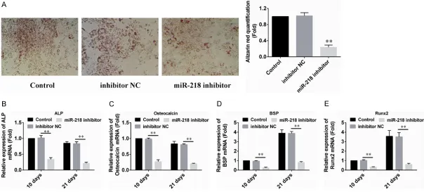
Knockdown of miR-218 inhibits osteogenic dif-ferentiation of PDLSCs
To further study the roles of miR-218 on the osteogenic differentiation of PDLSCs, we used the miR-218 inhibitor to transfect PDLSCs to retard the endogenous expression of miR- 218. As shown in Figure 3A, Alizarin red stain-ing and quantification results indicated that knockdown of miR-218 inhibited the forma- tion of mineralized nodules after induction. The mRNA expression of the osteogenic differen-
tiation markers ALP, OCN, BSP and Runx2 were all significantly decreased in PDLSCs trans- fected with miR-218 inhibitor (Figure 3B-E). To- gether, these experiments demonstrate the essential role of miR-218 for PDLSCs osteo- genic differentiation.
MiR-218 is a positive regulator of Wnt
signal-ing
To address the mechanism by which miR-218 regulates osteoblast differentiation, we exam-ined predicted targets of miR-218 relevant to bone formation. More evidences have found that SFRP2 is a direct target of miR-218, which is potentially involved in differentiation through the Wnt signaling pathways [24]. To examine whether miR-218 directly targets SFRP2 in PDLSCs, we performed dual-luciferase repor- ter assays to confirm that miR-218 targets the SFRP2 3’UTR (Figure 4A). The luciferase activity was significantly inhibited when SFR- P2-WT was co-transfected with miR-218 mim-ics compared with that after the mimic NC co-transfection, whereas the inhibitory effect was abolished when the SFRP2 3’UTR was muta- ted (Figure 4B).
To evaluate whether miR-218 regulated SFR- P2 expression, we detected the protein expres-sion level of SFRP2 in 218 mimic or miR-218 inhibitor infected cells. Western blot ana- lysis showed that miR-218 overexpression markedly decreased the protein level of SF- RP2 (Figure 4C), whereas miR-218 inhibition increased the protein expression of SFRP2 (Figure 4D). Together, these results indicated that SFRP2 is a direct target of miR-218 in PDLSCs, which suggested thatmiR-218 may regulate cell differentiation through activation of Wnt signaling by targeting SFRP2.
Discussion
Figure 3. Down-regulation of miR-218 inhibited PDLSC osteogenic differentiation. A. 10 days after induction, the cells were stained with alizarin red (pH = 4.1), and the results showed that PDLSCs transfected with miR-218 inhibitor formed fewer mineralized nodules than the control groups. B-E. PDLSCs were transduced with
miR-218 inhibitor. Total RNA was analyzed by real time qRT-PCR for mRNA expression profile of bone marker genes (ALP, OCN, BSP and Runx2) at d10 and d21. The
Several reports have demonstrated miRNAs were known to act as regulators in adipoge- nesis, myeloblasts differentiation and skeletal muscle development, and recently reported in regulating osteoblastogenesis. The function of miR-218 was recently reported. Zhang WB et al. found that miR-218 was up-regulated dur- ing osteogenic differentiation of Human adi-pose-derived stem cells (hASCs) and overex-pression of miR-218 enhanced osteogenic dif-ferentiation in vitro by directly targeting SFR- P2 and DKK2 [22]. Mohammad Q. Hassan et al. demonstrated that miR-218 stimulated the
[image:7.612.95.524.80.486.2]Wnt pathway by down-regulating three Wnt signaling inhibitors Sclerostin (SOST), Dickko- pf2 (DKK2), and secreted frizzled-related pro- tein2 (SFRP2) during the process of osteogen-esis [25]. Therefore, we concluded that miR-218 might regulate osteoblastic differentiation of PDLSCs. The results shown that miR-218 levels were increased in differentiating PDLSCs, and miR-218 overexpression promoted PDLSCs differentiation. When miR-218 function was blocked, the formation of mineralized nodules was inhibited and the mRNA expression of the osteogenic differentiation markers ALP, OCN, Figure 4. miR-218 directly binds and downregulates SFRP2. A. Schema of the firefly luciferase reporter constructs
for SFRP2, indicating the interaction sites between miR-218 and the 3’-UTRs of the SFRP2. B. Luciferase activities.
PDLSCs were co-transfected with firefly luciferase constructs containing the SFRP2 wild-type or mutated 3’-UTRs
BSP and Runx2 were all significantly decreased. However, there has been no research on the regulatory functions of miR-218 in the field of osteogenic differentiation of PDLSCs.
In our study, we identified that miR-218 tar- geted the SFRP2 gene, which was a Wnt sig- naling pathway antagonist [26, 27]. The re- porter assay showed that miR-218 was able to significantly repress luciferase contained SFRP2-3’-UTR expression. Western blot analy-sis also showed that miR-218 significantly inhibited the SFRP2 protein levels in differen- tiating PDLSCs cells. This result is in agree- ment with the report from Wei-Bing Zhang that SFRP2 is a direct target of endogenous miR-218 in hASCs [22]. sFRPs have been con-sidered antagonists of canonical Wnt signal- ing by binding to Wnt proteins and preventing signal transduction based on their sequence homology with the Wnt-binding domain of the Fz receptors [16, 17]. Recent study from Je- ffrey Schmeck peper et al. demonstrated that Sfrp2 treatment induced cardiac progenitor cells (CPCs) to exit the cell cycle and primed them for cardiac differentiation by inhibition of Wnt6 canonical signaling and activation of Wnt non-canonical pathways. In a pluripotent mouse embryonal carcinoma stem cell line, SFRP2 inhibits cardiomyogenic differentiation by regulating Wnt3a transcription [28]. Indeed, the expression of the negative regulators of Wnt signaling, sFRP2, is decreased in mature osteoblasts, providing a potential mechanism for increased Wnt signaling in more differenti-ated cells [29]. Therefore, it is possible that targeting of sFRP2 RNA by miR-218 is one mechanism contributing to PDLSCs differen- tiation. Because miR-218 is expressed in a wide array of tissues, it is likely that other miR-218 regulatory networks could exist [30, 31]. In conclusion, our data demonstrate that miR-218 regulates osteoblast differentiation by tar-geting SFRP2. Thus, miR-218 should be con- sidered an important candidate as a molecu- lar target of osteoblastic differentiation for the development of preventive or therapeutic agents against osteogenic disorders.
Acknowledgements
This study was supported by the Fundation from Wenzhou Medical University (Y201431- 298).
Disclosure of conflict of interest
None.
Address correspondence to: Yike Ma, Department
of Stomatology, The First Affiliated Hospital of
Wenzhou Medical University, 2 Fuxue Lane, Ouhai District, Wenzhou, Zhejiang Province, China. Tel: +86 13968817548; E-mail: mayikemyk@yeah.net
References
[1] Gronthos S, Mankani M, Brahim J, Robey PG
and Shi S. Postnatal human dental pulp stem cells (DPSCs) in vitro and in vivo. Proc Natl Acad Sci U S A 2000; 97: 13625-13630. [2] Miura M, Gronthos S, Zhao M, Lu B, Fisher LW,
Robey PG and Shi S. SHED: stem cells from
human exfoliated deciduous teeth. Proc Natl Acad Sci U S A 2003; 100: 5807-5812. [3] Seo BM, Miura M, Gronthos S, Bartold PM,
Batouli S, Brahim J, Young M, Robey PG, Wang
CY and Shi S. Investigation of multipotent post-natal stem cells from human periodontal liga-ment. Lancet 2004; 364: 149-155.
[4] Maeda H, Tomokiyo A, Fujii S, Wada N and Akamine A. Promise of periodontal ligament stem cells in regeneration of periodontium. Stem Cell Res Ther 2011; 2: 33.
[5] Feng F, Akiyama K, Liu Y, Yamaza T, Wang TM,
Chen JH, Wang BB, Huang GT, Wang S and Shi
S. Utility of PDL progenitors for in vivo tissue regeneration: a report of 3 cases. Oral Dis 2010; 16: 20-28.
[6] Gault P, Black A, Romette JL, Fuente F,
Sch-roeder K, Thillou F, Brune T, Berdal A and Wurtz T. Tissue-engineered ligament: implant con-structs for tooth replacement. J Clin Periodon- tol 2010; 37: 750-758.
[7] Sonoyama W, Liu Y, Fang D, Yamaza T, Seo BM,
Zhang C, Liu H, Gronthos S, Wang CY, Wang S
and Shi S. Mesenchymal stem cell-mediated functional tooth regeneration in swine. PLoS One 2006; 1: e79.
[8] Liu Y, Zheng Y, Ding G, Fang D, Zhang C, Bartold PM, Gronthos S, Shi S and Wang S. Periodontal
ligament stem cell-mediated treatment for periodontitis in miniature swine. Stem Cells 2008; 26: 1065-1073.
[9] Park JY, Jeon SH and Choung PH. Efficacy
of periodontal stem cell transplantation in the treatment of advanced periodontitis. Cell Transplant 2011; 20: 271-285.
de-fect model: a pilot study. J Periodontol 2009; 80: 1815-1823.
[11] Hassan MQ, Tare RS, Lee SH, Mandeville M, Morasso MI, Javed A, van Wijnen AJ, Stein JL,
Stein GS and Lian JB. BMP2 commitment to
the osteogenic lineage involves activation of Runx2 by DLX3 and a homeodomain transcrip-tional network. J Biol Chem 2006; 281: 40515-40526.
[12] Luu HH, Song WX, Luo X, Manning D, Luo J,
Deng ZL, Sharff KA, Montag AG, Haydon RC
and He TC. Distinct roles of bone morphoge-netic proteins in osteogenic differentiation of mesenchymal stem cells. J Orthop Res 2007; 25: 665-677.
[13] Canalis E. Growth factor control of bone mass.
J Cell Biochem 2009; 108: 769-777.
[14] Baron R and Rawadi G. Wnt signaling and the
regulation of bone mass. Curr Osteoporos Rep 2007; 5: 73-80.
[15] Boland GM, Perkins G, Hall DJ and Tuan RS.
Wnt 3a promotes proliferation and suppresses osteogenic differentiation of adult human mesenchymal stem cells. J Cell Biochem 2004; 93: 1210-1230.
[16] Kapinas K, Kessler C, Ricks T, Gronowicz G and
Delany AM. miR-29 modulates Wnt signaling in human osteoblasts through a positive feed-back loop. J Biol Chem 2010; 285: 25221-25231.
[17] Takada I, Kouzmenko AP and Kato S. Wnt and PPARgamma signaling in osteoblastogenesis and adipogenesis. Nat Rev Rheumatol 2009; 5: 442-447.
[18] Bodine PV, Billiard J, Moran RA, Ponce-de-Leon H, McLarney S, Mangine A, Scrimo MJ, Bhat
RA, Stauffer B, Green J, Stein GS, Lian JB and
Komm BS. The Wnt antagonist secreted friz-zled-related protein-1 controls osteoblast and osteocyte apoptosis. J Cell Biochem 2005; 96: 1212-1230.
[19] Filipowicz W, Bhattacharyya SN and Sonenberg N. Mechanisms of post-transcriptional regula-tion by microRNAs: are the answers in sight?
Nat Rev Genet 2008; 9: 102-114.
[20] Ambros V. The functions of animal microRNAs. Nature 2004; 431: 350-355.
[21] Mizuno Y, Yagi K, Tokuzawa Y, Kanesaki-Yatsuka Y, Suda T, Katagiri T, Fukuda T, Maruyama M, Okuda A, Amemiya T, Kondoh Y, Tashiro H and Okazaki Y. miR-125b inhibits osteoblastic differentiation by down-regulation of cell proliferation. Biochem Biophys Res Commun 2008; 368: 267-272.
[22] Zhang WB, Zhong WJ and Wang L. A
signal-amplification circuit between miR-218 and
Wnt/beta-catenin signal promotes human adi-pose tissue-derived stem cells osteogenic dif-ferentiation. Bone 2014; 58: 59-66.
[23] Luzi E, Marini F, Sala SC, Tognarini I, Galli G
and Brandi ML. Osteogenic differentiation of human adipose tissue-derived stem cells is modulated by the miR-26a targeting of the SMAD1 transcription factor. J Bone Miner Res 2008; 23: 287-295.
[24] Wang Y, Liu J, Cui J, Sun M, Du W, Chen T, Ming X, Zhang L, Tian J, Li J, Yin L, Liu F, Pu Z, Lv B, Hou J and Yu B. MiR218 Modulates Wnt Signaling in Mouse Cardiac Stem Cells by Promoting Proliferation and Inhibiting Differ- entiation through a Positive Feedback Loop. Sci Rep 2016; 6: 20968.
[25] Hassan MQ, Maeda Y, Taipaleenmaki H, Zhang
W, Jafferji M, Gordon JA, Li Z, Croce CM, van Wijnen AJ, Stein JL, Stein GS and Lian JB.
miR-218 directs a Wnt signaling circuit to pro- mote differentiation of osteoblasts and os- teomimicry of metastatic cancer cells. J Biol Chem 2012; 287: 42084-42092.
[26] Cui Y, Niziolek PJ, MacDonald BT, Zylstra CR, Alenina N, Robinson DR, Zhong Z, Matthes S, Jacobsen CM, Conlon RA, Brommage R, Liu Q, Mseeh F, Powell DR, Yang QM, Zambrowicz B,
Gerrits H, Gossen JA, He X, Bader M, Williams BO, Warman ML and Robling AG. Lrp5 func -tions in bone to regulate bone mass. Nat Med 2011; 17: 684-691.
[27] Oshima T, Abe M, Asano J, Hara T, Kitazoe K, Sekimoto E, Tanaka Y, Shibata H, Hashimoto T, Ozaki S, Kido S, Inoue D and Matsumoto T. Myeloma cells suppress bone formation by se-creting a soluble Wnt inhibitor, sFRP-2. Blood 2005; 106: 3160-3165.
[28] Deb A, Davis BH, Guo J, Ni A, Huang J, Zhang Z,
Mu H and Dzau VJ. SFRP2 regulates cardio-myogenic differentiation by inhibiting a posi-tive transcriptional autofeedback loop of Wnt3a. Stem Cells 2008; 26: 35-44.
[29] Kalajzic I, Staal A, Yang WP, Wu Y, Johnson SE, Feyen JH, Krueger W, Maye P, Yu F, Zhao Y, Kuo
L, Gupta RR, Achenie LE, Wang HW, Shin DG and Rowe DW. Expression profile of osteoblast lineage at defined stages of differentiation. J
Biol Chem 2005; 280: 24618-24626.
[30] Wang HT, Liu AG, Luo DS, Zhou ZN, Lin HG,
Chen RZ, He JS and Chen K. miR-218 expres-sion in osteosarcoma tissues and its effect on cell growth in osteosarcoma cells. Asian Pac J Trop Med 2014; 7: 1000-1004.
[31] Qu B, Xia X, Yan M, Gong K, Deng S, Huang G,