Original Article
MiR-124 inhibits
neural apoptosis in ischemic stroke
Kan Zhang1, Yangjun Zhu2, Ping Liu3, Renjie Ji3
1Department of Neurology, The First Affiliated Hospital of Medical School of Zhejiang University, Hangzhou,
Zhejiang, China; 2Department of Ultrasound, The First Affiliated Hospital of Medical School of Zhejiang University,
Hangzhou, Zhejiang, China; 3Department of Neurology and Brain Medical Center, The First Affiliated Hospital of
Medical School of Zhejiang University, Hangzhou, Zhejiang, China
Received July 7, 2016; Accepted August 31, 2016; Epub October 1, 2016; Published October 15, 2016
Abstract: Stroke is a common disease around the world, which causes cerebral tissue damage due to rupture or occlusion of cerebral vessels. Previous studies showed that a large number of microRNA was involved in post-stroke damage or regeneration of neural tissue, among which miR-124 was reported to play a pivotal role in neural dif-ferentiation. Our study was focused on the expression of miR-124 in ischemic stroke and the effect of miR-124 on neural apoptosis. 40 male C57BL6 mice were prepared for establishment of cerebral infarction model with suture method. Tissues of stroke lesion were extracted for examination of miR-124 expression by qRT-PCR and in situ molecular hybridization. Mice neurons were cultured in vitro and transfected with lentiviral expression vector of miR-124. Proliferation of neurocytes was assessed by MTT assays. Western-Blot was performed to examine the expression of Bcl-2, P53 and Caspase-3. Our results showed cerebral infarction model was successfully established as demonstrated by significant infarcted lesion. qRT-PCR and in situ molecular hybridization showed that, com -pared with normal tissues, tissues of stroke lesion had a higher expression of miR-124 (P<0.01). Overexpression of miR-124 up-regulated apoptosis-associated Bcl-2 expression (P<0.05), inhibited expression of activated caspase-3 (P<0.05), and enhanced proliferation of neurocytes (P<0.05). In conclusion, miR-124 was up-regulated in infarcted tissues after ischemic stroke. MiR-124 inhibited neural apoptosis and enhanced proliferation of neurocytes via apoptosis-associated pathways.
Keywords: MiR-124, ischemic stroke, neuron, proliferation, apoptosis
Introduction
Stroke is known as cerebral apoplexy, which is the leading cause of disability and death world-wide [1]. There are two main types of stroke, ischemic stroke and hemorrhagic stroke. Isch- emic stroke accounts for 85% of all stroke cases and always results from cerebral circula-tion insufficiency due to occlusion of the brain feeding arteries, including carotid artery and vertebral artery [2]. The mechanisms of isch-emic stroke are unclear. However, some stud-ies demonstrated that both nerve regeneration and neural apoptosis in infarcted tissues play important roles in neural functional recovery in ischemic stroke patients [3, 4]. Thus, it is of great significance to further study the regulato -ry mechanism underlying neural apoptosis in ischemic stroke.
microRNA is a kind of non-coding RNA with a length of 21 to 25 bp. Previous studies showed
that microRNA influenced multiple cellular ev-ents via RNA interference [5]. MiR-124 is highly expressed in neural tissues with high conserva-tion across different species [6]. MiR-124 is proved to play important roles in neural devel-opment and differentiation. MiR-124 regulates polypyrimidine tract-binding protein 1 (PTBP1), which changes alternative splicing patterns of multiple mRNA in neuron, and influences neural differentiation via targeting PTBP1 mRNA [7]. In addition, clinical trials showed that serum level of miR-124 was increased in ischemic stroke patients, suggesting miR-124 might be involved in the stroke occurrence and post-stroke nerve regeneration [8-10].
MiR-124 inhibits neural apoptosis in stroke
Materials and methods
Animal grouping and cerebral infarction model establishment
C57BL6 mice were purchased from Experime- ntal Animal Center of Medical School of Zhe- jiang University (Hangzhou, China). 40 male adult C57BL6 mice were randomly assigned into two groups with equal numbers. Cerebral infarction model was established with suture method. Detailed protocols for experimental group were as follows: Mice were fixed on the experiment table followed by shaving hair along the neck. Incise neck skin and find carotid artery. Ligate distal part of carotid artery with 5/0 sutures and proximal part with 2/0 sutures. Establish a slipknot in the middle part of carot-id artery with 0# sutures, clip a small hole above the slipknot and put 1 cm line embolism into vessels. Perform separation of vascular but not put the line embolism into vessels in control group. Euthanasia was performed to harvest tissues. All mice experiments were pro-cessed according to requirements of the Inter- national Council for Laboratory Animal Science. All procedures were approved by the Animal Ethics Committee of the First affiliated Hospital of Medical School of Zhejiang University.
qRT-PCR
[image:2.612.89.289.83.124.2]Cerebrum was harvested for RNA extraction. RNAprep pure Tissue Kit (QIAGEN) was used to extract RNA. Amplification was performed wi-th miR-124 primers (GeneBank NR_029668,
Table 1). mirVanat qRT-PCR miRNA detection kit (Ambion) was used to perform qRT-PCR. U6-RNA sequence was used as an internal con-trol. 2-ΔΔCt method was used to analyze the
results [11].
In situ hybridization
Paraffin section of cerebrum was performed. Antisense locked nucleic acid was used to mod-ify miR-124 oligonucleotide probe (5’-TGGCATT- CACCGCGTGCCTTAA-3’, underlined sequence was locked nucleic acid). Dewax paraffin sec
-cific binding sites after hydration. Add prehy -bridization solution after rinse, and incubate for 4 hours at 65°C. Add probe and incubate for 15 hours at 65°C. Block non-specific binding sites after SSC solution rinse. Add antibody and incu-bate for 30 minutes. Perform developing in dark place, and observe under a microscope [12].
Interpretation of the results: bluish violet parti-cles suggested positive expression of miR-124, and expression of miR-124 is positively corre-lated with the degree of color [13].
Culture of primary neuron
Suckling mice were prepared for extraction of primary neurons. Put harvested brain into cold PBS, and remove epencephalon, meninges and cerebral vascular tissues. Add trypsin and col-lect primary neurons. Primary neurons were cultured for 20 minutes at 37°C, 5% CO2, and remained trypsinization. Then, 10% fetal bovine serum was added to terminate trypsinization. Resuspend primary neurons with pipettor. Per- form cell counting after filter screening. Change medium and add 5-fluoro-deoxyuridine after 24 hours culture [10].
Cell transfection and overexpression of miR-124
Overexpression of miR-124 in primary neurons was performed with lentivirus infection. Len- tiviral expression vector with both green fluo -rescent protein and miR-124 was constructed followed by cell transfection with routine proto-cols. Replace culture medium with routine me- dium after 24 hours transfection. Analyze cell fluorescence with fluorescence microscope after 3 days’ culture. MiR-124 overexpressed primary neurons were screened in the medium with puromycin (1 mg/L).
MTT assays
MiR-124 overexpressed primary neurons were prepared for culture at different time points (20 h, 44 h, 68 h and 92 h). Remove the medium and add DMSO (150 μL). Cell proliferation was assessed according to the absorbance value (A) at 490 nm which was measured by a micro-plate reader. Growth curve was established with absorbance values.
Western blot
Primary neurons were cultured for 48 hours
fol-Table 1.Primers of miR-124
Name Sequence
miR-124-F 5’AGGCCTCTCTCTCCGTG3’
PAGE electrophoresis. Primary antibodies were rabbit-anti-mouse Bcl-2 antibody (Proteintech, 25850-1-AP), rabbit-anti-mouse P53 antibody (Proteintech, 10442-1-AP), rabbit-anti-mouse caspase-3 antibody (Proteintech, 19677-1-AP) and rabbit-anti- mouse β-actin antibody (Pro-teintech, 20536-1-AP). β-actin was used as an internal control. Gel imaging system was used to analyze the relative expression of specific protein bands [14].
Statistical analysis
SPSS 20.0 software was used for data pro-cessing. Measurement data were represented
as mean ± standard deviation (SD). One-Way ANOVA was performed for analysis of statistical significance. P value <0.05 was considered to be statistically significant.
Results
Establishment of cerebral infarction model
Cerebrum of mouse was harvested after 24 hours cerebral infarction. TTC stain was per-formed to determine the infarction of cerebrum [9]. As showed in Figure 1, cerebrum of control mice was totally stained (red), while cerebrum of experimental mice had a remarkable infarc-tion area, which was not stained (white). All staining results suggested the successful es- tablishment of cerebral infarction model.
Increased expression of miR-124 in infarction tissues
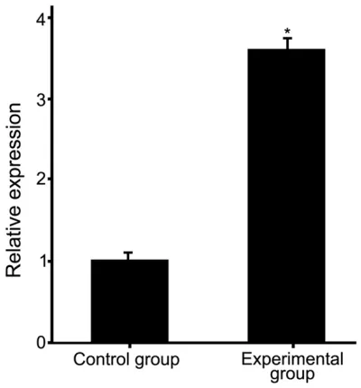
qRT-PCR showed that, compared with control group, experimental group had a higher expres-sion of miR-124 in infarcted tissues (3.5 fold increase) (Figure 2, P<0.01). Consistent with this, in situ hybridization also confirmed the increased expression of miR-124 in infarcted tissues as showed more tissues with more blu-ish violet particles in infarcted experimental group than that in control group (Figure 3).
Overexpression of miR-124 in cultured primary neurons
qRT-PCR was performed to examine the expres-sion of miR-124 in transfected primary
[image:3.612.90.521.74.238.2]neu-Figure 1. TTC staining to determine cerebral infarction.
[image:3.612.90.290.276.491.2]MiR-124 inhibits neural apoptosis in stroke
rons. Compared with normal primary neurons, transfected primary neurons had a higher ex- pression of miR-124 (Figure 4, P<0.05), sug-gesting overexpression of miR-124 was suc-cessfully established in primary neurons.
MiR-124 enhanced proliferation of neurocytes
MTT assays showed that all primary neurons proliferated gradually along with time, while the cell number of miR-124 transfected primary neurons was significantly higher than that of normal primary neurons after 48 hours’ culture
(Figure 5, P<0.05), suggesting overexpression of miR-124 enhanced the proliferation of neur- ocytes.
MiR-124 regulated expression of apoptosis-related proteins
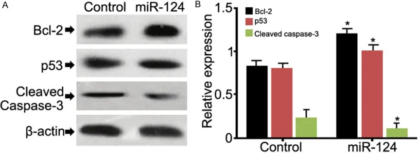
Western blot was performed to examine the expressions of apoptosis-related proteins, in- cluding Bcl-2, P53 and caspase-3. Over- expression of miR-124 significantly increased the expression of Bcl-2 and decreased the expression of caspase-3 (Figure 6, P<0.05), while no difference was observed in the expres-sion of P53.
Discussion
[image:4.612.91.523.72.247.2]As a kind of small non-coding RNA, microRNA regulates cellular events and metabolism [5]. Our mice cerebral infarction models showed
Figure 3. Image of in situ hybridization analysis of miR-124 expression. Red arrow refers to bluish violet particle, suggesting enhanced expression of miR-124.
[image:4.612.93.289.297.510.2]Figure 4. Analysis of the expression of miR-124 in pri-mary neurons. *P<0.05, versus control group (nor-mal primary neurons).
[image:4.612.326.524.299.416.2]that the expression of miR-124 was up-regulat-ed in infarctup-regulat-ed brain tissues, suggesting miR-124 might be involved in post-stroke neuropa-thology progress. Further experiment of trans-fection demonstrated that overexpression of miR-124 increased the expression of Bcl-2 in primary neurons, decreased the expression of caspase-3 and enhanced proliferation of neur- ocytes.
Plenty of current studies have elucidated that microRNA indeed regulates post-stroke neuron injury or regeneration, such as 125, miR-145, miR-181and miR-424 [12-15]. Dharap et al. found that miR-145 expression was incre- ased in infarcted brain tissues, while inhibition of miR-145 expression remarkable attenuated stroke lesion [12]. On the other hand, inhibition of miR-424 expression could exacerbate stroke [15]. All findings suggested that microRNA was a potential therapeutic target for stroke treat-ment. Our study firstly prove that miR-124 influ -enced post-stroke prognosis, and overexpres-sion of miR-124 had promising effect on the proliferation of neurocytes, which will enhance our understanding of how microRNA regulated stroke progression. However, the exact mecha-nism by how microRNA was regulated during the development of stroke remains unclear. There are some scholars believed that methyla-tion of microRNA transcripts promoter region influenced the expression of microRNA [16]. In addition, recent studies indicated that proteins encoded by microRNA target genes could re- versely regulate the expression of microRNA [17]. Further studies are required to perform to
investigate the molecular mechanisms underly-ing the changes of miR-124 expression in str- oke patients.
MiR-124 is specifically expressed in nerve tis -sues and involved in neural differentiation [18]. Our study found that miR-124 also regulated the proliferation of neurocytes and inhibited apoptosis via mediating the expression of apo- ptosis-related proteins, such as Bcl-2, cas-pase-3. Previous study proved that Bcl-2 inhib-ited apoptosis through directly inhibiting activa-tion of caspase-3 and other apoptosis-related proteins [19, 20]. Our study demonstrated that Bcl-2 expression was increased and caspase-3 expression was decreased after overexpres-sion of miR-124, which can explain the phe-nomena that miR-124 promoted proliferation possibly due to inhibited apoptosis.
Inflammation and neurological damage are two main pathological processes in stroke. Recent study demonstrated that neurological damage could be attenuated when apoptosis was redu- ced. Thus, study on the regulatory mechanism of apoptosis is of great significance to not only prognosis of stroke patients, but also improve-ment of treatimprove-ment and nervous system func-tion [21]. Collectively, our study confirmed miR-124 was up-regulated in stroke patients, and overexpression of miR-124 could be a potential therapeutic approach to improve post-stroke prognosis via enhancing proliferation and redu- cing apoptosis.
[image:5.612.93.524.74.232.2]In conclusion, miR-124 was up-regulated in in- farcted tissues after ischemic stroke. Overex-
MiR-124 inhibits neural apoptosis in stroke
pression of miR-124 reduced neural apoptosis and enhanced proliferation of neurocytes via increasing Bcl-2 expression and decreasing ca- spase-3 expression, suggesting miR-124 might be a new therapeutic target in the treatment of stroke.
Acknowledgements
Research supported by the Zhejiang Qianjiang talents plan Item (NO. 2013R10052)
Disclosure of conflict of interest
None.
Address correspondence to: Dr. Kan Zhang, Depart- ment of Neurology, The First Affiliated Hospital of Medical School of Zhejiang University, Hangzhou 310003, Zhejiang, China. Tel: +86-571-86026681; Fax: +86-571-86026681; E-mail: kanzhangasd@ sina.com
References
[1] Ciccone A, Valvassori L, Nichelatti M, Sgoifo A, Ponzio M, Sterzi R, Boccardi E; SYNTHESIS Expansion Investigators. Endovascular treat-ment for acute ischemic stroke. New Engl J Med 2013; 368: 904-913.
[2] Campbell BC, Mitchell PJ, Kleinig TJ, Dewey HM, Churilov L, Yassi N, Yan B, Dowling RJ, Parsons MW, Oxley TJ, Wu TY, Brooks M, Simpson MA, Miteff F, Levi CR, Krause M, Harrington TJ, Faulder KC, Steinfort BS, Priglinger M, Ang T, Scroop R, Barber PA, McGuinness B, Wijeratne T, Phan TG, Chong W, Chandra RV, Bladin CF, Badve M, Rice H, de Villiers L, Ma H, Desmond PM, Donnan GA, Davis SM; EXTEND-IA Investigators. Endova- scular therapy for ischemic stroke with perfu-sion-imaging selection. N Engl J Med 2015; 372: 1009-1018.
[3] Saver JL, Fonarow GC, Smith EE, Reeves MJ, Grau-Sepulveda MV, Pan W, Olson DM, Her- nandez AF, Peterson ED, Schwamm LH. Time to treatment with intravenous tissue plasmino-gen activator and outcome from acute isch-emic stroke. JAMA 2013; 309: 2480-2488. [4] Xia DY, Li W, Qian HR, Yao S, Liu JG, Qi XK.
Ischemia preconditioning is neuroprotective in a rat cerebral ischemic injury model through autophagy activation and apoptosis inhibition. Braz J Med Biol Res 2013; 46: 580-588. [5] van Rooij E. Developing microRNA
therapeu-tics for cardiovascular disease. Medicographia 2014; 36: 319-325.
[6] Neo WH, Yap K, Lee SH, Looi LS, Khandelia P, Neo SX, Makeyev EV, Su IH. MicroRNA miR-124 controls the choice between neuronal and as-trocyte differentiation by fine-tuning Ezh2 ex -pression. J Biol Chem 2014; 289: 20788-20801.
[7] Li M. Mir-124 Regulates The Metabolic State Of Vascular Adventitial Fibroblasts In Pulmon- ary Hypertension Through The Rna Splicing Factor Ptbp1. Am J Respir Crit Care Med 2014; 189: A3986.
[8] Leung LY, Chan CP, Leung YK, Jiang HL, Abrigo JM, Wang de F, Chung JS, Rainer TH, Graham CA. Comparison of miR-124-3p and miR-16 for early diagnosis of hemorrhagic and ischemic stroke. Clinica Chimica Acta 2014; 433: 139-144.
[9] Mannironi C, Camon J, De Vito F, Biundo A, De Stefano ME, Persiconi I, Bozzoni I, Fragapane P, Mele A, Presutti C. Acute stress alters amyg-dala microRNA miR-135a and miR-124 expres-sion: inferences for corticosteroid dependent stress response. PLoS One 2013; 8: e73385. [10] Chen Z, Lee H, Henle SJ, Cheever TR, Ekker SC,
Henley JR. Primary neuron culture for nerve growth and axon guidance studies in zebrafish (Danio rerio). PLoS One 2013; 8: e57539. [11] Shi XB, Xue L, Ma AH, Tepper CG,
Gandour-Edwards R, Kung HJ, deVere White RW. Tumor suppressive miR-124 targets androgen recep-tor and inhibits proliferation of prostate cancer cells. Oncogene 2013; 32: 4130-4138. [12] Dharap A, Bowen K, Place R, Li LC, Vemuganti
R. Transient focal ischemia induces extensive temporal changes in rat cerebral microRNA- ome. Cereb Blood Flow Metab 2009; 29: 675-687.
[13] Jia L, Hao F, Wang W, Qu Y. Circulating miR-145 is associated with plasma highsensitivity C reactive protein in acute ischemic stroke patients. Cell Biochem Funct 2015; 33: 314-319.
[14] Ouyang YB, Lu Y, Yue S, Xu LJ, Xiong XX, White RE, Sun X, Giffard RG. miR-181 regulates GRP- 78 and influences outcome from cerebral isch -emia in vitro and in vivo. Neurobiol Dis 2012; 45: 555-563.
[15] Zhao H, Wang J, Gao L, Wang R, Liu X, Gao Z, Tao Z, Xu C, Song J, Ji X, Luo Y. MiRNA-424 pro-tects against permanent focal cerebral isch-emia injury in mice involving suppressing mi-croglia activation. Stroke 2013; 44: 1706-1713.
[17] Ha M, Kim VN. Regulation of microRNA biogen-esis. Nat Rev Mol Cell Biol 2014; 15: 509-524. [18] Zhao WH, Wu SQ, Zhang YD. Downregulation
of miR-124 promotes the growth and invasive-ness of glioblastoma cells involving upregula-tion of PPP1R13L. Int J Mol Med 2013; 32: 101-107.
[19] Martinou JC, Youle RJ. Mitochondria in apopto-sis: Bcl-2 family members and mitochondrial dynamics. Dev Cell 2011; 21: 92-101.
[20] Ola MS, Nawaz M, Ahsan H. Role of Bcl-2 fam-ily proteins and caspases in the regulation of apoptosis. Mol Cell Biochem 2011; 351: 41-58.