Original Article
Epidemiological and pathobiological profiles
of
Clostridium perfringens
infections: review of
consecutive series of 33 cases over a 13-year period
Yuji Shindo1, Yoh Dobashi2, Toshiyasu Sakai3, Chie Monma4, Hiroyuki Miyatani1, Yukio Yoshida1
1Department of Gastroenterology, Saitama Medical Center, Jichi Medical University, Japan; 2Department of Pathology,
Saitama Medical Center, Jichi Medical University, Japan; 3Division of Bacteriology, Central Clinical Laboratory,
Saitama Medical Center, Jichi Medical University, Japan; 4Department of Microbiology, Tokyo Metropolitan Institute
of Public Health, Japan
Received November 10, 2014; Accepted December 24, 2014; Epub January 1, 2015; Published January 15, 2015
Abstract: Background: Although Clostridium perfringens (C. perfringens) is well known as the causative agent of several forms of enteric disease, precise epidemiological and pathobiological aspects are still unknown. Methods: We retrospectively reviewed the culture results of samples collected in our hospital from 2001 through 2013. In addition, for the detection and toxinogenic typing of C. perfringens, polymerase-chain-reaction amplification (PCR)-based rapid analysis was performed in 6 cases using DNA extracted from paraffin-embedded tissues. Results: A
total of 35 samples from 33 cases were positive for C. perfringens, representing an incidence of 0.017% (35/205,
114). Among 33 patients, 21 patients manifested sepsis and 7 patients had bacteremia. One of the septic cases
was complicated by fatal intravascular hemolysis and thus, the prevalence was estimated at 3.0% among C.
per-fringens infections (1/33). The direct causative disease or state for C. perfringens infection was identified in 18 pa -tients: surgery or intervention for cancers, 8 patients; chemotherapy for cancer, 2 patients; surgery or intervention for non-neoplastic disease, 6 patients; liver cirrhosis, 3 patients, etc. PCR-based toxinogenic typing of C. perfringens
detected the alpha-toxin gene only in tissue from a patient who died of massive hemolysis; none of the toxin genes
could be amplified in the other 5 cases examined. Conclusions: The prevalence of overt C. perfringens infection is low, but upon detection, infected patients should be carefully monitored for fatal acute hemolysis caused by type A
C. perfringens. Furthermore, PCR-based rapid detection of C. perfringens and toxinogenic typing by archival patho-logical material is applicable as a diagnostic tool.
Keywords:Clostridium perfringens, fatal acute hemolysis, alpha-toxin, polymerase-chain-reaction amplification
Introduction
Clostridium perfringens (C. perfringens), an anaerobic, gram-positive bacillus, is a normal inhabitant of the human bowel and genitouri-nary tract [1]. This bacterium is well known as a causative agent of several forms of enteric dis-ease, including food poisoning and fatal entero-toxemia[2, 3]. There are five types of C. perfrin-gens, A, B, C, D and E, classified according to
the production of four major exo-toxins (alpha, beta, epsilon and iota). The most commonly encountered type A (alpha-toxin-producing ty- pe) strain causes gas gangrene (myonecrosis) and food-borne illness in addition to enterotox-emia in humans [4]. Type B (alpha-, beta-, and
epsilon-toxin positive) and type D (alpha- and epsilon positive) strains are the causative agents of fatal enterotoxemia in animals and occasionally in humans [4]. Type C (alpha- and beta-toxin positive) also causes fatal enterotox-emiain humans [2, 5]. Type E (alpha- and iota-toxin positive) has rarely been isolated in hu- mans and thus, its pathogenicity remains unclear [2]. These species are sporadically iso-lated from samples, but such results are often interpreted as false positives due to environ-mental contamination.
and biochemical analysis [2, 6]. However, isola-tion from intestinal contents does not neces-sarily lead to the diagnosis of enterotoxemia, as this micro-organism is a normal inhabitant of the bowel even in healthy humans [7]. The
fur-We retrospectively reviewed the bacteriological records of the patients’ samples, including blood, bile, cystic content and
pleural/perito-neal fluid submitted and cultured in the Division
[image:2.612.88.399.83.624.2]of Bacteriology, Central Clinical Laboratory,
Table 1. Overall clinical profiles of 33 patients
Clinical factors Categories Summary measure
Age < 55 years 1 (3.0%)
[mean, 75.6 years; 41-98 years] 55 ≤, < 65 years 4 (12.1%)
65 ≤, < 75 years 8 (24.2%)
75 ≤, < 85 years 15 (45.5%)
85 years ≤ 5 (15.2%)
Sex Male 19 (57.6%)
Female 14 (42.4%)
Samples (35 samples) arterial blood 6 (18.2%)
venous blood 19 (57.6%)
bile 6 (18.2%)
peritoneal fluid 3 (9.1%)
surgical wound (pus) 1 (3.0%)
Clinical Diagnosis cholangitis/cholecystitis 6 (18.2%)
urinary tract infection 5 (15.2%)
peritonitis 5 (15.2%)
pneumonia 5 (15.2%)
sepsis 4 (12.1%)
liver abscess 2 (6.1%)
enterocolitis 2 (6.1%)
infectious endocarditis 1 (3.0%)
surgical site infection 1 (3.0%)
myonecrosis 1 (3.0%)
unknown 1 (3.0%)
Entry site respiratory tract 5 (15.2%)
urinary tract 5 (15.2%)
bowel 6 (18.2%)
biliary tract 8 (24.2%)
surgical wound 1 (3.0%)
unknown 8 (24.2%)
Clinical condition Sepsis (*SIRS) 21 (63.6%)
Bacteremia (without SIRS) 7 (21.2%)
Others 5 (15.2%)
Trigger operation or intervention 15 (45.5%)
cancer 9 (27.3%)
non-neoplastic disease 6 (18.2%)
Immunocompromised factor diabetes 4 (12.1%)
liver cirrhosis 3 (9.1%)
chemo (radiation) therapy 2 (6.1%)
interstitial pneumonia 2 (6.1%)
hemodialysis 2 (6.1%)
*SIRS, systemic inflammatory response syndrome.
ther toxinogenic typing of C. perfringens is perfor- med by the mouse neu-tralization test (MNT) af- ter the isolation of the micro-organism [6-8]. Ho- wever, since classic cul-ture procedures detect only live microorganisms, those conventional tech-niques are not applicable for the retrospective de- tection and further tox-inogenic genotyping of nonviable bacteria, such
as those in formalin-fixed, paraffin-embedded
(FF-PE) tissues. In this con-text, during the past few decades, polymerasech-
ain-reaction amplification
(PCR) techniques have been applied to detect and to type C. perfrin-gens [2, 7]. PCR is a well-accepted, rapid and sen-sitive technique for the detection of microbial pa- thogens, and moreover,
is efficient under circum -stances in which bacteria are present in low num- bers.
In the current study, we retrospectively examined the epidemiological, clini-cal, and pathological as- pects of the patients fr- om whose cultures C. per- fringens was isolated, in- cluding a case manifest-ing fatal acute hemoly- sis.
Materials and methods
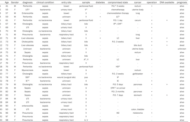
Table 2. Detail clinical profiles of 33 patients in the current series
Age Gender diagnosis clinical condition entry site sample diabetes compromised state cancer operation DNA available prognosis 1 41 M Peritonitis sepsis bowel peritoneal fluid - P.O.4) 3 weeks rectum + alive
2 55 F UTI1) bacteremia urinary tract V2) - chemotherapy uterine body - alive
3 57 M Enterocolitis sepsis bowel V - chemoradiation therapy lung - alive
4 59 M Peritonitis sepsis unknown V + LC5) - - alive
5 61 F Peritonitis no bacteremia bowel peritoneal fluid - P.O. 1 day cecum + + alive 6 67 M Cholangitis sepsis biliary tract bile - IP6), CHF7) - + alive
7 69 F UTI sepsis urinary tract V - - - + alive
8 70 M Cholangitis no bacteremia biliary tract bile + - - + alive 9 71 M Pneumonia bacteremia respiratory tract V - - lung - alive 10 71 M Liver abscess sepsis biliary tract V + LC liver + + dead 11 73 M Cholecystitis sepsis biliary tract bile - P.O. 3 weeks - + + alive 12 73 F Liver abscess sepsis biliary tract bile - - bile duct + + dead 13 73 F Unknown bacteremia unknown V - - uterine body - unknown
14 75 M Sepsis sepsis unknown V - - rectum + alive
15 75 F Cholangitis sepsis biliary tract bile - - - - alive 16 76 F Peritonitis sepsis unknown A3), V - LC liver + dead
17 76 F Pneumonia bacteremia respiratory tract V - - - - alive 18 76 M Peritonitis no bacteremia bowel peritoneal fluid - HD8) - - dead
19 76 F Myonecrosis sepsis bowel A - - rectum - alive
20 77 F Cholangitis sepsis biliary tract V - P.O. 3 weeks gallbladder + alive 21 78 M SSI9) no bacteremia wound (surgcal site) pus - IP - - alive
22 78 F IE10) bacteremia unknown A - IE - + alive
23 79 M Cholangitis no bacteremia biliary tract bile - P.O. 5 days pancreas + + alive 24 81 M Sepsis sepsis unknown A - CPA11) on arrival - - dead
25 82 M Sepsis sepsis unknown V - P.O. 2 months pancreas + alive 26 83 F Sepsis sepsis unknown V - P.O. 7 days stomach + + alive
27 84 M UTI bacteremia urinary tract A - - - - alive
28 84 M UTI bacteremia urinary tract V - - - - alive
29 90 F enterocolitis sepsis bowel V - - - - alive
30 93 M UTI sepsis urinary tract V - - colon, bladder - alive 31 96 M Pneumonia sepsis respiratory tract V - HD melanoma + alive 32 97 F Pneumonia sepsis respiratory tract V - - - - alive 33 98 F Pneumonia sepsis respiratory tract A, V + - - - alive
1)UTI, urinary tract infection; 2)V, verous blood; 3)A, arterial blood; 4)P.O., post operative state; 5)LC, liver cirrhosis; 6)IP, Interstitial pneumonia; 7)CHF, congestive heart failure; 8)HD, Hemodialysis; 9)SSI, surgical site infection; 10)IE, infectious
Saitama Medical Center, Jichi Medical Uni-
versity, between 2001 and 2013. The identifi
-cation of C. perfringens had been performed by Rap ID kit (ANA II System, Remel Inc. KS, U.S.A.) after culture in HK semi-solid medium and on Brucella HK agar plates (Kyokuto Pharma- ceutical Industrial. Ltd., Tokyo, Japan) [6-8]. This retrospective review detected 33 patients positive for C. perfringens (Table 1). Next, the medical records of these 33 patients were
reviewed for age, gender, clinical profile, under -lying disease, past medical events, radiograph-ic records, and laboratory data. Each of the
patients was classified into one of three cate
-gories: i) sepsis, defined as systemic inflamma -tory response syndrome (SIRS) caused by infec-tion; ii) bacteremia, without SIRS; and iii) other,
defined as no SIRS or bacteremia, according to
the guidelines of the ACCP/SCCM Consensus Conference Committee (Table 1) [9]. This study was approved by the Institutional Tissue Committees and written informed consent from each patient wasobtained prior to the molecu-lar analysis described below.
Samples used for PCR
Out of 33 cases, FFPE tissues were available in 9 cases, and informed consent for the molecu-lar analysis was obtained from 6 patients (Cases 5, 10-12, 23, and 26). These six cases included resected specimens for cancers of the cecum (Case 5), the pancreas (Case 23), the stomach (Case 26), cholecystitis (Case 11) and liver abscesses obtained by autopsy (Cases 10,
12). All the samples had been fixed in 15% buff -ered formalin (pH 7.4) for 24 to 48 hrs prior to
embedding in paraffin. The paraffin blocks had
been stored for periods between 6 months and 11 years. Microscopic examination of the
tis-sue sections from all, but one case (case 12)
revealed no identifiable gram-positive bacilli
morphologically suggestive of C. perfringens. Preparation of samples for PCR
Three 10-μm-thick sections were prepared from each paraffin block, using a new micro -tome blade for each block, and transferred into a microcentrifuge tube. The sections were
deparaffinized by two 30-min incubations in 1
mL xylene at 60°C followed by centrifugation at 1.5 × 10⁴g for 5 min. The resulting pellets were washed 3 times in 1 mL of absolute ethanol. In parallel, pure cultures of C. perfringens type A (NCTC8798) carrying alpha- and enterotoxin gene, type B (GTC15078) carrying alpha-, beta-, and epsilon-toxin gene and type E (GTC15081) carrying alpha- and iota-toxin, were used as positive controls for PCR [2, 7].
Total DNA was extracted using the DNeasy blood and tissue kit (Qiagen, Valencia, CA) according to the manufacturer’s instructions. In
brief, each sample was placed in 340 μL of buf
-fer ATL with 25 μl of proteinase K (50 mg/mL),
and reaction mixtures were incubated at 56°C overnight. The supernatants were obtained by
a brief centrifugation, mixed with 400 μL buffer
AL, and incubated at 70°C for an additional 20
min. After incubation, 400 μL of ethanol was
added, and the mixture was passed through a QIAamp spin column. DNA was eluted from the
column with 100 μL of buffer AE.
Primers for PCR
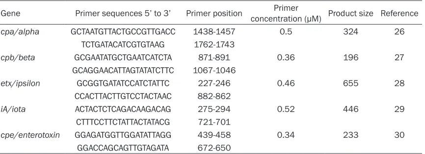
[image:4.612.92.525.84.241.2]The oligonucleotide primers specific for C. per-fringens loci were synthesized as previously described [10]. Sequences and locations of the
Table 3. PCR primers used in this study
Gene Primer sequences 5’ to 3’ Primer position concentration (μM)Primer Product size Reference
cpa/alpha GCTAATGTTACTGCCGTTGACC 1438-1457 0.5 324 26
TCTGATACATCGTGTAAG 1762-1743
cpb/beta GCGAATATGCTGAATCATCTA 871-891 0.36 196 27
GCAGGAACATTAGTATATCTTC 1067-1046
etx/ipsilon GCGGTGATATCCATCTATTC 227-246 0.46 655 28
CCACTTACTTGTCCTACTAAC 882-862
iA/iota ACTACTCTCAGACAAGACAG 275-294 0.52 446 29
CTTTCCTTCTATTACTATACG 721-701
cpe/enterotoxin GGAGATGGTTGGATATTAGG 439-458 0.34 233 30
primers for genes encoding alpha-, beta-, epsi-lon-, iota-toxin or enterotoxin are shown in
Table 3, and the predicted product sizes were 324, 196, 655, 446 and 233 bp, respectively [10].
PCR procedures
PCR was performed in a 50-μL scale composed
of 2 mM Tris-HCl (pH 8.0), 2 mM MgCl2, 10 mM
KCl, 0.2 mM of each dNTPs, 0.2 μM of each
primer, 20 ng of DNA, and 1.25 U TaKaRa Ex Taq Hot Start Version (TAKARA BIO INC. Otsu, Shiga, Japan). The reaction was performed in a thermal cycler (GeneAmp PCR system 9700; Applied Biosystems, CA, USA) for 35 cycles, with each cycle consisting of 1 min at 94°C, 1 min at 53°C, and 1 min at 72°C, and cycle was followed by 10 minutes at 72°C. PCR products and molecular size markers (Tracklt 100 bp DNA Ladder, Invitirogen CA, USA) were gel-elec-trophoresed in a 2.0% agarose gel and
visual-ized after staining with 0.5 μg/mL ethidium
bromide.
Results
Culture samples and patients’ profiles
All culture samples collected since 2001 have been monitored in our hospital, which is a ter-tiary care center with 608 beds. Among the total of 205, 114 samples, C. perfringens were isolated from 33 patients (0.017%), of whom 21 were categorized with “sepsis” (SIRS caused by infection), 7 with “bacteremia” (infection that was positive for C. perfringens in blood cul-ture, but was not complicated by SIRS), and 5 with “other” (Table 1). Clinicopathological
details of each patient are presented in Table 2.The number of patients presenting with iso-lates of C. perfringens generally increased with the patient’s age (younger than 55 years, 1 patient; 55-64 years, 4 patients; 65-74 years, 8 patients; 75-84 years, 15 patients; 85-97 years, 5 patients). Twenty eight patients (84.8%) were > 65 years old; mean age and standard deviation was 75 ± 12 years (range, 41-98 years). There were 19 male and 14 female patients. The samples were submitted from various wards: emergency room (8 patients), gastrointestinal medicine (8 patients), gastroin-testinal surgery (8 patients), cardiovascular medicine (2 patients), cardiovascular surgery (2 patients), gynecology (2 patients), urology (2 patients) and neurosurgery (1 patient).
As the most common underlying disease or state, surgery or intervention for cancer of the liver, of the hepatobiliary tract, or of the pan-creas was present in 6 patients: operation or intervention for pancreatic cancer, 2 patients; bile duct cancer, 1 patient; gallbladder cancer, 1 patient; transarterial infusion for hepatocel-lular carcinoma, 2 patients (both died). Other surgeries included those for cancer of the gas-trointestinal tract in 3 patients. Chemo (-radia-tion) therapy was performed in 2 patients: one case each for uterine and lung cancer. Surgery or intervention for non-neoplastic disease was performed in 6 patients: cholecystectomy for cholelithiasis (Case 6), nephrectomy for pyelo-nephritis (Case 7), partial hepatectomy for benign cyst (Case 8), ileocecal resection for ischemic enteritis (Case 11), colectomy for per-foration (Case 18), and mitral valve replace-ment (Case 22). Five patients were conserva-tively treated: lung cancer (Case 9), uterine can-cer (Case 13), brain abscess (Case 17), rectal cancer (Case 19), and colon cancer (Case 30). Viewed from the entry site, infections were
classified as follows: urinary tract, 5 patients;
bowel, including bacterial translocation, 6 patients; upper respiratory tract, 5 patients; biliary tract, 8 patients; surgical site, 1 patient; unknown sites, 8 patients.
In addition to the 15 patients who presented with infections after surgical intervention, 13
[image:5.612.89.288.71.202.2]patients were classified with an immunocom -promised or related condition: diabetes melli-tus, 4 patients (Cases 4, 8, 10, 38); chemo (radiation) therapy for cancer, 2 patients (Cases 2, 3); liver cirrhosis, 3 patients (Cases 4, 10, Figure 1. Radiographical imaging of the liver
16); interstitial pneumonia, 2 patients (Cases 6, 21); and hemodialysis, 2 patients (Cases 18,
31). A total of five patients (Cases 8, 10, 12, 13,
16) died of sepsis. The other 28 patients recov-ered from the infection. Among 21 patients with sepsis, one patient (Case 12) was a typical case complicated by fatal acute hemolysis
whose profile is presented below.
Report of a typical case
A 73-year-old female, who had a past history of pancreatoduodenectomy for bile duct carcino-ma at 59 years old, was admitted to our hospi-tal with chief complaint of general malaise and jaundice. She had suffered from fracture of the
right 6th and 7th ribs by traffic accident 7 days
prior to admission and had been managed conservatively.
The patient appeared confused at the time of admission, presenting with a temperature of 36.0°C, blood pressure 120/80 mmHg, and pulse rate of 110 bpm. Abdominal examination showed tenderness in the upper abdomen.
Laboratory findings showed severe anemia, massive hemolysis, liver dysfunction, inflam -matory reaction, and renal dysfunction: Clinical laboratory assays yielded the following: white blood cell count, 8500/mm3; red blood cell count, 210000/mm3; hemoglobin, 4.8 g/dL; hematocrit, 1.8%; platelet count, 440000/ mm3; albumin, 2.4 g/dL; total bilirubin, 10.34 mg/dL; direct bilirubin, 5.84 mg/dL; NH3, 1420
μg/dL (normal range; 30-86); cholinesterase,
118 U/L (normal range; 170-445); aspartate aminotransferase, 706 U/L; alanine amino-transferase, 168 U/L; lactate dehydrogenase, 6138 U/L (normal range; 110-220); creatine
kinase, 998 U/L (normal range; 6-142); alkaline
phosphatase, 740 U/L; γ-glutamyltransferase,
12 U/L; C-reactive protein, 9.21 mg/dL; urea, 77 mg/dL; creatinine, 1.44 mg/dL.
An abdominal enhanced computed tomograph-ic (CT) scan revealed a low-density area with gas formation in the right lobe of the liver (Figure 1). A diagnosis of liver abscess with possible rupture into the abdominal cavity was made.
Despite the fluid replacement therapy, the
patient’s hemodynamic condition was unsta-ble. A CT-guided percutaneous transhepatic drainage from the liver abscess was performed. After the drainage in spite of intensive treat-ment with massive blood transfusion, the patient expired twelve hours after admission. A culture of the liver abscess obtained through drainage tube revealed the presence of C. per-fringens and Klebsiella oxytoca. Autopsy was
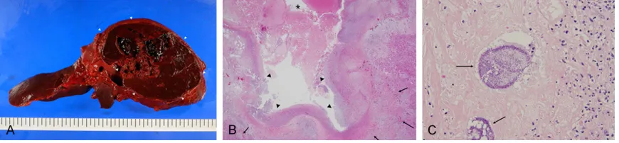
performed and the main finding was an 80-mm
liver abscess with perforation to the abdominal cavity (Figure 2A). Cavity was filled with blood
clot. Microscopically, inner surface was
cov-ered by inflammatory exudate and necrotic
material, and surrounded by viable hepatic tis-sue (Figure 2B). In the necrotic material, colo-nies of gram-positive bacillus were observed (Figure 2C). Moreover, microscopic aggregates
of red blood cells with fibrin were remarkable in
renal tubuli, and scattered fresh thrombi also were found in the glomeruli, suggesting dis-seminated intravascular coagulation.
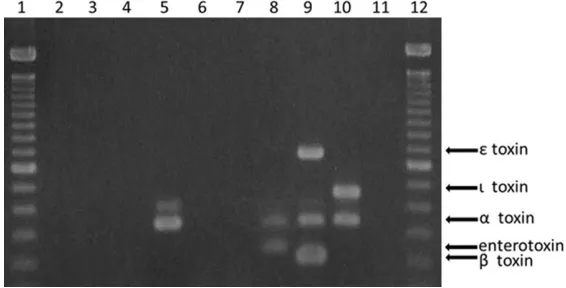
PCR amplification yielded the products of
[image:6.612.90.525.73.174.2]expected sizes for cpa (alpha-toxin, 324 bp), cpb (beta-toxin, 196 bp), etx (epsilon-toxin, 655 bp), iA (iota-toxin, 446 bp) and cpe (enterotoxin, Figure 2. A. Gross appearance of the liver obtained from the autopsy in Case 12. Cut surface revealed pseudocystic and partially necrotic lesion of 8 cm in the maximum dimension (arrow heads). B. Histological features of
233 bp) genes in the appropriate positive con-trols (Figure 3) [10]. Among the FFPE-derived samples obtained from 6 patients, the
alpha-toxin gene was amplified only from the Case-12
specimen; none of the other toxin genes were
amplified from any of the samples.
Discussion
Patients who contract C. perfringens infections exhibit various clinical symptoms including ali-mentary intoxication, necrotizing enteritis, liver abscess, gas gangrene in the soft tissue, and septic shock [11, 12].
Approximately, 0.5-2.0% of all isolates from blood cultures of septic patients are described as clostridial species, and among them, C. per-fringens is the most frequently identified
micro-organism, accounting for 20-50% [11, 13]. Clostridial sepsis often shows very poor prog-nosis, owing to the life-threatening combination of shock and acute massive hemolysis [14, 15]. Massive hemolysis is rare complication of C. perfringens infection, and thus, its prevalence has not been estimated accurately. Among 33 cases from whose samples C. perfringens was cultured in our hospital during the past 13 years, only one case manifested massive intra-vascular hemolysis, suggesting a prevalence of 3.0%. However, the mortality rate of clostridi-um-induced massive hemolysis is extremely
because in the early stages, infected patients
only manifest non-specific inflammatory symp
-toms and the slight gas formation as observed in the infected lesions by imaging studies [11]. In Case 12, the diagnosis could not be made when the patient was alive due to the fulminant clinical course. The results of the blood culture and hepatic drainage revealed C. perfringens as the pathogen after the patient died. In addi-tion to this patient, another four patients died, giving an infection-related mortality rate of 15.2%.
The presence of clostridial species in blood cul-tures are explained by multiple factors, includ-ing transient bacteremia (often from an un- known source), trauma, myonecrosis, surgery-related bowel leakage, and occasionally retro-grade biliary infections [12, 13], consistent with the examples of (emphysematous) cholecysti-tis, cholangicholecysti-tis, and liver abscess seen in our series. Cancer and immunosuppressive condi-tions are the main underlying diseases or states for C. perfringens bacteremia or sepsis [11]. In the present study, the most frequent underlying conditions were cancers of the hep-atobiliary system and the pancreas (6 cases). Bacteremia caused by C. perfringens often occurs in immunocompromised patients [12], and indeed, 15 of 33 patients (45.5%) in the current series were of this status, including Figure 3. Agarose gel electrophoresis of PCR amplicons obtained using
formalin-fixed paraffin-embedded colon tissues from 6 cases (lanes 2-7,
Case 5, 10-12, 23, and 25, respectively). Clostridium perfringens type A
(NCTC8798) carrying alpha- and entero-toxin gene (lane 8), and type B (GTC15078) carrying alpha-, beta-, and epsilon-toxin gene (lane 9), type E (GTC15081) carrying alpha- and iota-toxin gene (lane 10), negative control (lane 11), and molecular size marker (lanes 1 and 12). Lane 5 (Case 12)
showed amplified product of Clostridium perfringens type A-specific
alpha-toxin gene.
[image:7.612.88.371.75.219.2]being post-operative, post-intervention, or un-dergoing chemo (radiation) therapy for malig-nancy. Additional presumed risk factors noted include advanced age, diabetes mellitus, or past history of surgery for biliary and pancreatic cancers (e.g., for placement of choledocho-jeju-nostomy) as previously described [12].
The identification of bacterial type requires
laborious analysis in addition to the routine pro-cedures of bacterial culturing [6-8]. In the cur-rent retrospective study, residual samples from patients or bacterial colonies from growth on agar were not available, since most of the sam-ples had been collected more than a year previ-ously. Therefore, we tried to detect the pres-ence of the individual genes encoding each type of exotoxin, as well as the enterotoxin gene, by PCR with total DNA extracted from
FFPE tissues. Furthermore, we aimed to find a
difference among several categories of patho-logical lesions comprising gross liver abscess-es (2 casabscess-es), which were prabscess-esumed to have locally higher amounts of bacteria, and cancers of the stomach, cecum, and pancreas (one case each) as well as cholecystitis, which were expected to have locally lower levels of bacte-ria. We detected the presence of the alpha-tox-in-encoding gene in only one case (Case 12), obtained from a patient who showed liver
abscess and acute hemolysis, confirming that
the disease pathogen was type A.
To our knowledge, there is only one previous
report that describes the identification of tox -inogenic types of C. perfringens from human FFPE tissues. In that work, C. perfringens type C in a patient with emphysematous gastritis, and type B or D in a liver with gas gangrene was detected [2]. Case 12 in our series corresponds to a case complicated by fatal massive
hemoly-sis, which we believe represents the first report
of C. perfringens type A detected in FFPE human liver tissue.
Although negative PCR results cannot prove the absence of C. perfringens, the amount of DNA was below the level of detection threshold of this experimental setting. Since the tech-nique presented here is a qualitative rather than quantitative, it was not possible to esti-mate the amount of C. perfringens in the sam-ples. Moreover, considering the characteristics of the samples, which were FFPE tissues, and the presumed uneven distribution of bacteria
within infected tissue, quantification may be
impossible. Nonetheless, we speculated that the C. perfringens was locally concentrated and present at higher levels in the liver abscess of Case 12, since that specimen’s DNA was not expected to be well preserved: the tissue was partially necrotic, and the tissues had been left
in the paraffin block for a period longer than for
the other FFPE specimens. Therefore, the lesion presumably contained locally concen-trated C. perfringens, which could have been an inducer of the massive hemolysis observed in this patient. Although the diverse pathobio-logical mechanisms of C. perfringens remain to be elucidated, this method (PCR-based detec-tion of C. perfringens using DNA extracted from archival FFPE tissues obtained from a patholo-gy laboratory) could be widely utilized for diag-nosis and determination of toxinogenic types of C. perfringens in clinical specimens.
In conclusion, C. perfringens was detected in routine bacterial culture of patient samples at a frequency of 0.017%. Among the clostridial-infected patients, fatal massive intravascular hemolysis occurred at 3.0%, possibly due to the presence of a lesion containing localized high-density C. perfringens. In addition, deter-mination of toxigenic gene status by PCR with FFPE tissue may facilitate medical treatment strategies.
Acknowledgements
The authors wish to thank Professor Yoshikazu Hirai, Division of Bacteriology, Department of Infection and Immunity, Jichi Medical University for the helpful support, and the staff in the Department of Pathology, Saitama Medical Center, Jichi Medical University for their excel-lent technical help. This study was partially sup-ported by grants from the Japan Society for the Promotion of Science C26460438 (Y.D.) and from the Smoking Research Foundation (Y.D).
Disclosure of conflict of interest
None.
References
[1] van Bunderen CC, Bomers MK, Wesdorp E, Peerbooms P and Veenstra J. Clostridium per-fringens septicaemia with massive intravascu-lar haemolysis: a case report and review of the literature. Neth J Med 2010; 68: 343-346. [2] Wu J, Zhang W, Xie B, Wu M, Tong X, Kalpoe J
and Zhang D. Detection and toxin typing of
Clostridium perfringens in formalin-fixed, par
-affin-embedded tissue samples by PCR. J Clin
Microbiol 2009; 47: 807-810.
[3] Yoo HS, Lee SU, Park KY and Park YH. Mo- lecular typing and epidemiological survey of prevalence of Clostridium perfringens types by multiplex PCR. J Clin Microbiol 1997; 35: 228-232.
[4] Hatheway CL. Toxigenic clostridia. Clin Micro- biol Rev 1990; 3: 66-98.
[5] Petrillo TM, Beck-Sague CM, Songer JG, Abra- mowsky C, Fortenberry JD, Meacham L, Dean AG, Lee H, Bueschel DM and Nesheim SR. Enteritis necroticans (pigbel) in a diabetic child. N Engl J Med 2000; 342: 1250-1253. [6] Kadra B, Guillou JP, Popoff M and Bourlioux P.
Typing of sheep clinical isolates and identifica -tion of enterotoxigenic Clostridium perfringens strains by classical methods and by poly-merase chain reaction (PCR). FEMS Immunol Med Microbiol 1999; 24: 259-266.
[7] Warren AL, Uzal FA, Blackall LL and Kelly WR. PCR detection of Clostridium perfringens type
D in formalin-fixed, paraffin-embedded tissues
of goats and sheep. Lett Appl Microbiol 1999; 29: 15-19.
[8] Sterne M and Batty I. Diagnostic criteria for clostridial infections. Pathogenic Clostridia. London: Butherworth and Co.Ltd; 1975. pp. 79-84.
[9] Bone RC, Balk RA, Cerra FB, Dellinger RP, Fein AM, Knaus WA, Schein RM and Sibbald WJ.
Definitions for sepsis and organ failure and
guidelines for the use of innovative therapies in sepsis. The ACCP/SCCM Consensus Confe- rence Committee. American College of Chest Physicians/Society of Critical Care Medicine. Chest 1992; 101: 1644-1655.
[10] Meer RR and Songer JG. Multiplex polymerase chain reaction assay for genotyping Clostridium perfringens. Am J Vet Res 1997; 58: 702-705. [11] Fujita H, Nishimura S, Kurosawa S, Akiya I,
Nakamura-Uchiyama F and Ohnishi K. Clinical and epidemiological features of Clostridium perfringens bacteremia: a review of 18 cases over 8 year-period in a tertiary care center in metropolitan Tokyo area in Japan. Intern Med 2010; 49: 2433-2437.
[12] Rechner PM, Agger WA, Mruz K and Cogbill TH. Clinical features of clostridial bacteremia: a re-view from a rural area. Clin Infect Dis 2001; 33: 349-353.
[13] Onderdonk ABA, S.D. Clostridium. In: Murray
PR, Baron, E.J., Pfaller, M.A., Tenover, F.C., Yol- ken, R.H., editors. Manual of Clinical Micro- biology. 6th. ed. Washington, DC: ASM Press; 1995.
[14] Juntermanns B, Radunz S, Heuer M, Vernada- kis S, Reis H, Gallinat A, Treckmann J, Kaiser G, Paul A and Saner F. Fulminant septic shock due to Clostridium perfringens skin and soft tissue infection eight years after liver trans-plantation. Ann Transplant 2011; 16: 143-146. [15] Law ST and Lee MK. A middle-aged lady with a
pyogenic liver abscess caused by Clostridium perfringens. World J Hepatol 2012; 4: 252-255.