Original Article
Expression of exportin-1 in diffuse large B-cell
lymphoma: immunohistochemistry and TCGA analyses
Bin Luo1*, Lanshan Huang2*, Yongyao Gu2, Chunyao Li2, Huiping Lu2, Gang Chen2, Zhigang Peng1, Zhenbo
Feng2
Departments of 1Medical Oncology, 2Pathology, The First Affiliated Hospital of Guangxi Medical University,
Nan-ning, Guangxi Zhuang Autonomous Region, People’s Republic of China. *Equal contributors.
Received September 9, 2018; Accepted October 22, 2018; Epub December 1, 2018; Published December 15, 2018
Abstract: Exportin-1 (XPO1) is an essential nuclear export receptor that is involved in the pathogenesis of mul-tiple tumors. However, the role of XPO1 in diffuse large B-cell lymphoma (DLBCL) requires clarification. This study aims to detect XPO1 expression in DLBCL and to explore its relationships with clinicopathologic parameters and prognoses. Methods: A total of 131 cases of DLBCL and 30 cases of reactive lymphoid hyperplasia were selected for immunohistochemistry to examine XPO1 expression and analyze the relationships of XPO1 expression with clinicopathologic parameters and prognosis. DLBCL datasets downloaded from The Cancer Genome Atlas (TCGA) were used to analyze the mutations, expressions, and clinical values of XPO1 in DLBCL. Results: XPO1 expres-sion was markedly upregulated in DLBCL compared to the reactive lymphoid hyperplasia group (χ2 = 10.734, P =
0.001). High XPO1 expression was associated with an advanced clinical stage (χ2 = 4.036, P = 0.045) and a risky
International Prognostic Index (IPI) score (χ2 = 5.301, P = 0.025). Moreover, high XPO1 expression was associated
with a lower overall survival rate compared with low expression (P = 0.043). XPO1 was an independent prognostic factor for DLBCL (risk ratio, RR = 3.772, P = 0.006). Furthermore, XPO1 overexpression in DLBCL was correlated with a high IPI score (P = 0.024) in TCGA datasets. Conclusion: High XPO1 expression in DLBCL was related to an advanced clinical stage, poor IPI score, and poor prognosis. Thus, XPO1 may be useful for condition identification and prognostic assessment.
Keywords: Diffuse large B-cell lymphoma, exportin-1, clinicopathological significance, prognosis
Introduction
Diffuse large B-cell lymphoma (DLBCL) is the most common type of non-Hodgkin’s lympho-ma (NHL) [1-3]. Each DLBCL subtype has differ-ent pathological features, biological behaviors, clinical presentations, and prognoses [4, 5]. DLBCL is categorized into the germinal center B like (GCB) and non-germinal center B cell-like (non-GCB) subtypes based on immunohis-tochemistry tests [6-8]. The GCB subtype has a better prognosis [9]. Currently, rituximab plus cyclophosphamide, doxorubicin, vincristine, and prednisone (R-CHOP) is used as the first-line chemotherapy for DLBCL because this regi-men improves the prognosis and results in 70-80% complete remission (CR) and 50-60% progression-free survival (PFS) at 3-5 years [10-14]. However, one-third of patients with DLBCL are refractory to the treatment or
experi-ence recurrexperi-ence after treatment [15, 16]. Hence, providing treatment for patients who resist the first-line therapy or have early recur-rence is a pressing clinical issue. The identifica-tion of effective DLBCL markers for diagnosis and prognosis that will lead to accurate diag-nostic stratification and individualized treat-ment is of paramount importance.
ovar-ian cancer [21], multiple myeloma [17], and acute myeloid leukemia [22]. A number of stud-ies have shown that XPO1 can function as a target for oncotherapy [23-26], although stud-ies on its role in DLBCL are limited. The gene chip analyses performed by Kwiecinska [27], Scholtysik [28], and Trifonov [29] indicated that XPO1 was highly expressed in DLBCL tissues. Zhou [30] found that XPO1 was positively expressed in DLBCL. However, comprehensive research on the relationships between XPO1
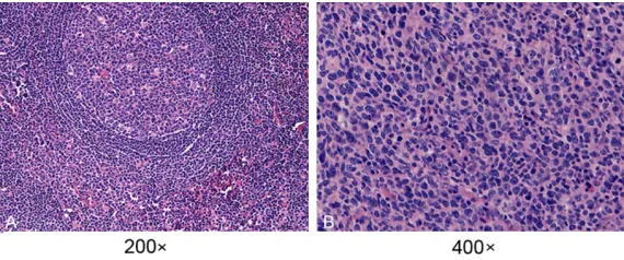
receive R-CHOP treatment (20 were in CR and 9 were in PR), while 10 patients preferred other forms of chemotherapy. Clinical staging was conducted based on the 2008 WHO Ann Arbor criteria, and the performance status of the patients was measured using the Eastern Cooperative Oncology Group (ECOG) and International Prognostic Index (IPI) scores. None of the patients received antineoplastic therapy prior to the biopsy or operation, and the patients had no medical history of immunodefi-Figure 1. Morphology of H&E-stained DLBCL and reactive lymph node
[image:2.612.90.375.71.190.2]hyper-plasia tissue sections. A. DLBCL tissues; B. Reactive lymph node hyperhyper-plasia tissue. Annotation: DLBCL, diffuse large B-cell lymphoma; H&E, hematoxylin-eosin.
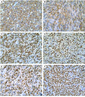
Figure 2. Detection of XPO1 protein expression by immunohistochemistry in DLBCL and reactive lymph node hyperplasia tissues. A: DLBCL tissues; B: Reactive lymph node hyperplasia tissues. Annotation: DLBCL, diffuse large B-cell lymphoma; XPO1, exportin-1.
expression, clinical parame-ters, and prognosis is lacking. This study aims to explore the correlation of XPO1 expres-sion in DLBCL with its clinico-pathological parameters and prognosis. Additionally, we analyze the biologic signifi-cance of XPO1 using TCGA datasets to provide evidence for the clinical diagnosis and treatment of DLBCL.
Materials and methods
Tissue samples
[image:2.612.90.375.261.535.2]ciency. The follow-up, which lasted between 14 and 65 months, ended on October 15, 2016. The overall survival (OS) and PFS were calcu-lated separately. Thirty cases of reactive hyper-plasia of the lymph node were selected as the controls. The study was approved by the Re- search Ethics Committees of the First Affiliated Hospital of Guangxi Medical University, China.
Reagents
The XPO1 antibody (sc-5595) was purchased from Santa Cruz and used at a 1:100 dilution. The ready-to-use Bcl-2 and Bcl-6 antibodies were acquired from Fuzhou Maixin Biotech. Co., Ltd. CD20, MUM1, CD10, and Ki67 were pur-chased from Beijing Zhongshan Golden Bridge Biotechnology Co., Ltd.
Immunohistochemistry
Paraffin-embedded tissues were sliced into 4-μm sections. Then, the slides were baked in
medium. Positive tissues were considered as the positive control group, and PBS was used in place of the primary antibody as the normal control group [31, 32].
Immunohistochemistry assessment
XPO1 expression was located in the nucleus and partly in the cytoplasm. Bcl-2, CD20, and CD10 were detected in the cytomembrane, and Bcl-6, MUM1, and Ki67 were found in the nucleus. The overall scoring system was based on the staining intensity of the tumor cells and the percentage of positive cells [33]. The stain color was compared against the background color in the same section. The scoring criteria for the staining intensity were as follows: 0 for colorless, 1 for light yellow, 2 for yellowish-brown, and 3 for dark brown. The scoring sys-tem for the percentages of positive cells was as follows: 0 for ≤ 5%, 1 for 6%-25%, 2 for 26%-50%, 3 for 51%-75%, and 4 for > 75%. The over-all score was achieved by multiplying the inten-Figure 3. Detection of CD20, CD10, Bcl-2, Bcl-6, MUM1, and Ki67 protein
expression in DLBCL tissues by immunohistochemistry. A. CD20; B. CD10; C. Bcl-2; D. Bcl-6; E. MUM1; F. Ki67 (400×). Annotation: DLBCL, diffuse large B-cell lymphoma.
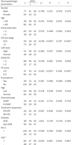
[image:3.612.89.375.71.399.2]Table 1. Relationships between XPO1 expression and clinicopath-ologic parameters in DLBCL patients
Clinicopathologic
parameters n +XPO1- χ2 P r P
Gender
Male 77 41 36 0.799 0.371 -0.078 0.375 Female 54 33 21
Age
> 60 49 29 20 0.231 0.631 0.042 0.634
≤ 60 82 45 37
Extranodal sites
< 2 92 50 42 0.576 0.448 -0.060 0.452
≥ 2 39 24 15
Clinical stage
III-IV 59 39 20 4.036 0.045* 0.176 0.045*
I-II 72 35 37
LDH level
High 51 26 25 0.382 0.537 -0.062 0.541 Normal 49 28 21
ECOG PS
< 2 98 56 42 0.158 0.691 0.035 0.694
≥ 2 32 17 15
IPI score
3-5 47 33 14 5.031 0.025* 0.196 0.025*
0-2 84 42 42
B symptoms
Yes 29 15 14 0.297 0.585 -0.048 0.589
No 101 58 43
Hgb
Low 64 38 26 0.378 0.539 0.055 0.542 Normal 63 34 29
Chemotherapy
CHOP 29 16 13 0.101 0.751 0.039 0.755 R-CHOP 39 20 19
Treatment responses
CR+PR 44 24 20 0.007 0.934 -0.010 0.935
SD+PD 27 15 12
Subtype
GCB 38 18 20 1.811 0.178 -0.118 0.181 Non-GCB 93 56 37
Bcl-2
+ 105 61 44 0.556 0.456 0.065 0.460
- 26 13 13
Ki67
+ 83 47 36 0.002 0.967 0.004 0.967
- 48 27 21
Annotation: XPO1, exportin-1; DLBCL, diffuse large B-cell lymphoma; LDH, lactate dehydrogenase; ECOG PS, Eastern Cooperative Oncology Group performance status; IPI, International Prognostic Index; Hgb, hemoglobin; CHOP, cyclophospha-mide, doxorubicin, vincristine, prednisone; R-CHOP, rituximab, cyclophosphacyclophospha-mide, daunorubicin, vincristine, prednisone; CR, complete remission; PR, partial remis-sion; SD, stable disease; PD, progressive disease; GCB, germinal center B cell-like; non-GCB, non-germinal center B cell-like; *P < 0.05.
sity score by the percentage score: 0-4 was considered negative (-), 5-8 positive (+), and 9-12 strongly positive (++). “Positive” and “strongly posi-tive” were classified as high expression. Additionally, a sample was considered posi-tive when more than 25% of the cells stained positive for Bcl-2, CD20, CD10, Bcl-6, and MUM1 and more than 70% of the cells stained positive for Ki67.
Analysis of TCGA datasets
Mutations in XPO1 in DLBCL (TCGA, provisional) were ana-lyzed with cBioPortal (www. cbioportal.org) [34, 35], which included amplification, mRNA upregulation, and missense mutations. OncoPrint in cBio-Portal was employed to draw the XPO1 mutation diagram for the 48 DLBCL cases. Addi- tionally, plot was applied to generate several pictures of the clinicopathological param-eters (Neoplasm American Jo- int Committee on Cancer Cli- nical Group Stage), OS, and DFS. The clinical data for XPO1 expression in DLBCL down-loaded from TCGA were used to analyze the correlations bet- ween XPO1 and the clinical parameters and prognosis. To obtain a better understanding of the relationships between XPO1 and other genes, the network of XPO1 and related genes was downloaded from cBioPortal.
Statistical analysis
SPSS 22.0 (IBM Corporation) was used for statistical analy-sis. Quantitative data were represented as the mean ± standard deviation, and cate-gorical data were analyzed with the χ2 test or Spearman’s
survival analysis, and comparisons between curves were tested with the log-rank test. A Cox proportional hazards model was adopted for analysis of the effects of multiple factors on the prognosis. Statistical significance was set at P
< 0.05.
Results
Morphology of the tissue sections
We observed HE-stained DLBCL and reactive lymphoid hyperplasia tissue sections using microscopy. The observation of typical DLBCL
[image:5.612.92.522.72.492.2]morphology showed that the tissues in or out-side of the lymph nodes were replaced by dif-fuse cells that contained nuclei that were approximately twice as large as the nuclei of small lymphocytes. The large cells had a rich endochylema, empty nucleus, and obvious nucleolus (Figure 1A). The reactive lymph node hyperplasia sections revealed the partial exis-tence of a normal structure with an increased number of lymphoid follicles, larger size, and expanded germinal center; additionally, the paracortical area was broadened, and the ves-sels were multiplied (Figure 1B).
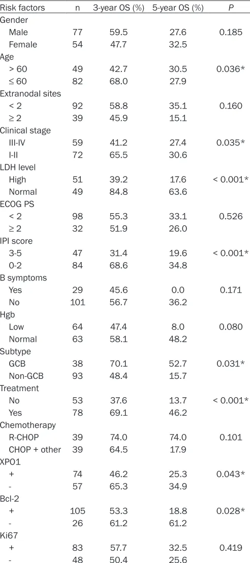
Table 2. Univariate prognostic analysis of clinicopathologic parameters in 131 DLBCL patients
Risk factors n 3-year OS (%) 5-year OS (%) P
Gender
Male 77 59.5 27.6 0.185
Female 54 47.7 32.5
Age
> 60 49 42.7 30.5 0.036*
≤ 60 82 68.0 27.9
Extranodal sites
< 2 92 58.8 35.1 0.160
≥ 2 39 45.9 15.1
Clinical stage
III-IV 59 41.2 27.4 0.035*
I-II 72 65.5 30.6
LDH level
High 51 39.2 17.6 < 0.001*
Normal 49 84.8 63.6
ECOG PS
< 2 98 55.3 33.1 0.526
≥ 2 32 51.9 26.0
IPI score
3-5 47 31.4 19.6 < 0.001*
0-2 84 68.6 34.8
B symptoms
Yes 29 45.6 0.0 0.171
No 101 56.7 36.2
Hgb
Low 64 47.4 8.0 0.080
Normal 63 58.1 48.2
Subtype
GCB 38 70.1 52.7 0.031*
Non-GCB 93 48.4 15.7 Treatment
No 53 37.6 13.7 < 0.001*
Yes 78 69.1 46.2
Chemotherapy
R-CHOP 39 74.0 74.0 0.101
CHOP + other 39 64.5 17.9 XPO1
+ 74 46.2 25.3 0.043*
- 57 65.3 34.9
Bcl-2
+ 105 53.3 18.8 0.028*
- 26 61.2 61.2
Ki67
+ 83 57.7 32.5 0.419
- 48 50.4 25.6
Annotation: DLBCL, diffuse large B-cell lymphoma; OS, overall survival; LDH, lactate dehydrogenase; ECOG PS, Eastern Cooperative Oncology Group performance status; IPI, International Prognostic Index; Hgb, he-moglobin; GCB, germinal center B cell-like; non-GCB, non-germinal center B cell-like; CHOP, cyclophosphamide, doxorubicin, vincristine, prednisone; R-CHOP, rituximab, cyclophosphamide, daunorubicin, vincristine, predni-sone; other chemotherapy including methotrexate or temozolomide; *P
< 0.05.
Immunohistochemical staining results
Positive XPO1 expression in the DLBCL sections was mostly detected in the nuclei of the tumor cells (Figure 2A). Additionally, some staining was detect-ed in the cytoplasm, with a positive percentage of 56.1% (74/131). XPO1 had extremely low or no expression in the reactive lymph node hyperplasia sections (Figure 2B). The majority of the expression was detected in the germinal center, while a small amount was seen in the medullary cord and medullary sinus. Positive expression was located in the cytoplasm and nucleus in 23.3% (7/30) of the sec-tions, which was significantly lower than the positive staining rate detect-ed in the DLBCL group (χ2 = 10.734, P
= 0.001). CD20 expression was posi-tive in all 131 cases, and Bcl-2 and Ki67 expression was positive in 80.2% (105/131) and 63.4% (83/131) of the cases, respectively. The staining results for CD20, Bcl-2, Bcl-6, MUM1, CD10, and Ki67 are shown in Figure 3.
Relationships between XPO1 expres-sion and clinicopathological param-eters in DLBCL patients
The positive percentages of XPO1 expression in the GCB subtype (CD- 10+; CD10-/Bcl-6+/MUM1-) and the non-GCB subtype (CD10-/Bcl-6+/MU- M1+; CD10-/Bcl-6-/MUM1+; CD10-/ Bcl-6-/MUM1-) were 47.4% (18/38) and 60.3% (56/93), respectively. The increased XPO1 expression was asso-ciated with an advanced clinical stage (χ2 = 4.036, P = 0.045) and a high IPI
score (χ2 = 5.301, P = 0.025) (Table 1). However, high XPO1 expression showed no relationship with the age, gender, extranodal involvement, B symptoms, subtypes, ECOG scores, hemoglobin (Hb), lactate dehydroge-nase (LDH), Bcl-2, or Ki67 parameters (P > 0.05).
Follow-up results and survival analy-sis
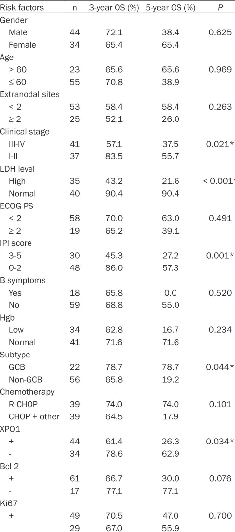
mortali-Table 3. Univariate prognostic analysis of clinicopatholog-ic parameters in 78 DLBCL patients with chemotherapy Risk factors n 3-year OS (%) 5-year OS (%) P
Gender
Male 44 72.1 38.4 0.625
Female 34 65.4 65.4
Age
> 60 23 65.6 65.6 0.969
≤ 60 55 70.8 38.9
Extranodal sites
< 2 53 58.4 58.4 0.263
≥ 2 25 52.1 26.0
Clinical stage
III-IV 41 57.1 37.5 0.021*
I-II 37 83.5 55.7
LDH level
High 35 43.2 21.6 < 0.001*
Normal 40 90.4 90.4
ECOG PS
< 2 58 70.0 63.0 0.491
≥ 2 19 65.2 39.1
IPI score
3-5 30 45.3 27.2 0.001*
0-2 48 86.0 57.3
B symptoms
Yes 18 65.8 0.0 0.520
No 59 68.8 55.0
Hgb
Low 34 62.8 16.7 0.234
Normal 41 71.6 71.6
Subtype
GCB 22 78.7 78.7 0.044*
Non-GCB 56 65.8 19.2 Chemotherapy
R-CHOP 39 74.0 74.0 0.101
CHOP + other 39 64.5 17.9 XPO1
+ 44 61.4 26.3 0.034*
- 34 78.6 62.9
Bcl-2
+ 61 66.7 30.0 0.076
- 17 77.1 77.1
Ki67
+ 49 70.5 47.0 0.700
- 29 67.0 55.9
Annotation: DLBCL, diffuse large B-cell lymphoma; OS, overall survival; LDH, lactate dehydrogenase; ECOG PS, Eastern Cooperative Oncology Group performance status; IPI, International Prognostic Index; Hgb, hemoglobin; GCB, germinal center B cell-like; non-GCB, non-germinal center B cell-like; CHOP, cyclophosphamide, doxorubicin, vincristine, prednisone; R-CHOP, rituximab, cyclophosphamide, daunorubicin, vincristine, prednisone; other chemotherapy including methotrexate or temozolomide; *P < 0.05.
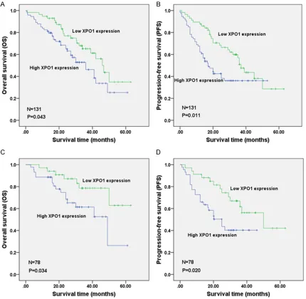
ty rate of 46.8% (7 were lost to follow-up). The three-year OS rate was 54.9%, and the 5-year OS rate was 29.4%, with a median survival time of 41.0 months (range 37.8-44.2 months). The univari-ate survival analysis revealed that higher XPO1 expression was correlated with a worse prognosis (OS P = 0.043, PFS P = 0.011; Figure 4A, 4B). Mo- reover, there was a close link between a worse prognosis and an elderly age, non-GCB subtype, high LDH level, Bcl-2 expression, Ann Arbor stages III-IV, and high IPI score (Table 2). The Cox regres-sion analysis indicated that the XPO1 protein expression and LDH levels were independent factors that influenced the OS of DLBCL patients and that the XPO1 and LDH levels, the clinical stage, and R-CHOP were independent factors that influenced the PFS of DLBCL patients. The risk ratios (RRs) of high XPO1 and LDH expression for OS were 3.772 (P = 0.006) and 10.375 (P < 0.001), respectively. The RRs of XPO1 and LDH expression, Ann Arbor stages III-IV, and R-CHOP treatment for PFS were 3.595 (P = 0.002), 5.474 (P < 0.001), 2.331 (P = 0.027), and 0.359 (P = 0.007), respectively. After analyz-ing the prognoses of the 78 patients who received chemotherapy for DLBCL, we found that high XPO1 expression was associated with a poor prognosis (OS P = 0.034, PFS P = 0.020; Figure 4C, 4D). Additionally, patients of the non-GCB subtype who had a high LDH level, high Bcl-2 expression, Ann Arbor stages III-IV, or a high IPI score had an undesirable prognosis (Table 3). The multiple factor analysis of the OS of the 78 patients who received chemothera-py suggested that the XPO1 (RR = 3.772, P = 0.006) and LDH (RR = 10.375, P < 0.001) expression levels were independent factors for the DLBCL prognosis, which was similar to the overall analysis results for the 131 patients with DLBCL.
TCGA data analysis
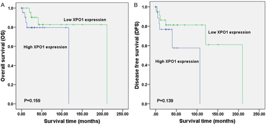
[image:7.612.93.332.96.637.2]No significant relationship was noted between XPO1 mRNA expression and the clinical stage (Figure 6). There was also no significant rela-tion found between the mutarela-tions and DFS (Figure 7A) or OS (Figure 7B) (P > 0.05). In TCGA, complete case information was obtained for the 47 DLBCL cases, which were divided into high and low expression groups using the median expression level (Table 4). High XPO1 expression levels were associated with an elderly age (χ2 = 7.982, P = 0.005) and risky IPI
score (P = 0.024), whereas no correlation was
[image:8.612.88.519.79.140.2]cells and that high FOXP1 expression was asso-ciated with the prognosis. Lu [37] discovered that high Myc and Bcl-2 expression and muta-tions were associated with a negative progno-sis. Additionally, metadherin (MTDH) prohibited proliferation of tumor cells and strengthened chemo-sensitivity to doxorubicin in DLBCL [38]. The NF-κB signaling pathway inhibited the apoptosis of DLBCL cells, thereby reducing the chemo-sensitivity of the tumor cells [39]. Due to the significant heterogeneity of DLBCL and the undesirable clinical treatment, identifying Figure 5. Genetic alterations of XPO1 in DLBCL (TCGA, provisional). Genetic alterations in XPO1, including amplifica -tion, mRNA upregula-tion, and missense mutations, were identified in DLBCL using OncoPrint from cBioPortal (www. cbioportal.org). Annotation: DLBCL, diffuse large B-cell lymphoma; XPO1, exportin-1; TCGA, The Cancer Genome Atlas.
Figure 6. Connection between the XPO1 mRNA level and the clinical stage in DLBCL (TCGA, provisional). The log2 mRNA expression levels from RNA Seq V2 RSEM were downloaded from TCGA data. The Plot figure was drawn using cBioPortal (www.cbioportal.org) to show the connection between mRNA ex-pression and the clinical stage (P > 0.05). Annotation: DLBCL, diffuse large B-cell lymphoma; XPO1, exportin-1; TCGA, The Cancer Genome Atlas.
found between XPO1 and gen-der, extranodal involvement, clinical stage, ECOG, or the therapeutic model (P > 0.05). The single factor analysis re- vealed that increased XPO1 expression was most likely a sign of a poor prognosis, but no statistical significance was observed (OS P = 0.159, DFS
P = 0.139, Figure 8). No inde-pendent factors that might affect the DLBCL prognosis were found in the Cox regres-sion analysis. The gene net-work illustrated the interac-tion between XPO1 and other genes in DLBCL (Figure 9). Moreover, the network showed the molecular targeting drug for XPO1 (Compound 7d-cis), which has not been approved by the FDA.
Discussion
[image:8.612.92.375.213.492.2]Figure 7. Relationship between XPO1 alterations and survival in DLBCL (TCGA, provisional). A. Overall survival (OS): two out of 9 cases with alterations resulted in death, and the median survival time was 116.72 months. Seven out of 38 cases without alterations resulted in death, and the median survival time was 211.07 months; B. Disease-free survival (DFS): one of the 8 cases with alterations relapsed, and the median time of DFS was 120.53 months, whereas 11 of the 35 cases without alterations relapsed. Survival was analyzed using the Kaplan-Meier estimate provided by cBioPortal (www.cbioportal.org). Annotation: DLBCL, diffuse large B-cell lymphoma; XPO1, exportin-1; TCGA, The Cancer Genome Atlas.
Table 4. Relationships between XPO1 expression and clinicopatho-logical parameters in DLBCL patients in TCGA datasets
Clinicopathological parameters n XPO1 χ2 P r P
+ -Gender
Male 21 10 11 0.180 0.671 -0.062 0.679
Female 26 14 12
Age
> 60 20 15 5 7.982 0.005* 0.412 0.004*
≤ 60 27 9 18
Extranodal sites
< 2 25 13 12 0.239 0.625 -0.139 0.413
≥ 2 12 8 4
Clinical stage
III-IV 17 9 8 0.034 0.853 0.029 0.857
I-II 24 12 12
ECOG PS
< 2 28 12 16 0.031 0.859 0.141 0.448
≥ 2 3 2 1
IPI score
3-5 6 6 0 - 0.024* 0.452 0.016*
0-2 22 10 12
Treatment
Chemotherapy 22 11 11 - 1.000 0.204 0.350 Chemotherapy + radiotherapy 1 0 1
Treatment responses
CR+PR 21 11 10 0.082 0.775 -0.167 0.425
SD+PD 4 3 1
Annotation: XPO1, exportin-1; DLBCL, diffuse large B-cell lymphoma; TCGA, The
Cancer Genome Atlas; ECOG PS, Eastern Cooperative Oncology Group performance status; IPI, International Prognostic Index; CR, complete remission; PR, partial remis-sion; SD, stable disease; PD, progressive disease; *P < 0.05.
key biological factors and seeking individualized treat-ment strategies are urgent issues.
[image:9.612.91.385.348.692.2]lym-Figure 8. Survival comparison between DLBCL patients from TCGA datasets with low and high XPO1 expression levels (TCGA, provisional). A: Overall survival (OS); B: Disease-free survival (DFS). Annotation: DLBCL, diffuse large B-cell lymphoma; XPO1, exportin-1; TCGA, The Cancer Genome Atlas.
[image:10.612.95.518.341.683.2]phoma showed great potential value for XPO1 research [43]. Inhibitors of XPO1 prohibited the proliferation of NHL cells and induced their apoptosis by down-regulating anti-apoptotic proteins, such as Survivin and NF-κB [42, 44]. However, research on the clinical significance and mechanism of XPO1 in DLBCL is lacking. To explore the clinical role of XPO1 in DLBCL, we applied immunohistochemistry to detect XPO1 expression. We found that XPO1 was overexpressed in DLBCL tissue, which was con-sistent with the results of Zhou’s study [30]. Additionally, XPO1 overexpression was related to an advanced clinical stage and a high IPI score. Currently, IPI is the most frequently used index for assessing the prognosis of DLBCL patients. The index is critical for the determina-tion of the prognosis and clinical therapeutics, although it has failed to completely assess patients’ statuses [11]. Univariate survival analysis indicated that high XPO1 expression was a poor prognostic factor for DLBCL because the OS and PFS were longer in the low XPO1 expression group. In the chemotherapy group, XPO1 exerted more remarkable effects on the prognosis, possibly because the patients had quicker progression and higher mortality with-out chemotherapy; thus, XPO1 expression had little impact on the prognosis. Conversely, anal-ysis of both groups (the 131 DLBCL patients and the 78 patients receiving chemotherapy) revealed that a poor prognosis was associated with a non-GCB subtype, high LDH level, high Bcl-2 expression, Ann Arbor stages III-IV, and a risky IPI score. Similar results were found in previous studies [9, 11], validating our research. The multivariate survival analysis revealed that XPO1 was an independent prognostic factor for DLBCL and has the potential of becoming a clinical prognostic indicator for DLBCL. In the TCGA datasets, high XPO1 expression in DLBCL was connected with an elderly age and a dan-gerous IPI score, while high XPO1 expression was a possible sign of a poor prognosis, although the association was not significant. Our results were in agreement with the TCGA data, but there were some minor differences that might be attributed to the small sample size or incomplete case information in the data. Moreover, fluorescent in situ hybridization (FISH) or polymerase chain reaction (PCR) were used for XPO1 mRNA detection in the TCGA, whereas in our study immunohistochemistry
was applied for protein detection. XPO1 might be a more powerful protein, but the clinical sig-nificance of XPO1 in DLBCL still needs to be demonstrated in larger numbers of studies. In the TCGA, we found XPO1 mutations in DLBCL, although they had no relationship with progno-sis. Camusa [45] employed second-generation sequencing and digital PCR to evaluate DLBCL tissues and peripheral blood-related cytokines and found abnormal XPO1expression. Similarly, Mareschal [46] used whole-exome sequencing for relapsed/refractory patients with DLBCL and observed XPO1 mutations. The XPO1 data in the TCGA and the network of related genes in DLBCL provide a basis for future research, although the mechanism of XPO1 in DLBCL needs to be ascertained experimentally.
Several studies have investigated the patho-genic mechanism of XPO1 in tumors. Kajiyama [47] demonstrated that XPO1 regulated the epithelial-mesenchymal transition (EMT) path-way in ovarian cancer, reducing cellular polarity and adhesion and thereby accelerating the infil-tration and transfer of tumor cells. An XPO1 inhibitor suppressed the activation of the IGF-1R/AKT pathway by upregulating insulin-like growth factor-binding protein 5 (IGFBP5) in lipo-sarcoma [48]. The tumor suppressor proteins FOXO, p53, and p27, which perform nucleo-cytoplasmic shuttling, play tumor-suppressive roles in the nucleus; a large proportion of tumor suppressor proteins were transported out of the nucleus when XPO1 was overexpressed, which promoted tumor cell proliferation [19, 40]. XPO1 inhibitors down-regulated NF-κB expression and upregulated p53 expression in NHL cells [42]. Additionally, XPO1 inhibitors pre-vented the nuclear export of NF-κB-IκBα and prohibited NF-κB signaling in primary mediasti-nal B-cell lymphoma (PMBCL), hence inducing tumor cell apoptosis [46]. Zhou [30] found that XPO1 participated in the occurrence and progression of DLBCL in cooperation with NF-κBp50. Moreover, XPO1 regulated the PI3K-Akt pathway, which facilitated the proliferation of mantle cell lymphoma and prevented apop-tosis [49]. However, because there has been limited research investigating XPO1 in DLBCL, determining its specific mechanism of action requires further exploration.
tissues. XPO1 overexpression was associated with an advanced clinical stage and a poor IPI score and was associated with a poor DLBCL prognosis. Additionally, the detection of XPO1 expression in DLBCL tissues could help achieve a better assessment of the patient’s condition and prognosis and provide evidence for the clinical diagnosis and treatment of DLBCL.
Acknowledgements
We would like to thank the Fund of the Key Programs of University Scientific Research of Guangxi Education Agency (No. ZD2014033), the Key Programs of Guangxi Natural Science Fund (No. 2015GXNSFDA139028), and the Promoting Project of Basic Capacity for Uni- versity Young and Middle aged Teachers in Guangxi (2017). The funders had no role in the study design, data collection and analysis, decision to publish, or preparation of the manu-script. We acknowledge the cBioPortal for Cancer Genomics site (http://www.cbioportal. org/) and the TCGA Research Network for gen-erating TCGA datasets (http://cancergenome. nih. gov/).
Disclosure of conflict of interest
None.
Address correspondence to: Zhigang Peng, De- partment of Medical Oncology, The First Affiliated Hospital of Guangxi Medical University, 6 Shuang- yong Road, Nanning 530021, Guangxi Zhuang Autonomous Region, People’s Republic of China. Tel: +86 771 5353121; Fax: +86 771 5353121; E-mail: [email protected]; Zhenbo Feng, Department of Pathology, The First Affiliated Hospital of Guangxi Medical University, 6 Shuangyong Road, Nanning 530021, Guangxi Zhuang Autonomous Region, People’s Republic of China. Tel: +86 771 5356534; Fax: +86 771 5356534; E-mail: fengzhenbo_ [email protected]
References
[1] Krause G, Hassenruck F and Hallek M. Copan-lisib for treatment of B-cell malignancies: the development of a PI3K inhibitor with consider-able differences to idelalisib. Drug Des Devel Ther 2018; 12: 2577-2590.
[2] Xu PP, Sun C, Cao X, Zhao X, Dai HJ, Lu S, Guo JJ, Fu SJ, Liu YX, Li SC, Chen M, McCord R, Ven-strom J, Szafer-Glusman E, Punnoose E, Kier-maier A, Cheng G and Zhao WL. Immune
char-acteristics of chinese diffuse large b-cell lymphoma patients: implications for cancer immunotherapies. EBioMedicine 2018; 33: 94-104.
[3] Gong QX, Lu TX, Liu C, Wang Z, Liang JH, Xu W, Li JY, Zhang ZH and Chen Q. Prevalence and clinicopathologic features of CD30-positive de novo diffuse large B-cell lymphoma in Chinese patients: a retrospective study of 232 cases. Int J Clin Exp Pathol 2015; 8: 15825-15835. [4] Board PDQPTE. Childhood non-hodgkin lym
-phoma treatment (PDQ(R)): health profession-al version. Edited by PDQ pediatric treatment editorial board. PDQ cancer information sum-maries [Internet]. Bethesda (MD): National Cancer Institute (US); 2002-2018 Aug 22. [5] Mu S, Ai L, Fan F, Qin Y, Sun C and Hu Y.
Prog-nostic role of neutrophil-to-lymphocyte ratio in diffuse large B cell lymphoma patients: an up-dated dose-response meta-analysis. Cancer Cell Int 2018; 18: 119.
[6] Gao HY, Wu B, Yan W, Gong ZM, Sun Q, Wang HH and Yang W. Microarray expression profiles of long non-coding RNAs in germinal center-like diffuse large B-cell lymphoma. Oncol Rep 2017; 38: 1363-1372.
[7] Yang JM, Jang JY, Jeon YK and Paik JH. Clinico-pathologic implication of microRNA-197 in dif-fuse large B cell lymphoma. J Transl Med 2018; 16: 162.
[8] Dobashi A. Molecular pathogenesis of diffuse large B-cell lymphoma. J Clin Exp Hematop 2016; 56: 71-78.
[9] Swerdlow SH, Campo E, Pileri SA, Harris NL, Stein H, Siebert R, Advani R, Ghielmini M, Salles GA, Zelenetz AD and Jaffe ES. The 2016 revision of the World Health Organization clas-sification of lymphoid neoplasms. Blood 2016; 127: 2375-2390.
[10] Zhong W, Xu X, Zhu Z, Yang L, Du H, Xia Z, Yuan Z, Xiong H, Du Q, Wei Y and Li Q. Increased in-terleukin-17A levels promote rituximab resis-tance by suppressing p53 expression and pre-dict an unfavorable prognosis in patients with diffuse large B cell lymphoma. Int J Oncol 2018; [Epub ahead of print].
[11] Seo S, Hong JY, Yoon S, Yoo C, Park JH, Lee JB, Park CS, Huh J, Lee Y, Kim KW, Ryu JS, Kim SJ, Kim WS, Yoon DH and Suh C. Prognostic sig-nificance of serum beta-2 microglobulin in pa -tients with diffuse large B-cell lymphoma in the rituximab era. Oncotarget 2016; 7: 76934-76943.
[13] Li J, Ding N, Wang X, Mi L, Ping L, Jin X, Liu Y, Ying Z, Xie Y, Liu W, Song Y and Zhu J. EP300 single nucleotide polymorphism rs20551 cor-relates with prolonged overall survival in dif-fuse large B cell lymphoma patients treated with R-CHOP. Cancer Cell Int 2017; 17: 70. [14] Yang S, Sheng L, Xu K, Wang Y, Zhu H, Zhang P,
Mu Q and Ouyang G. Anticancer effect of quacrine on diffuse large Bcell lymphoma via in-hibition of MSI2NUMB signaling pathway. Mol Med Rep 2018; 17: 522-530.
[15] Rhodes J and Landsburg DJ. Small-molecule inhibitors for the treatment of diffuse large B cell lymphoma. Curr Hematol Malig Rep 2018; 13: 356-368.
[16] Karmali R and Gordon LI. Molecular subtyping in diffuse large B cell lymphoma: closer to an approach of precision therapy. Curr Treat Op -tions Oncol 2017; 18: 11.
[17] Gandhi UH, Senapedis W, Baloglu E, Unger TJ, Chari A, Vogl D and Cornell RF. Clinical implica-tions of targeting XPO1-mediated nuclear ex-port in multiple myeloma. Clin Lymphoma My-eloma Leuk 2018; 18: 335-345.
[18] Fu SC, Fung HYJ, Cagatay T, Baumhardt J and Chook YM. Correlation of CRM1-NES affinity with nuclear export activity. Mol Biol Cell 2018; 29: 2037-2044.
[19] Ishizawa J, Kojima K, Hail N Jr, Tabe Y and An -dreeff M. Expression, function, and targeting of the nuclear exporter chromosome region main-tenance 1 (CRM1) protein. Pharmacol Ther 2015; 153: 25-35.
[20] Silva G, Marins M, Chaichanasak N, Yoon Y, Fachin AL, Pinhanelli VC, Regasini LO, Dos San-tos MB, Ayusso GM, Marques BC, Wu WW, Phue JN, Shen RF and Baek SJ. Trans-chalcone increases p53 activity via DNAJB1/HSP40 in-duction and CRM1 inhibition. PLoS One 2018; 13: e0202263.
[21] Shao WY, Yang YL, Yan H, Huang Q, Liu KJ and Zhang S. Phenethyl isothiocyanate suppresses the metastasis of ovarian cancer associated with the inhibition of CRM1-mediated nuclear export and mTOR-STAT3 pathway. Cancer Biol Ther 2017; 18: 26-35.
[22] Ranganathan P, Kashyap T, Yu X, Meng X, Lai TH, McNeil B, Bhatnagar B, Shacham S, Kauff -man M, Dorrance AM, Blum W, Sampath D, Landesman Y and Garzon R. XPO1 inhibition using selinexor synergizes with chemotherapy in acute myeloid leukemia by targeting dna re-pair and restoring topoisomerase iialpha to the nucleus. Clin Cancer Res 2016; 22: 6142-6152.
[23] Zhang W, Ly C, Ishizawa J, Mu H, Ruvolo V, Sha-cham S, Daver N and Andreeff M. Combinato-rial targeting of XPO1 and FLT3 exerts synergis -tic anti-leukemia effects through induction of
differentiation and apoptosis in FLT3-mutated acute myeloid leukemias: from concept to clin-ical trial. Haematologica 2018; 103: 1642-1653.
[24] Fabi F, Adam P, Vincent K, Demontigny F, Par-ent S, Joncas FH and Asselin E. Inhibition of CRM1 activity sensitizes endometrial and ovar-ian cell lines to TRAIL-induced cell death. Cell Commun Signal 2018; 16: 39.
[25] Camus V, Miloudi H, Taly A, Sola B and Jardin F. XPO1 in B cell hematological malignancies: from recurrent somatic mutations to targeted therapy. J Hematol Oncol 2017; 10: 47. [26] Turner JG, Dawson JL, Grant S, Shain KH, Dal
-ton WS, Dai Y, Meads M, Baz R, Kauffman M, Shacham S and Sullivan DM. Treatment of ac -quired drug resistance in multiple myeloma by combination therapy with XPO1 and topoisom-erase II inhibitors. J Hematol Oncol 2016; 9: 73.
[27] Kwiecinska A, Ichimura K, Berglund M, Dinets A, Sulaiman L, Collins VP, Larsson C, Porwit A and Lagercrantz SB. Amplification of 2p as a genomic marker for transformation in lympho-ma. Genes Chromosomes Cancer 2014; 53: 750-768.
[28] Scholtysik R, Kreuz M, Hummel M, Rosolowski M, Szczepanowski M, Klapper W, Loeffler M, Trumper L, Siebert R and Kuppers R. Charac -terization of genomic imbalances in diffuse large B-cell lymphoma by detailed SNP-chip analysis. Int J Cancer 2015; 136: 1033-1042. [29] Trifonov V, Pasqualucci L, Dalla Favera R and
Rabadan R. MutComFocal: an integrative ap-proach to identifying recurrent and focal ge-nomic alterations in tumor samples. BMC Syst Biol 2013; 7: 25.
[30] Zhou YQ, Chai L. NF-κBp50, JAB1 and CRM1 expression in diffuse large B cell lymphoma and their clinical significance. J of Wannan Medical College (In Chinese) 2012; 31: 92-95. [31] Ye ZH, Gao L, Wen DY, He Y, Pang YY and Chen
G. Diagnostic and prognostic roles of IRAK1 in hepatocellular carcinoma tissues: an analysis of immunohistochemistry and RNA-sequenc-ing data from the cancer genome atlas. Onco Targets Ther 2017; 10: 1711-1723.
[32] Mo CH, Gao L, Zhu XF, Wei KL, Zeng JJ, Chen G and Feng ZB. The clinicopathological signifi -cance of UBE2C in breast -cancer: a study based on immunohistochemistry, microarray and RNA-sequencing data. Cancer Cell Int 2017; 17: 83.
[34] Yang X, Deng Y, He RQ, Li XJ, Ma J, Chen G and Hu XH. Upregulation of HOXA11 during the pro-gression of lung adenocarcinoma detected via multiple approaches. Int J Mol Med 2018; 42: 2650-2664.
[35] Chen WJ, Tang RX, He RQ, Li DY, Liang L, Zeng JH, Hu XH, Ma J, Li SK and Chen G. Clinical roles of the aberrantly expressed lncRNAs in lung squamous cell carcinoma: a study based on RNA-sequencing and microarray data min-ing. Oncotarget 2017; 8: 61282-61304. [36] Dekker JD, Park D, Shaffer AL 3rd,
Kohlham-mer H, Deng W, Lee BK, Ippolito GC, Georgiou G, Iyer VR, Staudt LM and Tucker HO. Subtype-specific addiction of the activated B-cell subset of diffuse large B-cell lymphoma to FOXP1. Proc Natl Acad Sci U S A 2016; 113: E577-586. [37] Lu TX, Gong QX, Wang L, Fan L, Zhang XY, Chen
YY, Wang Z, Xu W, Zhang ZH and Li JY. Immuno-histochemical algorithm alone is not enough for predicting the outcome of patients with dif-fuse large B-cell lymphoma treated with R-CHOP. Int J Clin Exp Pathol 2015; 8: 275-286. [38] Li PP, Feng LL, Chen N, Lu K, Meng XH, Ge XL,
Lv X and Wang X. Metadherin interference in-hibits proliferation and enhances chemo-sen-sitivity to doxorubicin in diffuse large B cell lymphoma. Int J Clin Exp Med 2014; 7: 2081-2086.
[39] Turturro F. Constitutive NF-kappa B activation underlines major mechanism of drug resis-tance in relapsed refractory diffuse large b cell lymphoma. Biomed Res Int 2015; 2015: 484537.
[40] Inoue H, Kauffman M, Shacham S, Landes-man Y, Yang J, Evans CP and Weiss RH. CRM1 blockade by selective inhibitors of nuclear ex-port attenuates kidney cancer growth. J Urol 2013; 189: 2317-2326.
[41] Mendonca J, Sharma A, Kim HS, Hammers H, Meeker A, De Marzo A, Carducci M, Kauffman M, Shacham S and Kachhap S. Selective in-hibitors of nuclear export (SINE) as novel ther-apeutics for prostate cancer. Oncotarget 2014; 5: 6102-6112.
[42] Han X, Wang J, Shen Y, Zhang N, Wang S, Yao J and Shi Y. CRM1 as a new therapeutic target for non-Hodgkin lymphoma. Leuk Res 2015; 39: 38-46.
[43] Tabe Y, Kojima K, Yamamoto S, Sekihara K, Matsushita H, Davis RE, Wang Z, Ma W, Ishiza-wa J, Kazuno S, Kauffman M, Shacham S, Fu-jimura T, Ueno T, Miida T and Andreeff M. Ribo -somal biogenesis and translational flux in-hibition by the selective inhibitor of nuclear ex-port (SINE) XPO1 antagonist KPT-185. PLoS One 2015; 10: e0137210.
[44] Turner JG, Kashyap T, Dawson JL, Gomez J, Bauer AA, Grant S, Dai Y, Shain KH, Meads M, Landesman Y and Sullivan DM. XPO1 inhibitor combination therapy with bortezomib or carfil -zomib induces nuclear localization of Ikap-paBalpha and overcomes acquired protea-some inhibitor resistance in human multiple myeloma. Oncotarget 2016; 7: 78896-78909. [45] Camus V, Sarafan-Vasseur N, Bohers E, Dubois
S, Mareschal S, Bertrand P, Viailly PJ, Ruminy P, Maingonnat C, Lemasle E, Stamatoullas A, Pic-quenot JM, Cornic M, Beaussire L, Bastard C, Frebourg T, Tilly H and Jardin F. Digital PCR for quantification of recurrent and potentially ac -tionable somatic mutations in circulating free DNA from patients with diffuse large B-cell lym-phoma. Leuk Lymphoma 2016; 57: 2171-2179.
[46] Mareschal S, Dubois S, Viailly PJ, Bertrand P, Bohers E, Maingonnat C, Jais JP, Tesson B, Ru -miny P, Peyrouze P, Copie-Bergman C, Fest T, Jo Molina T, Haioun C, Salles G, Tilly H, Lecroq T, Leroy K and Jardin F. Whole exome sequencing of relapsed/refractory patients expands the repertoire of somatic mutations in diffuse large B-cell lymphoma. Genes Chromosomes Cancer 2016; 55: 251-267.
[47] Kajiyama H, Shibata K, Terauchi M, Yamashita M, Ino K, Nawa A and Kikkawa F. Chemoresis-tance to paclitaxel induces epithelial-mesen-chymal transition and enhances metastatic potential for epithelial ovarian carcinoma cells. Int J Oncol 2007; 31: 277-283.
[48] Garg M, Kanojia D, Mayakonda A, Said JW, Doan NB, Chien W, Ganesan TS, Chuang LS, Venkatachalam N, Baloglu E, Shacham S, Kauffman M and Koeffler HP. Molecular mech -anism and therapeutic implications of selinex-or (KPT-330) in liposarcoma. Oncotarget 2017; 8: 7521-7532.