Original Article
Advanced glycation end products
induced the epithelial-mesenchymal transition
in retinal pigment epithelial cells via ERK activation
Xiao-Li Chen*, Yu-Jing Bai*, Qin-Rui Hu, Lv-Zhen Huang, Xiao-Xin Li
Department of Ophthalmology, Peking University People’s Hospital, Key Laboratory of Vision Loss and
Restoration, Ministry of Education, Beijing Key Laboratory for The Diagnosis and Treatment of Retinal and Choroid Diseases, Beijing, China. *Equal contributors and co-first authors.
Received January 15, 2016; Accepted March 26, 2016; Epub April 1, 2016; Published April 15, 2016
Abstract: Proliferative diabetic retinopathy (PDR), an end-stage diabetic complication, accounts for blindness and low vision in most diabetic patients. Advanced glycation end products (AGEs) and the epithelial-mesenchymal tran-sition (EMT) of retinal pigment epithelial (RPE) cells are associated with the development of PDR via multifactorial mechanisms. However, whether AGEs can induce the EMT in RPE cells was previously unknown. In this study, we found that AGEs induced the EMT, accompanied by a decreased expression of the epithelial marker ZO-1, increased the expression of the mesenchymal marker fibronectin, elevated the production of EMT-related cytokines such as vascular endothelial growth factor (VEGF) and interleukin-6, and enhanced cell migration ability. Furthermore, the AGEs-induced EMT could be partly reversed by using an inhibitor of ERK activation, U0126. We also found that AGEs could regulate cell apoptosis and the cell cycle while promoting cell phenotype transformation from a typical cobblestone-like to a fibroblast-like morphology. Collectively, these data suggest that AGEs participate in the patho-genesis of PDR by inducing the EMT in an ERK-dependent pathway. Additional studies investigating the role of AGEs in the EMT may be promising for the prevention and treatment of PDR.
Keywords: Advanced glycation end products, epithelial-mesenchymal transition, proliferative diabetic retinopathy, ERK, RPE cells
Introduction
Diabetic retinopathy (DR) is a major cause of blindness and low vision in the elderly world-wide. Proliferative diabetic retinopathy (PDR), the advanced stage of DR and the most serious ocular complication of diabetes, can cause hemorrhages, retinal detachment and total blindness [1]. PDR is a wound healing-like response in which fibrotic epiretinal mem-branes are formed either on the surface of the retina or within the vitreous, accompanied by an influx of inflammatory cytokines and angio-genic factors into the retina [2]. The epiretinal membranes consist of various cell types, including retinal pigment epithelial (RPE) cells, which have a fibroblast-like morphology, under-go the epithelial-mesenchymal transition (EMT) and are considered to be contractile, leading to the contraction of epiretinal membranes and subsequently toretinal detachment [3, 4].
EMT is a process in which epithelial cells under-go a transition from their differentiated mor-phology to a mesenchymal-like phenotype and occurs during embryonic development, tumor metastasis, wound healing, and organ fibrosis [5]. This transition is characterized by a loss of cell-cell contacts, down-regulation of epithelial cell markers such as ZO-1, and up-regulation of mesenchymal markers such as fibronectin [6]. Additionally, EMT is associated with enhanced cell migration and the subsequent aggravation ofthe pathologic fibrosis process in the prolif-erative diseases of the eye, heart, kidney, liver, and lung [7]. In recent studies, increasing evi-dence has shown that the EMT of RPE cells con-tributes to the development of PDR [8, 9]; how-ever, the mechanism underlying the EMT is largely unknown.
diseases, including diabetic complications, aging and atherosclerosis [10]. Recently, a con-siderable number of studies have suggested that AGEs might play a pivotal role in diabetic retinopathy. Elevated levels of AGEs in the vitre-ous of patients with PDR were reported [11]. In vitro studies showed that AGEs induced the production of vascular endothelial growth fac-tor (VEGF) in cultured retinal Müller cells and RPE cells [12, 13]. An in vivo study showed that the injection of glycated albumin (Alb-AGE) into mice increases VEGF mRNA expression in the eyes, contributing to the development of DR [14]. Our study also found that the expression of the AGE receptor (RAGE) is increased in the retinas of type 2 diabetic rats and in high-glu-cose-treated ARPE-19 cells [15]. Although the EMT is crucial to the development and progres-sion of PDR, few reports have describedthe effect of AGEs on the EMT in RPE cells. Therefore, the present study aimed to investi-gate whether AGEs could induce the EMT in human RPE cells and the underlying molecular mechanisms.
Materials and methods Reagents and cell culture
AGEs were purchased from RD (Minneapolis, MN, US), and U0126 was purchased from Sigma (St. Louis, MO, US). A cell apoptosis and cell cycle kit was obtained from BD Biosciences (Bedford, MA, US). Anti-β-actin, anti-extracellu-lar signal-related kinase (ERK), anti-phospho ERK, zonula occludens-1 (ZO-1), and anti-fibronectin were obtained from Cell Signaling Technology (Danvers, MA, US). Goat anti-rabbit fluorescein isothiocyanate (FITC)-conjugated secondary antibodies were purchased from Molecular Probes (Invitrogen, Carlsbad, CA, US). A Cell Counting Kit-8 (CCK-8) and Transwell product were purchased from Dojindo (Shang- hai, China) and Corning (Tewksbury, MA, US), respectively.
The human RPE cell line ARPE-19 was obtained from the American Type Culture Collection (AT-CC, Mantissa, VA), and cells were cultured in in Dulbecco’s modified Eagle’s medium (DMEM)/ F12 medium (Hyclone, Grand Island, NY, US) supplemented with 10% fetal bovine serum (FBS; Gibco, Grand Island, NY, US) at 37°C under 5% CO2. The cells were used at passages 15-20, and all experiments were performed in serum-free medium.
Cell apoptosis and cell cycle assay
ARPE-19 cells (1×10⁶) were seeded in 6-well plates and treated with BSA-AGE (50 μg/ml, 100 μg/ml, and 200 μg/ml) for 48 h. The cells were detached using ethylene diamine tet-raacetic acid (EDTA), washed in ice-cold PBS (4°C), and treated with an FITC Annexin V Apoptosis Detection Kit or BD CycletestTMPlus DNA Reagent Kit according to the manufactur-er’s protocol. Samples were analyzed using a FACSCalibur cytometer (Becton Dickinson, US). All experiments were performed in triplicate.
Morphology observation and immunofluores -cencestaining
After ARPE-19 cells were treated with BSA-AGE (50 μg/ml, 100 μg/ml, and 200 μg/ml) for 48 h, cell morphology was observed and photo-graphed with an inverted phase-contrast micro-scope (Olympus, Tokyo, Japan). For immunocy-tochemistry, cells were washed and fixed in 4% (v/v) paraformaldehyde for 15 min at room tem-perature. Then, the cells were blocked in 10% goat serum with 0.1% Triton X-100, followed by incubation with primary antibodies against fibronectin diluted to 1:100 at 4°C overnight. After being washed three times with PBS, the cells were incubated with FITC-conjugated sec-ondary antibodies (1:500) at room temperature for 1 h. Nuclei were counterstained with 4’, 6’ -diamidino-2-phenylindole hydrochloride (DAPI) for 5 min. Images were acquired witha fluores-cence microscope (Leica).
Cell proliferation assay
Cells were seeded onto 96-well microplates (2×10³ cells per well). The CCK-8 assay was used to measure cell viability at 0 h, 12 h, 24 h, 48 h, and 72 h according to the manufacturer’s instructions. Ten microliters of CCK-8 solution was added to each well, and cells were further incubated for 2.5 h. The optical density was measured at 450 nm with a microplate reader (Finstruments Multiskan Models 347; MTX Lab Systems, Inc., Vienna, VA). All experiments were performed in triplicate.
Quantitative real-time PCR analysis
(Bio-Rad, Hercules, CA). The following primers were used: p-ERK forward, 5’-CTG AAA TGC GCA CAG TTG CT-3’, and reverse, 5’-CCT GTC AGT TCA GCC AAC CT-3’; ZO-1 forward, 5’-GTG TTG TGG ATA CCT TGT-3’, and reverse, 5’-GAT GAT GCC TCG TTC TAC-3’; fibronectin forward, 5’-AGC GGA CCT ACC TAG GCA AT-3’, and reverse, 5’-GGT TTG CGA TGG TAC AGC TT-3’; glyceralde-hyde phosphate dehydrogenase (GAPDH) for-ward, 5’-GAA GGT GAA GGT CGG AGT C-3’, and reverse, 5’-GAA GAT GGT GAT GGG ATT TC-3’ (Sangon. Shanghai, China). The amplification and thermo-cycling procedures were conduct-ed according to the manufacturer’s instruc-tions. GAPDH served as the reference gene for quantity control. Experiments were performed in triplicate and repeated at least three times. Western blot analysis
Cell proteins were prepared with a total protein extraction kit (Amsbio, Abingdon, UK), followed by protein concentration measurements with a BCA Protein Assay Kit (Pierce, Rockford, IL, US). Equal amounts of protein were separated by 10% sodium dodecyl sulfate polyacrylamide gels, transferred to polyvinylidene difluoride fil-ters, and visualized with enhanced chemilumi-nescence detection reagents (Pierce). Bands were quantified using Quantity One software (Bio-Rad, Richmond, CA, US) and normalized to that of β-actin. All immunoblot analyses were repeated three times, and similar results were obtained.
Cytometric bead array
The concentrations of interleukin 1β, 6, 8, 10, and 12p (IL-1β, IL-6, IL-8, IL-10, and IL-12p) and tumor necrosis factor-alpha (TNF-α) in ARPE-19 cell supernatants were measured with a cyto-metric bead array (CBA, No. 552932; BD Bioscience, San Jose, CA, US) as previously described [16]. Briefly, cell supernatants were collected and centrifuged at 12,000×g at 4°C. Then 50 μl of the supernatant sample was used for each test and measured with flow cytometry (BD FACSCalibur, BD Bioscience). The concen-tration of each cytokine was calculated accord-ing to the protocol of the CBA kit.
Cell migration assay
The ARPE-19 cell migration assay was per-formed using a Transwell chamber. Briefly,
2×10⁴ cells in 200 μl serum-free medium were placed in the upper chamber, whereas 600 μl DMEM with 10% FBS was in the bottom cham-ber. All migration assays were conducted for 6 h at 37°C. Then, cells were fixed in 4% parafor-maldehyde and stained with DAPI for 15 min. After removal of the non-migrating cells with a cotton swab, the membrane was imaged and cells from five random fields of view were count-ed. Each experiment was repeated three times. Statistical analysis
Data were expressed as the mean ± standard deviation (SD). Statistical analysis was con-ducted using Prism 5 (GraphPad Software, Inc., San Diego, CA, US). Differences were evaluated using a one-way analysis of variance (ANOVA) followed by Tukey’s multiple comparison test or using an unpaired Student t-test. P<0.05 was considered statistically significant.
Results
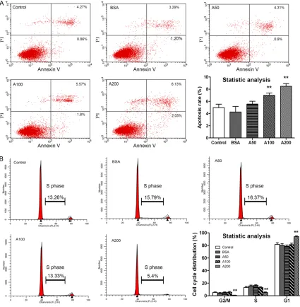
Effects of AGEs on cell apoptosis and the cell cycle
We first evaluated the effects of AGEs on ARPE-19 cell apoptosis (early and late apoptosis) and cell cycle arrest (G2/M, S and G1 phase). AGEs induced cell apoptosis and cell cycle arrest in a dose-dependent manner. As shown in Figure 1A, cells treated with 100 μg/ml and 200 μg/ ml AGEs displayed a significant increase in cell apoptosis compared to controls, whereas there was no significant difference among 50 μg/ml AGEs, BSA and control groups. Additionally, AGEs at a dose of 200 μg/ml resulted in a sig-nificant accumulation of ARPE-19 cells in the G1 phase and a reduction of cells in the G2/M and S phase compared to controls (P<0.01; Figure 1B). However, there was no significant difference among 100 μg/ml AGEs, 50 μg/ml AGEs, BSA and control groups in the cell cycle test (P>0.05; Figure 1B).
Effects of AGEs on cell morphology and fibro -nectin expression
a typical cobblestone-like epithelial morpho- logy. As the concentrations of AGEs increa- sed, cells gradually changed to a spindle fibro-blast-like morphology (Figure 2A). In addition, fibronectin protein expression was enhan- ced with the increased AGEs concentration (Figure 2B). In contrast, BSA-treated cells retained their epithelial morphology and dis-played slightly strengthened fibronectin expres-sion (Figure 2).
AGEs induced the EMT in ARPE-19 cells via ERK phosphorylation
[image:4.612.92.517.72.506.2]Because we had observed an association among AGEs, phenotypic transition and fibro-nectin expression in ARPE-19 cells, we next sought to examine whether AGEs could induce the EMT. Cells were treated with various doses of AGEs for 12 h, 24 h, 48 h and 72 h. As shown in Figure 3A, cell proliferation was significantly Figure 1. Effects of AGEs on RPE cell apoptosis and the cell cycle. A. Apoptosis of RPE cells treated with BAS or dif-ferent concentrations of AGEs. Samples were taken 48 h after treatment, and cells were stained with Annexin V and PI for flow cytometry analysis. B. AGEs (200 μg/ml) induced cell cycle arrest in ARPE-19 cells. Samples were taken 48 h after BSA or AGEs treatment, and DNA content was analyzed with PI staining. The percentage of cells in the G2/M, S and G1 are indicated. Data are the mean ± SD of results from three independent experiments. **P<0.01
decreased with AGEs treatment of all doses at 48 h (P<0.01) and with 200 μg/ml of AGEs treatment at 72 h (P<0.05) compared to con-trols. However, there was no significant differ-ence in cell proliferation between cells exposed to BSA and controls at all checked time-points (P>0.05).
Then, we explored the expression of certain epithelial and mesenchymal markers in ARPE-19 cells treated with 100 μg/ml AGEs for 48 h.
[image:5.612.88.524.69.498.2]phorylation (Figure 3C, P<0.01) and attenuated AGEs-caused protein expression changes of ZO-1 and fibronectin (Figure 3D, P<0.01). Furthermore, the CBA method was used to determine the concentrations of secreted IL-1β,
[image:6.612.89.519.72.518.2]However, there was no significant difference in the secretion of other cytokines examined among those three groups, such as IL-1β and TNF-α (data not shown). Collectively, these results suggested that ARPE-19 cells had undergone EMT in response to AGEs treatment via ERK activation.
AGEs promoted ARPE-19 cell migration
Because enhanced migration is an important characteristic of the EMT, we next examined the migration of ARPE-19 cells. As shown in
AGEs for further experiments. Our results showed that doses of AGEs higher than 100 μg/ml led to significant cell apoptosis and cell cycle arrest at the G1 phase (Figure 1). Next, we explored the appropriate doses of AGEs for ARPE-19 cell phenotypic changes and fibronec-tin expression. As the concentrations of AGEs increased, RPE cells gradually changed from the cobblestone-like shape to a spindle fibro-blast-like morphology and gradually displayed intensive expression of fibronectin (Figure 2). Based on our results, we chose 100 μg/ml AGEs for further studies, which was similarto Figure 4. Effects of AGEs on ARPE-19 cell migration. Cells were left
un-treated or un-treated with 100 μg/ml BSA, or 100 μg/ml AGEs in the pres-ence or abspres-ence of a pre-treatment of 30 min with U0126 (10 μM) for 48 h, and then cell migratory activity was determined with a Transwell assay. A. Representative images of cells migrated through the filter of the chamber. B. Statistical analysis based on the number of migrated cells. AGEs promoted cell migration significantly, whereas U0126 im-paired the migration activity in the presence of AGEs (100 μg/ml). Data are shown as the mean ± SD, n=4 experiments. **P<0.01. Controls were
[image:7.612.90.360.70.443.2]set at 100%.
Figure 4, cellular migration activi-ty was increased by approximately 2-fold in the AGEs group com-pared with that of the control and BSA groups (P<0.01). As expect-ed, the migration capability of ARPE-19 cells in the AGEs group was significantly compromised by U0126 treatment (P<0.01). How- ever, no significant difference was observed between the control and BSA groups.
Discussion
In the present study, we have for the first time, to our knowledge, provided direct evidence that AGEs induced an EMT in ARPE-19 cells, which was characterized by a phenotype transition to a mes-enchymal-like appearance, decre- ased ZO-1 expression, increased fibronectin expression, strength-ened EMT-related cytokine pro-duction and enhanced cell migra-tion capability. Our data also showed the effect of AGEs on EMT through ERK activation, as U0126 partly blocked those EMT chang-es. Furthermore, we demonstrat-ed that higher concentrations of AGEs could result in apoptosis and cell cycle arrest in ARPE-19 cells.
the doses used in many otherin vitro studies [18, 19].
Because AGEs induced EMT phenotype chang-es in ARPE-19 cells, we next examined the effect of AGEs on the expression of EMT mark-ers and cellular migration activity. Tamiya et al. [20] strongly suggested that a loss of cell-cell adhesion was responsible for initiating the EMT and the proliferation of RPE cells. ZO-1 is key to the maintenance of an epithelial phenotype, and a loss of ZO-1 is considered a hallmark of the EMT. We also demonstrated the down-regu-lation of ZO-1 by AGEs (100 μg/ml) treatment for 48 h. The loss of ZO-1 has also been report-ed in EMTs occurring in other organic diseases, such as pulmonary fibrosis [21]. When epitheli-al cells initiate the EMT, they lose epitheliepitheli-al pro-teins while elevating the synthesis of cytoskel-etal proteins [22]. The expression of fibronec-tin, a mesenchymal marker, was also up-regu-lated by AGEs treatment in ARPE-19 cells. Similarly, the effect of AGEs on EMT has been reported in diabetic nephropathy [19].
RPE cells secrete inflammatory or fibrosis-relat-ed cytokines, such as TNF-α, IL-6, VEGF and TGF-β, which trigger EMT changes [23]. The vit-reous fluid of PDR patients contains a higher concentration of cytokines, including TGF-β, pentosidine (a sensitive marker for all AGEs) and IL-6, compared to that of non-diabetic patients [24, 25]. TGF-β is a potent chemoat-tractant in transforming RPE cells into mesen-chymal fibroblastic cells [26]; thus, it was ratio-nal to use TGF-β as a positive control in our study and Zhu and co-workers’ study [27]. IL-6 is a multifunctional cytokine, and Nakumura et al. [25] demonstrated that IL-6 levels in the vit-reous of PDR patients were correlated with the severity of the disease. Cohen et al. [28] report-ed that IL-6 may increase the expression of VEGF. In addition, it has been reported that AGEs were involved in the development of dia-betic retinopathy by enhancing the production of IL-6 and VEGF both in vivo and in vitro [14, 25, 29]. Similarly, our study found that AGEs increased the production of IL-6, IL-8 and VEGF in ARPE-19 cells.
The molecular mechanisms that govern EMT are complicated, with cross talk among signal-ing pathways such as the Smad pathway, Wnt pathway and Notch pathway [30]. Because AGEs are reported to be involved in the ERK
pathways [31], we also used U0126, the inhibi-tor of ERK activation, to investigate whether AGEs induced EMT through the ERK pathway. Our data showed that U0126 partly blocked the EMT induced by AGEs in ARPE-19 cells by up-regulating the protein levels of ZO-1, down-reg-ulating the protein expression of fibronectin, preventing the release of EMT-related factors, and impairing the cellular migration ability. The limitations of this study should be noted. One limitation is that only in vitro experiments were carried out, as the animal model for PDR is not well established. Another limitation is that we focused on the ERK pathway in the present study, although other pathways, such as the Wnt pathway, should also be studied. Further studies are needed to elucidate all the possible pathways involved and their cross talk.
In conclusion, we have demonstrated that AGEs induced the EMT in ARPE-19 cells, at least in part through ERK activation. Moreover, we found that AGEs regulate cell apoptosis and the cell cycle in ARPE-19 cells while simultaneously inducing the EMT. Therefore, AGEs may be tar-geted for the treatment of human PDR. Acknowledgements
This work was supported by the National Basic Research Program of China (973 Program, 2011CB510200) and the National Natural Science Foundation of China (Grant 81570858). The funders had no role in the study design, data collection and analysis, decision to pub-lish or preparation of the manuscript.
Disclosure of conflict of interest
None.
Address correspondence to: Dr. Xiao-Xin Li, De- partment of Ophthalmology, Peking University Peo- ple’s Hospital, 11 South Avenue, Xizhimen, Xicheng District, Beijing 10044, China. Tel: +86 10 8832-5413; Fax: +86 10 68312393; E-mail: drlixiaox-in@163.com
References
[2] Walshe R, Esser P, Wiedemann P, Heimann K. Proliferative retinal diseases: myofibroblasts cause chronic vitreoretinal traction. Br J Ophthalmol 1992; 76: 550-552.
[3] Hiscott P, Hagan S, Heathcote L, Sheridan CM, Groenewald CP, Grierson I, Wong D, Paraoan L. Pathobiology of epiretinal and subretinal mem-branes: possible roles for the matricellular pro-teins thrombospondin 1 and osteonectin (SPARC). Eye (Lond) 2002; 16: 393-403. [4] Hiscott PS, Grierson I, McLeod D. Retinal
pig-ment epithelial cells in epiretinal membranes: an immunohistochemical study. Br J Oph- thalmol 1984; 68: 708-715.
[5] Kalluri R, Weinberg RA. The basics of epitheli-al-mesenchymal transition. J Clin Invest 2009; 119: 1420-1428.
[6] Lee H, O’Meara SJ, O’Brien C, Kane R. The role of gremlin, a BMP antagonist, and epithelial-to-mesenchymal transition in proliferative vitreo-retinopathy. Invest Ophthalmol Vis Sci 2007; 48: 4291-4299.
[7] Wynn TA, Ramalingam TR. Mechanisms of fi-brosis: therapeutic translation for fibrotic dis-ease. Nat Med 2012; 18: 1028-1040.
[8] Morescalchi F, Duse S, Gambicorti E, Romano MR, Costagliola C, Semeraro F. Proliferative vit-reoretinopathy after eye injuries: an overex-pression of growth factors and cytokines lead-ing to a retinal keloid. Mediators Inflamm 2013; 2013: 269787.
[9] Chen X, Xiao W, Liu X, Zeng M, Luo L, Wu M, Ye S, Liu Y. Blockade of Jagged/Notch pathway abrogates transforming growth factor beta2-induced epithelial-mesenchymal transition in human retinal pigment epithelium cells. Curr Mol Med 2014; 14: 523-534.
[10] Wei Q, Ren X, Jiang Y, Jin H, Liu N, Li J. Advanced glycation end products accelerate rat vascular calcification through RAGE/oxidative stress. BMC Cardiovasc Disord 2013; 13: 13.
[11] Pachydaki SI, Tari SR, Lee SE, Ma W, Tseng JJ, Sosunov AA, Cataldergirmen G, Scarmeas N, Caspersen C, Chang S, Schiff WM, Schmidt AM, Barile GR. Upregulation of RAGE and its li-gands in proliferative retinal disease. Exp Eye Res 2006; 82: 807-815.
[12] Wang J, Xu X, Elliott MH, Zhu M, Le YZ. Muller cell-derived VEGF is essential for diabetes-in-duced retinal inflammation and vascular leak-age. Diabetes 2010; 59: 2297-2305.
[13] Ma W, Lee SE, Guo J, Qu W, Hudson BI, Schmidt AM, Barile GR. RAGE ligand upregulation of VEGF secretion in ARPE-19 cells. Invest Oph- thalmol Vis Sci 2007; 48: 1355-1361.
[14] Treins C, Giorgetti-Peraldi S, Murdaca J, Van Obberghen E. Regulation of vascular endothe-lial growth factor expression by advanced gly-cation end products. J Biol Chem 2001; 276: 43836-43841.
[15] Chen XL, Zhang XD, Li YY, Chen XM, Tang DR, Ran RJ. Involvement of HMGB1 mediated sig-nalling pathway in diabetic retinopathy: evi-dence from type 2 diabetic rats and ARPE-19 cells under diabetic condition. Br J Ophthalmol 2013; 97: 1598-1603.
[16] Yoshida S, Kubo Y, Kobayashi Y, Zhou Y, Nakama T, Yamaguchi M, Tachibana T, Ishikawa K, Arita R, Nakao S, Sassa Y, Oshima Y, Kono T, Ishibashi T. Increased vitreous con-centrations of MCP-1 and IL-6 after vitrectomy in patients with proliferative diabetic retinopa-thy: possible association with postoperative macular oedema. Br J Ophthalmol 2015; 99: 960-966.
[17] Border WA, Noble NA. Transforming growth fac-tor beta in tissue fibrosis. N Engl J Med 1994; 331: 1286-1292.
[18] Wang XL, Yu T, Yan QC, Wang W, Meng N, Li XJ, Luo YH. AGEs Promote Oxidative Stress and Induce Apoptosis in Retinal Pigmented Epithelium Cells RAGE-dependently. J Mol Neurosci 2015; 56: 449-460.
[19] Bai YH, Wang JP, Yang M, Zeng Y, Jiang HY. SiRNA-HMGA2 weakened AGEs-induced epi-thelial-to-mesenchymal transition in tubular epithelial cells. Biochem Biophys Res Commun 2015; 457: 730-735.
[20] Tamiya S, Liu L, Kaplan HJ. Epithelial-mesenchymal transition and proliferation of retinal pigment epithelial cells initiated upon loss of cell-cell contact. Invest Ophthalmol Vis Sci 2010; 51: 2755-2763.
[21] Willis BC, Liebler JM, Luby-Phelps K, Nicholson AG, Crandall ED, du Bois RM, Borok Z. Induction of epithelial-mesenchymal transition in alveo-lar epithelial cells by transforming growth fac-tor-beta1: potential role in idiopathic pulmo-nary fibrosis. Am J Pathol 2005; 166: 1321-1332.
[22] Zeisberg M, Neilson EG. Biomarkers for epithe-lial-mesenchymal transitions. J Clin Invest 2009; 119: 1429-1437.
[23] Holtkamp GM, Kijlstra A, Peek R, de Vos AF. Retinal pigment epithelium-immune system in-teractions: cytokine production and cytokine-induced changes. Prog Retin Eye Res 2001; 20: 29-48.
[24] Abcouwer SF. Angiogenic Factors and Cytokines in Diabetic Retinopathy. J Clin Cell Immunol 2013; Suppl 1.
[26] Chung EJ, Chun JN, Jung SA, Cho JW, Lee JH. TGF-beta-stimulated aberrant expression of class III beta-tubulin via the ERK signaling pathway in cultured retinal pigment epithelial cells. Biochem Biophys Res Commun 2011; 415: 367-372.
[27] Zhu L, Li X, Chen Y, Fang J, Ge Z. High-mobility group box 1: a novel inducer of the epithelial-mesenchymal transition in colorectal carcino-ma. Cancer Lett 2015; 357: 527-534.
[28] Cohen T, Nahari D, Cerem LW, Neufeld G, Levi BZ. Interleukin 6 induces the expression of vascular endothelial growth factor. J Biol Chem 1996; 271: 736-741.
[29] Hirata C, Nakano K, Nakamura N, Kitagawa Y, Shigeta H, Hasegawa G, Ogata M, Ikeda T, Sawa H, Nakamura K, Ienaga K, Obayashi H, Kondo M. Advanced glycation end products in-duce expression of vascular endothelial growth factor by retinal Muller cells. Biochem Biophys Res Commun 1997; 236: 712-715.
[30] Lovicu FJ, Shin EH, McAvoy JW. Fibrosis in the lens. Sprouty regulation of TGFbeta-signaling prevents lens EMT leading to cataract. Exp Eye Res 2016; 142: 92-101.