Original Article
Whole exomes sequencing for
neurofibromatosis type I patients
complicated with gastrointestinal stromal tumors
Han-Xing Tong1*, Quan Jiang1*, Jing Xu1*, Yu-Hong Zhou2, Ying-Yong Hou3, Jing-Lei Li1, Jiong-Yuan Wang1, Ming Li4, Yong Zhang1, Wei-Qi Lu1
Departments of 1General Surgery, 2Oncology, 3Pathology, Zhongshan Hospital, Fudan University, Shanghai, China; 4Department of Dermatology, Xinhua Hospital, Shanghai Jiaotong University School of Medicine, Shanghai, China. *Equal contributors.
Received July 10, 2016; Accepted July 20, 2016; Epub September 1, 2016; Published September 15, 2016
Abstract: Gastrointestinal stromal tumors (GISTs) are the most common mesenchymal neoplasms of the gastroin-testinal tract. 85% GISTs carry mutations in the KIT or PDGFRA and the rest 15% cases which present without KIT or PDGFRA mutation are called wild type (WT) GISTs. Patients with neurofibromatosis type I (NF1) have higher risk of developing GISTs and all the cases belong to the WT GISTs. Compared with the somatic cases, NF1-associated GISTs are characterized with unique clinical manifestations and are not responding to Imatinib-the first line drug for GISTs. This study was designed to identify the central somatic mutations in NF1-associated GISTs by conducting
whole exomes sequencing (WES). We sequenced the tumor and peripheral blood exomes of two patients who de-veloped KIT/PDGFRA wild-type GISTs concurrent with NF1. Three novel somatic mutated genes (PRSS and FOLR3, and TAS2R43) were identified in GISTs but not NF1 tumors. And we further test the function of the mutated genes
(PRSS3, FOLR3) in GIST cells and prove the oncogenic roles they play in NF-1 associated GIST. This is the first report, to our knowledge, that WES is applied to study the genomic profile of tumors and peripheral blood exomes in NF1 patients. And it is also the first time that PRSS3 and FOLR3 are revealed to be associated with the development of
NF1-associated GISTs.
Keywords: Gastrointestinal stromal tumor, neurofibromatosis type I, whole exome sequencing, somatic mutation
Introduction
Neurofibromatosis type 1 (NF1), or von Reck- linghausen disease, is the most commonly seen genetic syndrome with an incidence of 1 in 2500 to 3000 and a prevalence of 1 in 4000 to 5000 [1]. As a hereditary disease with an autosomal dominant pattern of inheritance, NF1 is characterized with dermatologic, neuro-logic and orthopedic manifestations. It is reported that one half of the NF1 cases pre-sented with complete penetrance and the oth- er turned out to be sporadic cases [2, 3]. Diagnosis of NF1 is generally based on the clini-cal criteria developed by the National Institutes of Health Consensus Development Conference in 1988, where two or more criteria were enough to make the diagnosis [1]. The pheno-types of NF1 patients vary. Some patients were
affected severely by the manifestations of the disease while others may live their whole life without any of the complaining. The phenome-non reminds us the necessity of supervising those who were detected with a family history [4]. Though the clinical manifestations of NF1 patients vary, the reasons remain still unknown [5]. As a genetic disease, NF1 patients were predisposed to both benign and malignant tumors of neurogenic and nonneurogenic ori-gin. Of all the tumors, gastrointestinal stromal tumors (GISTs) have attracted great attention for its unique clinical manifestations compared with the common GISTs which harbored c-KIT or
PDGFRA mutation [6].
caused by oncogenic mutations in c-KIT or
PDGFRA. Despite that, about 15% of GISTs do not harbor any mutations in the KIT or PDGRA, and this kind of GIST is called wild type GIST (WT GIST) [7, 8]. According to current studies, GIST is believed to arise from the Cajal cells in the alimentary tract. Cajal cells act like auto-nomic nerve-related gastrointestinal pacemak-er cells that regulate gastrointestinal motility. 70 percent of the sporadic cases occur in the stomach while the rest develop in the intestine or extra gastrointestinal tract. This clinical observation, on the other hand, reflects the reli-ability of the Cajal cells as the origin of GIST because stomach has been the place where most of the Cajal cells locate [5, 9].
As for GIST associated with NF1, all the cases were proved to be wild type. So far, intestine tract is the only occurring site reported for NF1-associated GIST and most of the cases were reported to be multifocal. Patients who were diagnosed most frequently presented with gas-trointestinal bleeding. However, a better prog-nosis was observed. The unique clinical mani-festations indicate that the development mech-anism could be different compared to GIST with
c-KIT or PDGFRA mutation. And Imatinib, the first line drug for GIST, was proved to be ineffi-cient for NF1-associated GIST [5, 6, 10]. So far, the mechanism still remains unknown. It no doubt leads to the lack of treatment option for this kind of disease. It is urgent to dig out the central event of NF1-associated GIST. Therefore we try to find out the somatic gene mutation existing in GIST but not in neurofibroma for the same patient. That could provide a hint for our further study.
PRSS3 and FOLR3 proteins have been report-ed to be associatreport-ed with tumors. For example, clinical significance and expression of the PRSS3 were revealed to be related to the early development of ovarian cancer [11]. PRSS3 has also been regarded as a potential thera-peutic target for metastatic prostate cancer [12]. FOLR3 has also been reported to play a role in lung cancer and uterine leiomyosarcoma [13, 14]. However, no study has indicated the possible link between GIST and the two genes. By conducting a whole exome sequencing for the comparison of expression profile difference in GIST lesion, neurofibroma lesion and periph-eral blood in two NF1 patients, we find the pos-sible relationship the two proteins present with
the development of NF1-associated GIST. Furthermore, we conduct an experiment in the GIST T1 cells to confirm the vitality of the two proteins.
Materials and methods
Patients
This study was approved by the Institutional Review Board of the Zhongshan Hospital, Fudan University.
Case #1 (174568): A 57-year-old Chinese wo- man was admitted to the hospital for treatment of a submucosal tumor in duodenum, revealed by gastroscopy. There were no special symp-toms relating to the tumor. The history of the patient, however, was complex, beginning with the diagnosis of NF1 at the age of 15 years for the presence of multiple neurofibromas and café-au-lait spots covering the whole body. No close predecessor presented any of the classi-cal cutaneous characteristics, but her only son presented the cutaneous characteristics of NF1. She received a partial small bowel resec-tion for perforaresec-tion of the small intestine caused by a tumor; the pathologic diagnosis was small-bowel GIST. After resection, no adju-vant therapy was performed. At the time of the recent hospitalization, the patient underwent distal gastrectomy, resection of part of descending duodenum, and Billroth II style reconstruction. The main tumor, 2.0 cm in diameter, was on the wall of the duodenal bulb; three additional submucosal tumors with diam-eters of 0.3-0.5 cm were found in the duode-num. The histological diagnosis of these tumors was GIST. The tumors were composed of spin-dle cells, but there were ≤5 mitoses in 50 high-power fields. Immunohistochemical staining showed that the tumor cells expressed CD117, CD34, and DOG-1. As determined by PCR and by direct sequencing of the PCR products, there were no mutations in exons 9, 11, 13, and 17 in the KIT gene or in exons 12 and 18 in the PDGFRA gene. For this case, the GIST, NF1, and the blood samples were obtained after written consent was signed by the patient prior to adju-vant therapy.
was dull pain in upper abdomen over the previ-ous two weeks. Blood analysis showed moder-ate anemia (hemoglobin level, 8.0 g/dL). An abdominal CT scan revealed a lesion, 20 cm in diameter, in the upper abdominal cavity. The lesion had a cystic center and a solid marginal area. The diagnosis, GIST, was proved by nee-dle core biopsy and pathology assay. The case history of the patient was not complex, begin-ning with the diagnosis of NF1 at the age of 20 years for the presence of multiple neurofibro-mas and café-au-lait spots covering the whole body. No close relatives presented any of the classical cutaneous characteristics. Physical examination confirmed the presence of multi-ple neurofibromas, café-au-lait spots, and skin fold freckling. Based on these findings, it was diagnosed as a GIST associated with NF1. The patient first received imatinib (400 mg/d) as neoadjuvant therapy. But one month later, she developed a severe adverse reaction, and a CT image showed that the tumor had enlarged. Sub- sequently, an operation was performed, and the tumor, along with part of the small intes-tine, was resected. The postoperative course was favorable, without any major complica-tions. The main tumor, on the wall of the intes-tine, was 20 cm in diameter; multiple submuco-sal tumors with diameters of 1-2 cm were present in the intestine. The histological diag-nosis of these tumors was GISTs. The tumors were composed of spindle cells, with nuclear palisading. Mitoses were rare, as determined in 50 high-power fields. Immunohistochemical ass- ays revealed that the tumor cells expressed CD117, CD34, and DOG-1. As determined by PCR and direct sequencing of the PCR prod-ucts, there were no mutations in exons 9, 11, 13, and 17 in the KIT gene or in exons 12 and 18 of the PDGFRA gene. The blood sample and the GIST tissue samples were obtained during the operation, which was performed after the first round of imatinib therapy. Written consent was obtained from the patient.
Exome sequencing and copy number analyses
Genome DNA was extracted with the QIAamp DNA Mini Kit (Qiagen), and genomic DNA librar-ies were constructed following the protocols recommended by Illumina (Illumina, San Diego, CA). Whole-exome enrichment was conducted using a TruSeq Exome Enrichment Kit (Illumina).
Captured DNA libraries were sequenced using the Illumina HiSeq2000 Genome Analyzer, which yielded 200 (2 X 100) base pairs from the final library fragments. Genomic DNA iso-lated from tumors was amplified and fragment-ed using a core SNP 6 reagent kit and DNase I (Affymetrix). Then the sample is hybridized onto Affymetrix GeneChip® genome-wide human SNP array 6.0 arrays and the arrays were scanned on an Affymetrix GeneChip® scanner 3000 7G. Data was initially analyzed on the Affymetrix Genotyping Console™ and subject to further in-house analysis. Raw CEL data of SNP 6.0 arrays was processed on the Gen- otyping Console (version 4.1.1.834, Affymetrix). Segment summary of each sample was gener-ated by the Genotyping Console with default configuration. Then all summaries were pro-cessed by a proprietary algorithm to retrieve HUGO gene symbols of genes that are located within the segments regions, with copy number status and gene annotations. The resulting reads were aligned to the hg19 reference genome using Burrows-Wheeler Aligner (BWA). The Genome Analysis Toolkit (GATK) was applied for base quality score recalibration, insertion and/or deletions (indels) realignment, and removal of duplicates. Somatic single-nucleotide variants (SNVs) were detected using MuTect, and somatic indels were detected using Pindel. Results for SNVs and indels were combined and compared to the COSMIC da- tabase (http://cancer.sanger.ac.uk/cancerge-nome/projects/cosmic/) to identify novel tions. Potential functional effects for the muta-tions were predicted using SnpEff, PolyPhen and PROVEAN.
Cell culture and siRNA transfection
PRSS3-240: CCU CCU ACC CUG GAA AGA UTT, AUC UUU CCA GGG UAG GAG GTT; PRSS3-825: GAC AGC UCC AAG GAG UUG UTT, ACA ACU CCU UGG AGC UGU CTT; FOLR3-244: GGA CUG AUC UCC UCA AUG UTT, ACA UUG AGG AGA UCA GUC CTT; FOLR3-547: GGA UGU GCC CUU AUG CAA ATT, UUU GCA UAA GGG CAC AUC CTT; FOLR3-684: GCA CCU UUG AGU CCU ACU UTT, AAG UAG GAC UCA AAG GUG CTT.
Result
To identify the genetic lesions in KIT/PDGFRA
wild-type, NF1-assocated GISTs, genome DNA was extracted from GIST tissue, NF1 tissue, and peripheral blood from one patient (case #1). The DNA preparations were used to per-form whole exome, next-generation sequenc-ing, which allows identification of the genetic alterations in genes. For another patient (case #2), with NF1 and GIST, only GIST tissue and peripheral blood were obtained. From the three tumor samples and two peripheral blood sam-ples, 79,143 potential variants were obtained when aligned with the hg19 reference genome sequence. 5,382 candidate variants, including 4,726 base substitutions and 656 small somat-ic insertion and/or deletions (indel) variants were identified (Figure 1A). Through a compari-son of the sequencing results for tumor
sam-ording to sequence changes in gene coding regions. Together, there were 91 (GIST#1), 98 (NF1#1), and 77 (GIST#2) non-synonymous somatic gene mutations in DNA of the tumors (Figure 1A).
For non-synonymous somatic SNVs, the C: G>T: A substitution was most frequent, and the T: A>A: T transversion was most rare, in both GIST and NF1 tumors (Figure 1C). However, the prev-alence of base-pair mutations in the two types of tumors showed different patterns. T: A muta-tions were more likely to be found in the GIST tumor genome, whereas NF1 tumors showed more C: G mutations (Figure 1C). Our results suggest that, in NF1-associated GIST tumors, predominant C: G>T: A substitution, accompa-nied by T: A mutations, is a characteristic of the mutation process. The distributions of non-syn-onymous somatic SNVs in chromosomes are shown in Figure 1D. Of the non-synonymous somatic mutations found in three paired tumor samples from two patients, 105 genes were identical (Figure 1E), whereas 51 genes (49%) were detected only in GIST samples (Table 1 and Supplementary Table 1), 13 genes (12%) were detected only in NF1 tumors (Supp- lementary Table 2), and 41 genes (39%) were detected in both tumor types (Supplementary
Figure 1. A. Flowchart for the process of screening and identifying somatic mutated genes in GIST and NF1 tumors
after exome sequencing; B. The distribution of somatic SNVs in gene regions; C. The spectra of somatic non-synon-ymous mutations in different samples; D. The distribution of somatic non-synonnon-synon-ymous SNVs in chromosomes; E.
The distribution of somatic mutated genes in GIST and NF1 tumors. The numbers of mutations are indicated in the
corresponding circles.
Table 1. Somatically mutated genes in the exome sequences of two KIT and PDGFRA wild-type, NF1-associated GISTs
Gene
name Gene ID coordinateGenomic Patient mutationAllele Amino acid mutation NF1 4763 chr17: 29585454 GIST#1 T>A Y1067*
chr17: 29657440 GIST#2 T>A S1557R PRSS3 5646 chr9: 33798574 GIST#1 G>A S175N
GIST#2
FOLR3 2352 chr11: 71847122 GIST#1 C>T H40Y GIST#2
TAS2R43 259289 chr12: 11244261 GIST#1 G>C L190V GIST#2
chr12: 11244321 GIST#1 T>G S170R GIST#2
chr12: 11244323 GIST#1 T>C K169R GIST#2
Table 3), suggesting that the two types of tumors shared most mutations but the tumor-specificity also existed.
Of the 51 GIST-specific, non-silent mutated genes, in addition to the NF1 gene, PRSS3,
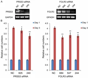
FOLR3, and TAS2R43 genes were found in GIST tissues of both patients (Table 1). To further investigate the influence of these proteins on the progression of GISTs, specific siRNAs were used to inhibit their expressions in GIST-T1 cells (Figure 2A). As shown in Figure 2B, after endog-enous PRSS3 or FOLR3 was knocked down, the growth of GIST-T1 cell was significantly reduced. However, when TAS2R43 was knocked down, the growth of GIST-T1 cell was not significantly affected. So the result was not listed. These results suggest that PRSS3 and FOLR3 play oncogenic roles in GIST tumorigenesis.
Discussion
As a tumor suppressor gene located on chro-mosome 17q11.2, NF1 is responsible for a pro-tein called neurofibromin. Neurofibromin is involved in the downregulation of the rat sarco-ma viral oncogene homologue (RAS)-mitog-
that regulate gastrointestinal motility. On the other hand, its function is nerve-related. That prompts us to think about whether the aberrant expression of neurofibromin in NF1 patients contributes to the development of GIST. After reviewing the previous studies, we found no direct evidences were available. However, some indirect evidences were detected. Johanna analyzed the 50 GISTs derived from 15 NF1 patients microscopically and find hyperplastic foci (diffuse and focal) of the interstitial cells of Cajal that were often associated with micro-scopic GIST in the surrounding intestinal mus-cle wall [16]. Study based on autopsy indicated a higher frequency of GIST occurrence in NF1 patients compared with somatic GIST. 3 of 12 patients proved to develop GIST according to the pathology test [17]. More involving studies are urgently needed.
The first case associated with gastrointestinal neoplasms resembling leiomyomas in NF-1 patients could be dated back to 1969, when GIST has not yet been well elucidated and defined as one kind of tumor [18]. As is known to us, GIST is the most commonly seen
mesen-Figure 2. A. After transfected with different siRNA (50 nM) for 72 h, GIST-T1 cells were harvested for mRNA extraction; and the expression of PRSS3 and
FOLR3 was determined by RT-PCR; B. GIST-T1 cells were transfected with PRSS3 or FOLR3 siRNA, and cell growth was evaluated by the CCK-8 assay.
*P<0.05; **P<0.01 compared with NC group (mean ± SD, Student t test).
en activated protein kinase (MAPK) pathway [15]. Though the most frequent detected disease in NF1 is generally derived from nervous system, NF1 patients could some-times be accepted into clinic for non-nervous system dis-eases such as GIST, phaeo-chromocytoma and even bre- ast cancer [5]. Of all the non-nervous system diseases, GIST has attracted the most attention for doctors majored in general surgery.
[image:6.612.90.374.72.326.2]chymal tumor. The most important index for diagnosing GIST is the positive immunohisto-chemistry staining for CD117 and DOG-1. C-KIT
or PDGFRA mutation has been recognized as the central event for the development of GIST. Of all the detected mutation, exon 11 leads as the most frequently affected site (60%) and exon 9 follow (15%) [2]. Besides the mutation analysis, GIST was also characterized as soli-tary tumor classified into three types: spindle cell-type (70%), epithelioid-type (20%) and mixed-type (10%) [2, 8]. Most of the primary tumors were solitary lesion distributing along the alimentary tract, with the stomach as the most common site (60%), followed by the small intestine (25%) [2, 15]. Though demonstrated CD117 and DOG-1 expression, NF1-associated GIST were different from the sporadic cases in many aspects [19, 20]. Based on the existing studies, all of the NF1-associated cases were detected without c-KIT or PDGFRA mutation, indicating that the molecular pathogenesis for NF1-associated cases were different from the sporadic cases [21]. NF1-associated cases could only been found in intestine. No gastric origin cases have been reported yet [20]. Primary cases tended to be multifocal and mixed-type cases were more frequently seen [20, 21]. The deep cause of the differences still remains unknown.
As a member of serine protease family, trypsin is encoded by PRSS1, PRSS2 and PRSS3. The relationship between PRSS3 and cancers has been highlight recently, especially in the study of lung cancer and ovarian cancer [12, 22, 23]. Though mainly expressed by pancreas, PRSS3 could be found in some epithelial cancers and cancer cell lines [23]. Besides, overexpression of PRSS3 has been reported to be associated with progression of cancers [22, 23]. Some studies have even put forward the opinion that over expression of PRSS3 can not only promote the cell proliferation and invasion in ovarian cancer but also act as a potential marker for predicting the metastasis. FOLR3 belongs to the folate receptor gene family and participate in the transport and binding of folate and the naturally occurring form of folic acid [24]. It is a secretory molecular expressed predominantly in hematopoietic cells [25]. Most of the studies before have focused on its function on meta-bolic pathway. Seldom has a study relieved how the aberrant FOLR3 could affect the
occur-rence of cancer, letting along the NF1-associated GIST. Our study has selected PRSS3 and FOLR3 as the keys in the development of NF1-associated GIST for the statistical analysis results derived from WES for the two NF1 patients. The cell experiment proved that those two genes played oncogenic roles in the GIST T1 cells. Our results give a primary impression that PRSS3 and FOLR3 are potential treatment targets for NF1-associated GIST. That evidence is still not enough. Our group has successfully established the PDTX models, relevant studies are underwent now in our lab [26]. More telling evidence is urgently needed.
Conclusion
In summary, through next-generation sequenc-ing of the exome of DNA from GIST and NF1 tis-sue of the patients with KIT/PDGFRA wild-type GISTs concurrent with NF1, we identified three potential novel driver mutation genes in GISTs, which may reveal the underlying etiology of the
KIT/PDGFRA wild-type GISTs developing in NF1 patients. The genes could be targets for inter-vention and/or therapy for GISTs that develop from NF1.
Acknowledgements
This study was supported by Grants from the Ministry of Health of the China, No. W2012R- Q02; Shanghai Science and Technology Com- mittee, No. 12NM0501402; and Shanghai Education Committee, No. 120311. Thanks to the technical help provided by Wuxi AppTec Co, Ltd. Waigaoqiao Free Trade Zone, Shanghai, China. This study is part of the program achieve-ment of Shanghai Municipal Commission of Health and Family Planning, Key developing dis-ciplines, 2015ZB0201.
Disclosure of conflict of interest None.
Address correspondence to: Yong Zhang and Wei- Qi Lu, Department of General Surgery, Zhongshan
Hospital, Fudan University, Shanghai 200032,
China. E-mail: [email protected] (YZ); lu. [email protected] (WQL)
References
[1] Ferner RE. Neurofibromatosis 1 and neurofi
[2] Valencia E, Saif MW. Neurofibromatosis type 1
and GIST: is there a correlation? Anticancer Res 2014; 34: 5609-5612.
[3] Ferner RE, Huson SM, Thomas N, Moss C, Willshaw H, Evans DG, Upadhyaya M, Towers R,
Gleeson M, Steiger C, Kirby A. Guidelines for the diagnosis and management of individuals
with neurofibromatosis 1. J Med Genet 2007;
44: 81-88.
[4] Rasmussen SA, Friedman JM. NF1 gene and neurofibromatosis 1. Am J Epidemiol 2000;
151: 33-40.
[5] Brems H, Beert E, de Ravel T, Legius E. Mechanisms in the pathogenesis of malignant
tumours in neurofibromatosis type 1. Lancet
Oncol 2009; 10: 508-515.
[6] Huss S, Elges S, Trautmann M, Sperveslage J,
Hartmann W, Wardelmann E. Classification of KIT/PDGFRA wild-type gastrointestinal stromal
tumors: implications for therapy. Expert Rev Anticancer Ther 2015; 15: 623-628.
[7] Janeway KA, Albritton KH, Van Den Abbeele AD, D’Amato GZ, Pedrazzoli P, Siena S, Picus J,
Butrynski JE, Schlemmer M, Heinrich MC,
Demetri GD. Sunitinib treatment in pediatric patients with advanced GIST following failure of imatinib. Pediatr Blood Cancer 2009; 52: 767-771.
[8] Agaram NP, Laquaglia MP, Ustun B, Guo T, Wong GC, Socci ND, Maki RG, DeMatteo RP,
Besmer P, Antonescu CR. Molecular character-ization of pediatric gastrointestinal stromal tu-mors. Clin Cancer Res 2008; 14: 3204-3215. [9] Jiang Q, Zhang Y, Zhou YH, Hou YY, Wang JY, Li
JL, Li M, Tong HX, Lu WQ. A novel germline
mu-tation in SDHA identified in a rare case of gas -trointestinal stromal tumor complicated with renal cell carcinoma. Int J Clin Exp Pathol 2015; 8: 12188-12197.
[10] Mussi C, Schildhaus HU, Gronchi A,
Ward-elmann E, Hohenberger P. Therapeutic conse-quences from molecular biology for gastroin-testinal stromal tumor patients affected by
neurofibromatosis type 1. Clin Cancer Res
2008; 14: 4550-4555.
[11] Azizmohammadi S, Safari A, Seifoleslami M, Rabati RG, Mohammadi M, Yahaghi H,
Azizmohammadi S. Clinical significance and expression of the PRSS3 and Wiskott-Aldrich
syndrome protein family verprolin-homologous protein 1 for the early detection of epithelial ovarian cancer. Tumour Biol 2016; 37: 6769-6773.
[12] Hockla A, Miller E, Salameh MA, Copland JA, Radisky DC, Radisky ES. PRSS3/mesotrypsin
is a therapeutic target for metastatic prostate cancer. Mol Cancer Res 2012; 10: 1555-1566.
[13] Corrigan A, Walker JL, Wickramasinghe S, Hernandez MA, Newhouse SJ, Folarin AA, Lewis CM, Sanderson JD, Spicer J, Marinaki
AM. Pharmacogenetics of pemetrexed com- bination therapy in lung cancer: pathway anal-ysis reveals novel toxicity associations. Pharmacogenomics J 2014; 14: 411-417. [14] Davidson B, Abeler VM, Forsund M, Holth A,
Yang Y, Kobayashi Y, Chen L, Kristensen GB, Shih I, Wang TL. Gene expression signatures of primary and metastatic uterine leiomyosarco-ma. Hum Pathol 2014; 45: 691-700.
[15] Agaimy A, Vassos N, Croner RS. Gastrointesti-
nal manifestations of neurofibromatosis type 1 (Recklinghausen’s disease): clinicopathologi -cal spectrum with pathogenetic consider-ations. Int J Clin Exp Pathol 2012; 5: 852-862. [16] Andersson J, Sihto H, Meis-Kindblom JM,
Joensuu H, Nupponen N, Kindblom LG.
NF1-associated gastrointestinal stromal tumors have unique clinical, phenotypic, and genotyp-ic characteristgenotyp-ics. Am J Surg Pathol 2005; 29: 1170-1176.
[17] Ghrist TD. Gastrointestinal involvement in
neu-rofibromatosis. Arch Intern Med 1963; 112:
357-362.
[18] Lukash WM, Johnson RB. Gastrointestinal neo
-plasms in von Recklinghausen’s disease.
South Med J 1969; 62: 1237-1239.
[19] Badache A, Muja N, De Vries GH. Expression of
Kit in neurofibromin-deficient human Schwann
cells: role in Schwann cell hyperplasia
associ-ated with type 1 neurofibromatosis. Oncogene
1998; 17: 795-800.
[20] Maertens O, Prenen H, Debiec-Rychter M,
Wozniak A, Sciot R, Pauwels P, De Wever I,
Vermeesch JR, de Raedt T, De Paepe A,
Speleman F, van Oosterom A, Messiaen L,
Legius E. Molecular pathogenesis of multiple
gastrointestinal stromal tumors in NF1 pa -tients. Hum Mol Genet 2006; 15: 1015-1023. [21] Miettinen M, Lasota J. Gastrointestinal stromal
tumors: pathology and prognosis at different sites. Semin Diagn Pathol 2006; 23: 70-83. [22] Ma R, Ye X, Cheng H, Ma Y, Cui H, Chang X.
PRSS3 expression is associated with tumor progression and poor prognosis in epithelial ovarian cancer. Gynecol Oncol 2015; 137: 546-552.
[23] Cohen I, Kayode O, Hockla A, Sankaran B, Radisky DC, Radisky ES, Papo N. Combinatorial
protein engineering of proteolytically resistant mesotrypsin inhibitors as candidates for can-cer therapy. Biochem J 2015; 473: 1329-1341.
genes of primates. Genomics 2014; 103: 40-47.
[25] Sadasivan E, Cedeno MM, Rothenberg SP. Characterization of the gene encoding a folate-binding protein expressed in human placenta.
Identification of promoter activity in a G-rich SP1 site linked with the tandemly repeated
GGAAG motif for the ets encoded GA-binding protein. J Biol Chem 1994; 269: 4725-4735.
Supplementary Table 1. Somatically mutated genes in the exome sequences of GIST
Gene name Gene ID coordinateGenomic Patient mutationAllele Amino acid mutation PRAMEF22 653606 1:13036736 GIST#1 T>A Y270N RGPD3 653489 2:107049425 GIST#1 A>C L570R
DSPP 1834 4:88537088 GIST#1 A>G N1092D
4:88537261 GIST#1 A>T E1149D ZDHHC11 79844 5:848742 GIST#1 C>T V86I FAM153A 285596 5:177163580 GIST#1 A>G F145L
HLA-DRB1 3123 6:32549582 GIST#1 G>T T135N RAET1L 154064 6:150343211 GIST#1 A>G M85T TAS2R30 259293 12:11286214 GIST#1 C>G Q210H PARP4 143 13:25021201 GIST#1 G>A L1080F
MUC16 94025 19:9002587 GIST#1 C>T R13410Q 19:9002597 GIST#1 C>T D13407N FRG1B 284802 20:29628251 GIST#1 A>G N85D RBMXL3 139804 X:114425136 GIST#1 A>G S378G PRAMEF16 654348 1:13497572 GIST#2 G>A C290Y 1:13497584 GIST#2 C>T P294L MST1P9 11223 1:17085006 GIST#2 C>T G459E RBMXL1 494115 1:89449237 GIST#2 T>A R91S OR2T29 343563 1:248722611 GIST#2 C>G S61T FER1L5 90342 2:97361378 GIST#2 T>C S1319P SNED1 25992 2:242011113 GIST#2 G>C E1238Q SORCS2 57537 4:7735143 GIST#2 G>A G1068D
ALB 213 4:74279140 GIST#2 G>T D133Y
FHDC1 85462 4:153896049 GIST#2 A>C T536P
BDP1 55814 5:70848945 GIST#2 T>A F407I
OR12D3 81797 6:29342440 GIST#2 A>G F209L
SASH1 23328 6:148664232 GIST#2 G>A G10E PRSS1 5644 7:142460339 GIST#2 G>A C121Y
MLL3 58508 7:151970840 GIST#2 C>T S321N
PABPC1 26986 8:101727750 GIST#2 T>C I150V CBWD3 445571 9:70912519 GIST#2 A>C E232D TAS2R31 259290 12:11183475 GIST#2 G>A R154W 12:11183484 GIST#2 C>G E151Q 12:11183485 GIST#2 T>A K150N PTPRO 5800 12:15669792 GIST#2 T>A Y561N GXYLT1 283464 12:42512971 GIST#2 T>C Y106C FAM186A 121006 12:50745822 GIST#2 T>G E1598A EP400 57634 12:132445256 GIST#2 A>C H31P
12:132445273 GIST#2 T>C S37P POTEM 641455 14:20007593 GIST#2 T>C I365M
GALC 2581 14:88452853 GIST#2 T>C N115S
GOLGA6B 55889 15:72954595 GIST#2 A>G K284E FURIN 5045 15:91424716 GIST#2 C>T H665Y CCDC165 23255 18:8786031 GIST#2 G>A R610H COL5A3 50509 19:10078030 GIST#2 C>T G1484D ANGPTL6 83854 19:10206822 GIST#2 C>A G140W SIN3B 23309 19:16987383 GIST#2 G>T E951D ZNF285 26974 19:44901381 GIST#2 C>T G16S
NEFH 4744 22:29885644 GIST#2 C>A A672E
Supplementary Table 2. Somatically mutated genes in the exome sequences of NF1
Gene name Gene ID coordinateGenomic Patient mutationAllele Amino acid mutation
FLG 2312 1:152280685 NF1#1 C>A G2226V
KCNN3 3782 1:154842250 NF1#1 G>T P159Q 1:154842253 NF1#1 G>T P158H POTEF 728378 2:130832185 NF1#1 G>A R954W LRP1B 53353 2:141625828 NF1#1 G>A L1330F
HSPD1 3329 2:198363406 NF1#1 C>T G56E ADAM29 11086 4:175899129 NF1#1 C>T T818M RP1L1 94137 8:10467448 NF1#1 C>T G1387E HOMEZ 57594 14:23744820 NF1#1 A>T D539E GOLGA6L10 647042 15:82637166 NF1#1 T>C E307G TPSAB1 7177 16:1291545 NF1#1 C>T T115I
16:1291598 NF1#1 G>A V133I 16:1291608 NF1#1 A>G H136R CYP2A7 1549 19:41381671 NF1#1 A>G I420T ZSCAN18 65982 19:58600105 NF1#1 C>G S168T FAM182A 284800 20:26061945 NF1#1 A>G G40
Supplementary Table 3. Somatically mutated genes in the exome sequences in both GISTs and NF1
Gene name Gene ID coordinateGenomic Patient mutationAllele Amino acid mutation
FTH1 2495 11:61735080 GIST#1, NF1#1 G>T S1*
GOLGA6L6 727832 15:20740252 GIST#1, NF1#1 C>A E500*
15:20740292 GIST#1, NF1#1 G>C H486Q
15:20740293 GIST#1, NF1#1 T>C H486R
15:20740294 GIST#1, NF1#1 G>T H486N
OTOA 146183 16:21747639 GIST#1, NF1#1 G>T E196*
GRIK3 2899 1:37319270 GIST#1, NF1#1 C>A L386F
HRNR 388697 1:152188569 GIST#1, NF1#1 C>T G1846S
IGFN1 91156 1:201178667 GIST#2, NF1#1 C>G T1549R 1:201178849 GIST#2, NF1#1 G>A E1610K 1:201179068 GIST#1, GIST#2, NF1#1 G>A G1683R 1:201180208 GIST#1, NF1#1 G>C G2063R 1:201180214 GIST#1, NF1#1 G>A V2065M 1:201180217 GIST#1, NF1#1 A>G N2066D
HLX 3142 1:221053600 GIST#1, NF1#1 A>C Q134P
REG3A 5068 2:79385823 GIST#1, NF1#1 T>G H50P ANKRD36 375248 2:97820417 GIST#1, NF1#1 T>G L400R
2:97820434 GIST#1, NF1#1 T>G F406V
PRR21 643905 2:240982172 GIST#1, NF1#1 C>T M76I
MUC4 4585 3:195505816 NF1#1 G>C T4212S
3:195505829 GIST#1, NF1#1 G>A P4208S 3:195505855 GIST#1, NF1#1 G>T S4199Y
3:195507323 NF#1 T>C T3710A
3:195507433 NF#1 G>A A3673V
3:195507982 NF#1 T>G H3490P
3:195508451 NF#1 G>T P3334T
3:195508835 GIST#1, GIST#2, NF1#1 C>T A3206T
3:195510457 NF#1 A>G V2665A
3:195510731 GIST#1, NF1#1 C>T A2574T 3:195510755 GIST#1, NF1#1 T>C T2566A 3:195510779 GIST#1, NF1#1 C>T A2558T
3:195511283 NF#1 C>G A2390P
3:195511285 NF#1 T>C D2389G
3:195511286 NF#1 C>T D2389N
3:195511465 GIST#1, NF1#1 G>A A2329V
3:195513446 NF#1 T>C S1669G
3:195513515 NF#1 C>T A1646T
3:195515008 NF#1 C>G G1148A
CRIPAK 285464 4:1388656 GIST#1, NF1#1 C>T R103C
4:1388817 GIST#1, NF1#1 C>G P173R
FRG1 2483 4:190876293 GIST#1, GIST#2, NF1#1 C>A P140Q
4:190878569 GIST#2, NF1#1 T>C L150S
4:190878571 GIST#2, NF1#1 G>A A151T
PCDHA13 56136 5:140263173 GIST#1, NF1#1 C>A S440R
MUC12 10071 7:100636551 NF#1 C>G P1046A
7:100645731 NF#1 G>A E3963K LRRC4 64101 7:127670327 GIST#1, NF1#1 C>G A123P C9orf150 286343 9:12775880 GIST#2, NF1#1 T>G C56G
9:12775883 GIST#2, NF1#1 A>G S57G
CYLC2 1539 9:105767469 GIST#1, NF1#1 A>G K186E PAPPA-AS1 493913 9:119160776 GIST#1, NF1#1 G>A S97F
AGAP7 653268 10:51465138 GIST#1, NF1#1 C>T E440K
MUC6 4588 11:1017169 GIST#1, NF1#1 G>A P1878S
11:1017591 GIST#1, NF1#1 C>G R1737P
11:1018182 GIST#1, NF1#1 G>T T1540N
11:1018186 GIST#1, NF1#1 G>T P1539T
11:1018192 GIST#1, NF1#1 C>T V1537I PRDM10 56980 11:129801109 GIST#1, NF1#1 T>G E161D KRT75 9119 12:52822136 GIST#1, NF1#1 C>G R429P
KRT8 3856 12:53298675 GIST#2, NF1#1 A>C S109A
CR383656.1 14:19378189 GIST#1, NF1#1 G>T R199L POTEG 404785 14:19563409 GIST#1, NF1#1 C>T A308V TEP1 7011 14:20876580 GIST#1, GIST#2, NF1#1 G>A H7Y
14:20876588 GIST#1, GIST#2, NF1#1 A>G L4P TPSB2 64499 16:1279438 GIST#1, NF1#1 C>T A85T ZFPM1 161882 16:88599698 GIST#1, NF1#1 G>C E444D NCOR1 9611 17:16068377 GIST#1, NF1#1 C>G K178N CDC27 996 17:45234432 GIST#1, NF1#1 G>T S169Y MAP1S 55201 19:17837593 GIST#1, NF1#1 C>G P441R ZNF208 7757 19:22155725 GIST#1, NF1#1 C>G W604S
19:22156826 NF#1 C>A G337V
ZNF676 163223 19:22363701 GIST#1, NF1#1 G>A A273V
19:22363702 GIST#1, NF1#1 C>T A273T
ZNF492 57615 19:22836805 GIST#1, NF1#1 G>A A40T ZNF579 163033 19:56089870 GIST#1, NF1#1 G>C A379G POM121L1P 25812 22:22985728 GIST#1, NF1#1 C>T V277I ZXDB 158586 X:57618845 GIST#1, GIST#2, NF1#1 G>A E122K
X:57618849 GIST#1, GIST#2, NF1#1 A>C E123A FMR1 2332 X:147010263 GIST#1, GIST#2, NF1#1 A>C K119N