Original Article
Expression of chemokine receptor CXCR7 and its effects
on gastric cancer tissues and cell lines
Jing Yang1, Xianbin Huang1, Xiangyong Hao2, Li Wei3, Wutang Jing1, Tiankang Guo1
1Department of First General Surgery, Gansu Provincial Hospital, Lanzhou, Gansu 730000, China; 2Department
of Fourth General Surgery, Gansu Provincial Hospital, Lanzhou, Gansu 730000, China; 3Dean’s Office, Gansu
Provincial Hospital, Lanzhou, Gansu 730000, China
Received October 29, 2015; Accepted December 25, 2015; Epub February 1, 2016; Published February 15, 2016
Abstract: Gastric cancer (GC), the most common and malignant tumor of the digestive system, exhibits high inva-sive capacity that escapes immune attack. Chemokine receptor CXCR7 plays an important role in the development of cancer. Therefore, the aim of the present study is to investigate the expression of CXCR7 in GC tissues and to evaluate the role of CXCR7 in tumor growth, apoptosis, and invasion of GC cells. The expression status of CXCR7 was detected in 65 primary GC tissues by immunohistochemistry. The correlation of CXCR7 expression with the clinicopathologic parameters and prognostic factors of GC was analyzed. Further, the effects of CXCR7 knock-down by CXCR7-shRNA lentiviral vector on the proliferation, apoptosis, and invasion of GC cells were explored in vitro. MTT
assay, FCM analysis, and transwell chamber test were employed. The expression of CXCR7 was significantly higher
in GC tissues than in normal tissues. CXCR7 expression was correlated with the degree of differentiation, depth of invasion, and lymph node metastasis. Transfection of MKN-45 cells with CXCR7-shRNA lentiviral vector resulted in a
significantly decreased expression of CXCR7 at an mRNA and protein level. Tumor invasiveness was suppressed in vitro by silencing of CXCR7 in MKN-45 cells. However, no obvious suppression in cell proliferative activity and apop-tosis were observed. The expression of CXCR7 could be used as a biomarker to predict prognosis and metastasis of GC. LV-CXCR7-shRNA constructs effectively inhibited the expression of CXCR7 RNA, thus reducing the migration and invasion capacity of human GC cells in vitro.
Keywords: Gastric cancer, chemokine receptor CXCR7, invasion, lentivirus-mediated CXCR7 gene silencing
Introduction
Gastric cancer (GC) is currently the third cause of cancer-related deaths worldwide, and is par-ticularly prevalent in Asia [1]. Local and distant metastases after curative gastrectomy are crit-ical events affecting the prognosis of GC patients [2]. Since the molecular mechanisms underlying metastasis and recurrence of GC
have not been defined, an effective approach
towards prevention and cure is not yet avail-able. Therefore, it is necessary to elucidate the molecular mechanisms related to progression of the malignant potential of GC. CXC chemo-kine ligand CXCL12, also called stromal cell-derived factor (SDF)-1, is a member of the CXC sub-family and mediates hematopoiesis [3], vascular formation [4], and neurogenesis [5] through interaction with receptor CXCR4. It has recently been discovered that CXCL12 also
binds to the receptor CXCR7 with high affinity.
CXCR7 plays a crucial role in several cancers, including colon cancer [6], liver cancer [7], and bladder cancer [8]. In addition, studies have shown that CXCL12-CXCR7 axis promotes tumor aggressiveness and metastasis [9]. The importance of CXCR7 in cancer is further
clarified by the fact that malignant tumors (in
involving interventions of CXCR7 and related signaling pathways may offer new therapeutic opportunities in cancer. In a previous study, CXCR7 was found to be highly expressed in patients with GC, which was additionally asso-ciated with lifestyle risk factors, such as alco-hol consumption [13]. Although the role of CXCR4 in the promotion of GC invasive growth
is well documented, the significance of CXCL12/
CXCR7 axis in regulating GC biological behavior has not been completely described. We, there-fore, hypothesized the involvement of CXCR7 in the malignant properties of GC. Herein, we investigated the expression of CXCR7 in prima-ry GC by immunohistochemistprima-ry. Furthermore, we explored the effects of CXCR7 knock-down by CXCR7-shRNA lentiviral vector on the biologi-cal behavior, including proliferation, apoptosis, and invasion of human GC cells.
Materials and methods
Ethics
The Ethics Committee of Gansu Provincial Hospital, Lanzhou, China approved the protocol of the present study. Informed written consent was obtained from all patients for the acquisi-tion and use of patient tissue samples and ano-nymized clinical data.
Patients and samples
The present study included 65 patients with primary GC, all of whom underwent gastrecto-my between 2011 and 2013 at the Department of General Surgery, Gansu Provincial Hospital. Gastric cancer was pathologically diagnosed after operation in the cancer group. This group included 51 men and 14 women ranging from 35 to 74 years of age (mean, 55 years). No patient was treated with local ablative therapy, chemotherapy, or immunotherapy prior to sur-gery. Control tissues were obtained from 20 benign gastric ulcer patients, who received gastrectomy. Human gastric tissue samples
were formalin-fixed and paraffin-embedded.
According to AJCC and UICC criteria, there were 7 (10.8%), 15 (23.1%), 40 (61.5%), and 3 (4.6%) cases at stage I, II, III, and IV, respectively. The median follow-up period was 11 months (range, 2-24). Pathological characteristics were
collect-ed and classificollect-ed. All specimens were patho -logical and were reassessed independently by two gastroenterology pathologists blinded to the clinical data.
Immunohistochemistry
To assess the CXCR7 protein expression in gas-tric cancer and normal tissue, monoclonal rab-bit anti-human CXCR7 (P25106; Abgent, San
Diego, CA, USA) was used. Paraffin-embedded sections (5-µm thick) were fixed in freshly pre -pared 10% paraformaldehyde for 5 min. After blocking the endogenous peroxidase activity with 0.3% hydrogen peroxide in TBS for 15 min, the sections were immersed in horse serum (diluted 1:10 in TBS) for 30 min to reduce any
nonspecific binding, and were subsequently
incubated with the primary antibody (1:100) overnight at 4°C after washing with TBS. The sections were then incubated with biotinylated goat anti-rabbit IgG for 30 min, and avidin-bio-tin-peroxidase complex for 30 min. After each step in the staining procedure, the sections were given three 5-min washes in TBS. Immunoreactivity (IR) was visualized using 1 mg/ml diaminobenzidine (DAB) as chromogen and 0.01% hydrogen peroxide as substrate. The peroxidase reaction was stopped after 5 min with distilled water, and the sections were counter-stained with Toluidine blue, dehydrat-ed, and then mounted with Entellan. Negative controls were obtained by substituting the pri-mary antibody with phosphate-buffered saline. Slides were evaluated under a light microscope
(× 400 magnifications). For digital image analy -sis, Adobe Photoshop software version 7.0 was used. Results were scored by two independent
pathologists. The proportion score reflected
the fraction of positive staining cells (score 0, <25%; score 1, 25-50%; score 2, 50-75%; and score 3, >75%), and the intensity score repre-sented the staining intensity (score 0, no stain-ing; score 1, weak positive; score 2, moderate positive; and score 3, strong positive). The two scores were averaged. Based on the analysis, CXCR7 was regarded as negatively expressed in GC tissues if the score was <2, and positively
expressed with the score ≥2.
Synthesis of siRNA
GCCTGG ATATTCACCCag-5’ (antisense); sequ- ence D (siCXCR7-D): 5’-ctGCACACACAAC GAA- CAGTT-3’ (sense) and 3’-AACTGTTCGTTGTGTG- TGCag-5’ (antisense). In addition, the negative control siCXCR7 (CON049) was synthesized by Shanghai Genechem Co. Ltd. (Shanghai, China)
for monitoring the influence of exogenous
genes.
Construction and identification of LV-shCXCR7
Vector GV118 was digested with restriction enzymes (HpaI/XhoI) and siRNA was inserted into the vector. The sequencing of LV-shCXCR7 was carried out by the Shanghai Genechem Co. Ltd. The packing of lentiviral vector was done according to the instructions provided with the Lenti-X Bicistronic Expresion System. Then, the LV-shCXCR7 was tittered using a kit based on the manufacturer’s instructions.
Culture of MKN-45 cells and transfection
If not indicated otherwise, all materials were purchased from Gibco (Carlsbad, CA, USA). MKN-45 cells were cultured in RPMI-1640 medium supplemented with 10% fetal bovine
after transfection, the medium was removed and serum containing growth medium was added. On day 3 post-transfection, transfection
efficiency was measured by the frequency of green fluorescent protein (GFP)-positive cells.
Positive cells (>80%) were collected and 1 ml of Trizol reagent was added, and quantitative real time PCR was performed to detect the mRNA levels.
Quantitative real-time PCR
Total RNA was extracted from MKN-45 cells using Trizol reagent (Invitrogen, Carlsbad, CA,
USA). Using a first strand cDNA synthesis kit
(Promega, Beijing, China), cDNA was generated with Oligo dT primer. Primers for CXCR7 (136 bp) were synthesized by Sangon Inc. (Shanghai, China) - F: AGCATCAAGGAGTGGCT GAT; R: TG- TGCTTCTCCTGGTCACTG. GAPDH was used as an endogenous control. The reaction mixture contained 1.0 µl cDNA, 0.5 µl forward primer, 0.5 µl reverse primer, 10 µl SYBR Master Mix, and 8.0 µl RNase-Free H2O (Takara, Dalian, China). Reaction mixtures were incubated at
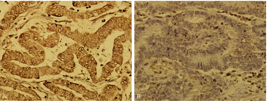
[image:3.629.99.532.79.243.2]95°C for 5 min, and then amplified for 45 cycles Figure 1. Immunohistochemistry for CXCR7 in gastric cancer tissues and normal gastric tissues (magnification ×
400). A. CXCR7 is strongly detected in the membranes and cytoplasm of cancer cells; B. CXCR7 is not detected in normal gastric cells.
Table 1. Expression of CXCR7 protein in GC tissues
Variables Groups Cases (N)
Expression levels
(n) Positive
rate (%) χ2 value P-Negative Positive
CXCR7 GC 65 10 55 84.62% 38.55 <0.01
NC 20 18 2 10%
GC, gastric cancer; NC, normal control tissue.
serum and seeded on 6-well plates at a density of 5 × 10⁴
cells/well. At 30% confluence,
[image:3.629.96.375.318.385.2]of 15 s at 95°C, 30 s at 60°C, and 30 s at 72°C annealing temperature. Data were analyzed using the comparative Ct method (2-ΔΔCt). All
experiments were performed in triplicate. Cells transfected with LV-shCXCR7-B showed the lowest expression of CXCR7, and were there-fore used in the following experiment.
Western blot
Nuclear protein extracts of MKN-45 cells were prepared in SDS lysis buffer containing prote-ase inhibitors. Cell extracts were denatured in laemmli buffer (10% glycerol, 2% SDS, 0.1 m dithiothreitol, 50 mm Tris, 0.01 mg/ml
bromo-Cell proliferation assay
Cell proliferation was analyzed with the MTT
assay. Briefly, cells infected with LV-shCXCR7
were incubated in 96-well plates at a density of 1 × 10⁵ cells per well. Cells were treated with 20 µl MTT dye (5 mg/ml). The color reaction was measured at 490/570 nm with enzyme immunoassay analyzer (Biotek, Winooski, VT, USA). The proliferation activity was calculated for each clone.
Cell apoptosis assay
[image:4.629.100.406.105.541.2]To detect cell apoptosis, cells were trypsinized, washed with PBS, and re-suspended in 1 × Table 2. Relationship between CXCR7 expression and clinical
character-istics in patients with gastric cancer
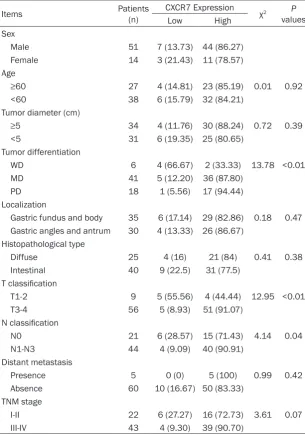
Items Patients (n) CXCR7 Expression χ2 P
values
Low High
Sex
Male 51 7 (13.73) 44 (86.27)
Female 14 3 (21.43) 11 (78.57)
Age
≥60 27 4 (14.81) 23 (85.19) 0.01 0.92
<60 38 6 (15.79) 32 (84.21)
Tumor diameter (cm)
≥5 34 4 (11.76) 30 (88.24) 0.72 0.39
<5 31 6 (19.35) 25 (80.65)
Tumor differentiation
WD 6 4 (66.67) 2 (33.33) 13.78 <0.01
MD 41 5 (12.20) 36 (87.80)
PD 18 1 (5.56) 17 (94.44)
Localization
Gastric fundus and body 35 6 (17.14) 29 (82.86) 0.18 0.47 Gastric angles and antrum 30 4 (13.33) 26 (86.67)
Histopathological type
Diffuse 25 4 (16) 21 (84) 0.41 0.38
Intestinal 40 9 (22.5) 31 (77.5)
T classification
T1-2 9 5 (55.56) 4 (44.44) 12.95 <0.01
T3-4 56 5 (8.93) 51 (91.07)
N classification
N0 21 6 (28.57) 15 (71.43) 4.14 0.04
N1-N3 44 4 (9.09) 40 (90.91)
Distant metastasis
Presence 5 0 (0) 5 (100) 0.99 0.42
Absence 60 10 (16.67) 50 (83.33)
TNM stage
I-II 22 6 (27.27) 16 (72.73) 3.61 0.07
III-IV 43 4 (9.30) 39 (90.70)
phenol blue; pH 6.8) at 90°C for 5 min, and were separated by SDS-PAGE. Separated pro-teins were transferred onto a PVDF membrane in Trans-blot wet buffer (Bio-Rad Laboratories, Hercules, CA, USA). The membranes were blo- cked with 5% nonfat milk powder. The pri- mary antibody against CXCR7 (P25106; Ab- gent, USA) was diluted according to the instru- ctions and incubated overnight at 4°C. Ho- rseradish peroxidase-conjugated goat anti-rabbit IgG were then added for 1 h at room temperature, and wash- ed with 1 × TBS three times. Membranes we- re treated with ECL plus Western blotting detection kit (Transgen, Beijing, China), and bands were detected using STORM 840v20- 05 with ImageQuant software (GE, Pittsbur- gh, PA, USA). The rela-tive protein level in dif-ferent groups was
binding buffer according to the instructions pro-vided in the apoptosis kit (eBioscience, San Diego, CA, USA). 10 µl Annexin V-APC was
added to the fixed cells for 15 min in darkness
at room temperature. Subsequently, apoptosis
in treated cells was analyzed by flow cytometry
(Guava easyCyte HT, Germany) with Cellquest software (BD Biosciences-Pharmingen, San Diego, CA, USA). Three experiments were per-formed for each clone.
Transwell invasion assay
Harvested cells (1 × 10⁵) in 100 µl serum-free DMEM were added into the upper chamber.
The lower chamber was filled with 600 µl condi
-variate analysis. Statistical significance was set
at P<0.05. Results
Expression of CXCR7 protein was upregulated in human GC tissues
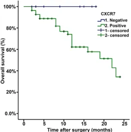
[image:5.629.101.372.81.353.2]The expression of CXCR7 protein was evaluated using immunohistochemical (IHC) staining. The positive expression of CXCR7 protein was detected in the cytoplasm and cell membranes of GC tissues. CXCR7 in cancer cells stained more intensely as compared to the normal gas-tric cells (Figure 1). The positive rates of CXCR7 expression were detected in 84.62% (55/65) of Figure 2. Analysis of overall survival according to the expression of
CXCR7 in gastric cancer patients.
Figure 3. Lenti-CXCR7-siRNA infected MKN-45 cells shows green fluores -cence (× 100).
tioned medium derived from MKN-45 cells as a source of che-moattractant. After 6 h of incuba-tion at 37°C with 5% CO2, the medium was removed from the upper chamber. The non-invaded cells on the upper side of the chamber were scraped off with a cotton swab. Migrated cells were dyed with 400 µl Giemsa for 20 min. Then, the color reaction was measured at 570 nm with enzy- me immunoassay analyzer (Bio- tek). MTS method measured migration of cells according to the instructions given in the CellTiter 96 AQueous One So- lution Cell Proliferation Assay kit (Promega, China). The transfer rate (OD 570/MTTOD 490) was calculated. Each assay was repeated three times.
Statistical analysis
SPSS 19.0 was used for the
[image:5.629.98.371.409.488.2]multi-GC tissues, while the normal tissues displayed positive staining in 10% (2/20) of the cases;
and a significant difference was observed between the two (χ2=38.55, P<0.01; Table 1). These observations suggested that CXCR7 expression is increased in GC tissues as com-pared with normal tissues.
Relationship between CXCR7 expression and clinicopathologic characteristics of GC patients
IHC staining for CXCR7 levels was statistically analyzed to determine their relationship with various clinicopathologic features in all the 65 gastric cancer patients. As shown in Table 2,
there were no significant differences in age,
gender, primary tumor location, tumor size, and TNM stage. However, CXCR7 tended to be high-er in patients with poor diffhigh-erentiation, deephigh-er invasion, and lymphatic metastasis in compari-son to patients with well differentiated tu- mor, shallow invasion, and no lymphatic metastasis.
Association of CXCR7 expression with overall survival of GC patients
Cancer-specific survival was estimated based
on the CXCR7 expression using Kaplan-Meier
shCXCR7-D, and NC groups, the level of CXCR7 mRNA was observed to be 0.85±0.44, 0.21± 0.08, 1.34±0.64, 0.67±0.24, and 1.02±0.02, respectively. The expression of CXCR7 mRNA was the lowest in the LV-shCXCR7-B group and
differed significantly from the NC group
(P=0.0015; Figure 4A). CXCR7 mRNA levels were reduced by 79.4% in LV-shCXCR7-B group, when compared with the control cells. Similar to RT-PCR results, the expression levels of CXCR7 proteins were markedly downregulated in the CXCR7shRNA group in comparison to the CON and NC groups (Figure 4B).
Effect of CXCR7 silencing on cell proliferation
To examine the effect of CXCR7 on the growth of GC cells, we investigated the proliferative activities of MKN-45 cells by MTT assay. Silencing of CXCR7 gene did not decrease the proliferative activities of MKN-45 cells in a time dependent manner as compared to the NC groups (Figures 5A and 5B).
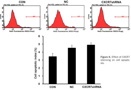
Effect of CXCR7 silencing on cell apoptosis
[image:6.629.100.380.78.336.2]Cell apoptosis was monitored by flow cytomet -ric analysis. As compared with that of the NC group and control group cells, apoptosis rate in Figure 4. The expression of CXCR7 mRNA and proteins in different
LV-shCX-CR7 groups and NC group.
survival analysis. Patients with high CXCR7 expression showed a reduced two-year survival rate as compared to the patients with low CXCR7 expression (34.2% vs. 100%), as was depicted in the survival curve (P=0.0124; Figure 2).
LV-shCXCR7 construction and transfection
Purified LV-shCXCR7 was used
for the transfection of MKN-45 cells. The transfection out-come was detected using a
fluorescence microscope 72 h
after the experimental treat-ment. All infected cells showed
green fluorescence (Figure 3).
Inhibition of CXCR7 shRNA on CXCR7 mRNA and proteins
the LV-shCXCR7 group cells showed no signifi -cant increase (Figure 6).
Effect of CXCR7 silencing on cell migration and invasion
To determine the effect of CXCR7 on cell migra-tion and invasion, transwell assay was per-formed. The invasive potential was determined on the basis of the ability of cells to invade a matrix barrier. Representative micrographs of
transwell filters have been presented in Figure 7A. The invasive potential of MKN-45 cells was
significantly reduced in LV-shCXCR7 group,
compared to the NC group (P<0.05; Figure 7B).
Discussion
As chemoattractant cytokines, chemokines can help in cell activation, differentiation, and
[image:7.629.102.530.81.237.2]trafficking. CXCR7, also known as a chemokine Figure 5. Effect of CXCR7 silencing on cell proliferation. Note: OD490/fold refers to times of OD490 on days 1-5 of
all experimental groups compared to the first day, indicating proliferation multiples on different day points.
[image:7.629.100.532.291.587.2]receptor, mediates biological effects through seven trans-membrane domain G protein cou-pled receptors (GPCRs). Additionally, it regu-lates fundamental biological processes includ-ing proliferation, cell survival, and adhesion, which contribute to essential functions of the receptor in multiple aspects of normal develop-ment and physiology [14]. CXCR7 also plays a role in cell growth, metastasis, and an- giogenesis of certain malignant tumors [15]. The expression of CXCR7 has been found to be correlated with solid tumor size, differentiation, and lymph node status, thereby suggesting its potential function in worse tumor prognosis [16]. Therefore, in the present study, we exam-ined the expression of the chemokine receptor CXCR7 in gastric cancer tissues using IHC and analyzed the relationship between their expres-sion and clinicopathologic features of GC patients. CXCR7, mainly localized in the
cyto-plasm and membranes, was significantly upreg -ulated in GC tissues as compared to the normal gastric tissues. The positive expression of CXCR7 was observed to be associated with poor differentiation, deeper invasion, and lym-phatic metastasis, suggesting its involvement in tumor aggression and differentiation. In
addition, we identified high CXCR7 expression
as a poor prognostic factor for 2-year overall survival following gastrectomy in GC patients.
colorectal cancer, and glioma [18-20]. Silencing
of CXCR7 expression significantly inhibited
hepatocellular carcinoma cells invasion, adhe-sion, and angiogenesis [21]. Furthermore, downregulation of CXCR7 expression caused inhibition of trans-endothelial migration of can-cer cells and lymph node metastasis [22], thus implying that CXCR7 could be used as a new targeted therapy for patients with GC. However, functionality of CXCR7 has long been a source of controversy. Burns et al. reported that ligand activation of CXCR7 does not induce typical chemokine responses, such as cell migration and calcium mobilization [14]. In the present study, the function of CXCR7 in GC was mani-fested using a recombinant lentiviral RNA inter-ference vector of the CXCR7 gene in highly
aggressive MKN-45 cells, and a significant
repression of the invasion ability of MKN-45 cells was noted in vitro. Nevertheless, both pro-liferation and apoptosis abilities of MKN-45 cells were not affected. These results were
con-sistent with the findings of Zheng et al., which
showed that CXCR7 mediates chemotaxis of cancer cells towards CXCL12 [21]. Hence, it is possible to prevent the development of GC metastasis through inhibition of CXCR7. The mechanism of growth, invasion, and metasta-sis of chemokine CXCL12/CXCR7 axis may involve direct activation of the ERK1/2 signal-Figure 7. Migration experiment: MKN-45 group (CON); NC group;
CXCR7shR-NA group (× 100). The number of migrated cells was significantly lower in
the lenti-shCXCR7 group than in the MKN-45 group and NC group. Note: KD group refers to normal cells, and cells infected with target genes, in accor-dance with the CXCR7shRNA group in text.
[image:8.629.100.387.79.292.2]ing pathway in cancer cells [23]. Another report suggested that CXCR7 can induce phosphoryla-tion of MAPK p42/44 and AKT in human rhab-domyosarcoma [24]. We thereby speculated that the different biological effects elicited by CXCR7 may depend on the cell types. However, in this study, we did not describe the molecular mechanisms through which CXCR7 regulates the invasion of GC cells. Therefore, further in-depth studies need to be undertaken to eluci-date the role of CXCR7 in invasion and signaling pathways activated by CXCL12/CXCR7 axis. In conclusion, lymphatic metastasis of GC is a prevalent and serious problem, and an effec-tive therapy is required. Upregulation of CXCR7 expression is associated with poor differentia-tion, deeper invasion, and lymph node metas-tasis; whereas silencing of CXCR7 gene repress-es the migration of gastric cancer cells. This study, therefore, suggested the potential appli-cation of lentiviral mediated CXCR7 RNAi as a therapeutic target for the treatment of gastric cancer.
Disclosure of conflict of interest
None.
Address correspondence to: Tiankang Guo, De- partment of The First General Surgery, Gansu Provincial Hospital, Lanzhou 730000, Gansu, China.
Tel: +86-18794838392; Fax: +86-21-64085875; E-mail: xinghewangwei@sina.com
References
[1] Siegel R, Naishadham D and Jemal A. Cancer statistics, 2013. CA Cancer J Clin 2013; 63: 11-30.
[2] Isobe Y, Nashimoto A, Akazawa K, Oda I, Hayashi K, Miyashiro I, Katai H, Tsujitani S, Kodera Y, Seto Y and Kaminishi M. Gastric can-cer treatment in Japan: 2008 annual report of the JGCA nationwide registry. Gastric Cancer 2011; 14: 301-316.
[3] Nagasawa T, Hirota S, Tachibana K, Takakura N, Nishikawa S, Kitamura Y, Yoshida N, Kikutani H and Kishimoto T. Defects of B-cell lymphopoiesis and bone-marrow myelopoiesis in mice lacking the CXC chemokine PBSF/SDF-1. Nature 1996; 382: 635-638.
[4] Tachibana K, Hirota S, Iizasa H, Yoshida H, Kawabata K, Kataoka Y, Kitamura Y, Matsushima K, Yoshida N, Nishikawa S, Kishimoto T and Nagasawa T. The chemokine receptor CXCR4 is essential for vascularization
of the gastrointestinal tract. Nature 1998; 393: 591-594.
[5] Zou YR, Kottmann AH, Kuroda M, Taniuchi I
and Littman DR. Function of the chemokine receptor CXCR4 in haematopoiesis and in cer-ebellar development. Nature 1998; 393: 595-599.
[6] Guillemot E, Karimdjee-Soilihi B, Pradelli E, Benchetrit M, Goguet-Surmenian E, Millet MA, Larbret F, Michiels JF, Birnbaum D, Alemanno P, Schmid-Antomarchi H and Schmid-Alliana A. CXCR7 receptors facilitate the progression of colon carcinoma within lung not within liver. Br J Cancer 2012; 107: 1944-1949.
[7] Monnier J, Boissan M, L’Helgoualc’h A,
Lacombe ML, Turlin B, Zucman-Rossi J, Theret
N, Piquet-Pellorce C and Samson M. CXCR7 is up-regulated in human and murine
hepatocel-lular carcinoma and is specifically expressed
by endothelial cells. Eur J Cancer 2012; 48: 138-148.
[8] Hao M, Zheng J, Hou K, Wang J, Chen X, Lu X,
Bo J, Xu C, Shen K and Wang J. Role of chemo-kine receptor CXCR7 in bladder cancer pro-gression. Biochem Pharmacol 2012; 84: 204-214.
[9] Sun X, Cheng G, Hao M, Zheng J, Zhou X, Zhang J, Taichman RS, Pienta KJ and Wang J.
CXCL12/CXCR4/CXCR7 chemokine axis and cancer progression. Cancer Metastasis Rev 2010; 29: 709-722.
[10] Schrevel M, Karim R, ter Haar NT, van der Burg SH, Trimbos JB, Fleuren GJ, Gorter A and Jordanova ES. CXCR7 expression is associated
with disease-free and disease-specific survival
in cervical cancer patients. Br J Cancer 2012; 106: 1520-1525.
[11] Ierano C, Santagata S, Napolitano M, Guardia F, Grimaldi A, Antignani E, Botti G, Consales C, Riccio A, Nanayakkara M, Barone MV, Caraglia M and Scala S. CXCR4 and CXCR7 transduce through mTOR in human renal cancer cells. Cell Death Dis 2014; 5: e1310.
[12] Ehrlich A, Ray P, Luker KE, Lolis EJ and Luker GD. Allosteric peptide regulators of chemokine receptors CXCR4 and CXCR7. Biochem Pharmacol 2013; 86: 1263-1271.
[13] Shi A, Dong L, Shi H, Jia M, Guo X, Jiang J and Qin B. [Expression of chemokine receptor CXCR7 in gastric cancer tissues and cell lines]. Nan Fang Yi Ke Da Xue Xue Bao 2014; 34: 1780-1784.
[14] Burns JM, Summers BC, Wang Y, Melikian A,
Berahovich R, Miao Z, Penfold ME, Sunshine
tu-mor development. J Exp Med 2006; 203: 2201-2213.
[15] Singh S, Sadanandam A and Singh RK. Chemokines in tumor angiogenesis and me-tastasis. Cancer Metastasis Rev 2007; 26: 453-467.
[16] Liang JJ, Zhu S, Bruggeman R, Zaino RJ, Evans DB, Fleming JB, Gomez HF, Zander DS and
Wang H. High levels of expression of human stromal cell-derived factor-1 are associated with worse prognosis in patients with stage II pancreatic ductal adenocarcinoma. Cancer Epidemiol Biomarkers Prev 2010; 19: 2598-2604.
[17] D’Alterio C, Consales C, Polimeno M, Franco R,
Cindolo L, Portella L, Cioffi M, Calemma R,
Marra L, Claudio L, Perdona S, Pignata S, Facchini G, Carteni G, Longo N, Pucci L, Ottaiano A, Costantini S, Castello G and Scala S. Concomitant CXCR4 and CXCR7 expression predicts poor prognosis in renal cancer. Curr Cancer Drug Targets 2010; 10: 772-781. [18] Guo JC, Li J, Yang YC, Zhou L, Zhang TP and
Zhao YP. Oligonucleotide microarray identifies
genes differentially expressed during tumori-genesis of DMBA-induced pancreatic cancer in rats. PLoS One 2013; 8: e82910.
[19] Li XX, Zheng HT, Huang LY, Shi DB, Peng JJ,
Liang L and Cai SJ. Silencing of CXCR7 gene represses growth and invasion and induces apoptosis in colorectal cancer through ERK and beta-arrestin pathways. Int J Oncol 2014; 45: 1649-1657.
[20] Esencay M, Sarfraz Y and Zagzag D. CXCR7 is
induced by hypoxia and mediates glioma cell migration towards SDF-1alpha. BMC Cancer 2013; 13: 347.
[21] Zheng K, Li HY, Su XL, Wang XY, Tian T, Li F and
Ren GS. Chemokine receptor CXCR7 regulates the invasion, angiogenesis and tumor growth of human hepatocellular carcinoma cells. J Exp Clin Cancer Res 2010; 29: 31.
[22] Zabel BA, Lewen S, Berahovich RD, Jaen JC
and Schall TJ. The novel chemokine receptor CXCR7 regulates trans-endothelial migration of cancer cells. Mol Cancer 2011; 10: 73. [23] Ray P, Mihalko LA, Coggins NL, Moudgil P,
Ehrlich A, Luker KE and Luker GD. Carboxy-terminus of CXCR7 regulates receptor localiza-tion and funclocaliza-tion. Int J Biochem Cell Biol 2012; 44: 669-678.
[24] Grymula K, Tarnowski M, Wysoczynski M, Drukala J, Barr FG, Ratajczak J, Kucia M and
Ratajczak MZ. Overlapping and distinct role of