Original Article
miR-148a overexpression inhibits cell proliferation and
induces cell apoptosis by suppressing the
Wnt/β-catenin signal pathway in
breast cancer MCF-7 cells
Fei Li1,2, Wei Liu3, Zhangjun Song2, Dongmin Chang4
1Department of Galactophore, The First Affiliated Hospital of Xi’an Jiaotong University, Xi’an 710061, Shaanxi,
China; 2Department of Galactophore, Shaanxi Provincial Tumor Hospital, Xi’an 710061, Shaanxi, China;
3Department of General Surgery, Shaanxi Provincial Tumor Hospital, Xi’an 710061, Shaanxi, China; 4Department
of Surgical Oncology, The First Affiliated Hospital of Xi’an Jiaotong University, Xi’an 710061, Shaanxi, China
Received November 16, 2015; Accepted January 12, 2016; Epub March 1, 2016; Published March 15, 2016
Abstract: Increasing evidence shows that pathogenesis for breast cancer remains complicate and recent study refers that the down-regulated miR-148a is associated with the diagnosis for breast cancer. This study was aimed to investigate the molecular correlation between miR-148a and Wnt signal pathway in breast cancer development. The expression of miR-148a in normal breast Hs578Bst cells and MCF-7 cells were detected using the RT-PCR. Cell viability and apoptosis were analyzed using the MTT assay and Annexin V-FITC assay respectively. Furthermore, the Wnt/β-catenin signal pathway-related protein expression was measured using the western blotting analysis. Compared to the normal Hs578Bst cells, miR-148a expression was decreased in MCF-7 cells. The MCF-7 cell vi-ability was significantly decreased while the percentage of apoptosis was promoted by the overexpressed miR-148a. Also, the mRNA and protein levels for Wnt1, β-catenin, and C-myc were decreased by the overexpressed miR-148a. Taken together, our study revealed that the overexpressed miR-148a might function as a suppressor in the develop-ment of breast cancer by suppressing the activation of Wnt/β-catenin signal pathway.
Keywords: Breast cancer, miR-148a, Wnt/β-catenin signal pathway, cell proliferation, cell apoptosis
Introduction
Breast cancer remains to be one of the most common malignancies among females globally, which has a high morbidity and with -465,000 mortalities from breast cancer annually world-wide [1]. In recent decades, various studies have devoted efforts to the pathogenesis and treatment methods exploration for breast can-cer [2-4]. However, the cure methods for breast cancer including either drug or surgery still
remain insufficient due to its complicate patho -gen mechanism [5, 6]. Hence, it is necessary to explore several novel and a useful therapeutic target for breastcancer treatment.
Previous evidences show that early diagnosis reduces the rate of mortality from breast can-cer [7-9]. microRNAs (miRNAs) are some endog-enous, highly conserved non-coding RNAs 20- to 22-nt in length that function in a variety
kinds of biological processes at the transcrip-tional or post-transcriptranscrip-tional level via targeting the 3’-UTR of genes [10]. Increasing evidence refers that various miRNAs are involved in the development or biological processes for breast cancer, such as 10b, 21, and miR-125b [11-13]. Also, miR-148a down-regulation has been reported to be involved in many kinds of cancers including urothelial carcinoma, gas-tric cancer, and breast cancer through ingas-tricate mechanisms [14, 15]. Luo et al report that miR-148a is down-regulated in breast cancer and
acts as a specific tool for diagnosis of breast
cancer [16].
Wnt is a proto-oncogene that isolated from the mammary gland of mice, functioning as a regu-lator in cell proliferation and apoptosis under the regulation of many factors in cells [17, 18]. The Wnt signal pathway is consist of Wnt,
Cell proliferation ability was assessed using the 3-(4, 5-dimethyl-2-thiazolyl)-2, 5-diphenyl-tetrazolium bromide (MTT) assay as previously
described [24]. Briefly, after transfection for 24
h, cells adjusted to 5×10³ cells were used for the injection onto the 96-well plates. After 24 h, cells were centrifuged at 12,000 rpm, and then
supernatant was removed. Then 20 μL MTT
was added into the cells and then cultured for
another 4 h. Finally, 150 μL dimethylsulfoxide
(DMSO) was mixed with cells for 10 min to stop the reactions. Absorbance of cells in each well was observed at 570 nm under an absorption spectrophotometer (Olympus, Japan).
Cell apoptosis assay
The apoptosis cells were measured using the
flow cytometry with the Annexin V-FITC cell
apoptosis kit (Invitrogen, USA) according to the
manufacturer’s protocol. Briefly, after transfec -tion for 36 h, cells were cultured in the fresh serum-free RPMI 1640 medium. Then total cells were harvested and washed 3 times with the PBS buffer, followed with the resuspended
in the staining buffer. After that, 5 μL of annex
-in-V-FITC and 5 μL of propidium iodide (PI) were
added into the cells at room temperature for 10 min. Mixtures were analyzed using the FACScan
flow cytometry. Annexin V-positive and propidi -um iodide-negative cells were considered to be apoptotic cells.
Real time (RT)-PCR
Total RNA extraction from the cells was per-formed using the TRIzol Reagent (Invitrogen) according to manufacturer’s protocol. The extracted RNA was treated with RNse-free Dnase I (Promega Biotech, USA) to remove the other proteins [19]. The combination between
the nomadic β-catenin and Tcf/Lef caused by the dysfunction of β-catenin degradation, which
results in the activation of the downstream pro-teins including cyclin D1 and C-myc, has been considered as the pivotal step for Wnt signal pathway in tumorigenesis [20]. Studies have reported that the dynamic expression of Wnt family protein regulates the cell proliferation and differentiation in breast mammary gland development [21, 22]. Recently, several stud-ies report that some miRNAs can directly regulate Wnt signal pathway and function in breast cancer, including miR-148a [23]. However, the molecular mechanism still remains incomplete.
In the current study, we analyzed the expres-sion of miR-148a in breast cancer MCF-7 cells
and further investigated the influence of
miR-148a expression on breast cancer cell prolifer-ation and apoptosis. Besides, the effects of
miR-148a expression on the Wnt/β-catenin sig -nal pathway-related protein expression were analyzed. This study was aimed to investigate
the correlation between miR-148a and
Wnt/β-catenin signal pathway in breast cancer and to reveal its action. Out study may provide basis for the possible application of miR-148a in breast cancer therapeutic treatment.
Materials and methods Cell lines and transfection
Human normal breast Hs578Bst cells and human breast cancer MCF-7 cells (obtained from American Type Culture Collection) were cultured in RPMI 1640 medium (Sigma, USA) containing 10% fetal bovine serum (FBS; Sigma)
in a humidified atmo -sphere of 5% CO2 at 37°C. The overexpressed vec- tor for miR-148a (Sangon Biotech, Shanghai, China) was transferred into the breast MCF-7 cells using the Lipofectamine 2000 protocol (Life Technolo- gies, USA). Cells transfect-ed with the scramble miR-148a was used as the controls.
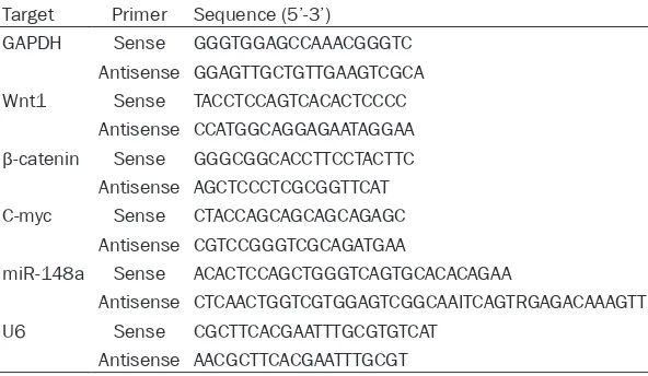
[image:2.629.100.396.94.266.2]Cell proliferation assay Table 1. Primers used for targets amplification in this study
Target Primer Sequence (5’-3’)
GAPDH Sense GGGTGGAGCCAAACGGGTC Antisense GGAGTTGCTGTTGAAGTCGCA Wnt1 Sense TACCTCCAGTCACACTCCCC
Antisense CCATGGCAGGAGAATAGGAA β-catenin Sense GGGCGGCACCTTCCTACTTC
Antisense AGCTCCCTCGCGGTTCAT C-myc Sense CTACCAGCAGCAGCAGAGC
Antisense CGTCCGGGTCGCAGATGAA
miR-148a Sense ACACTCCAGCTGGGTCAGTGCACACAGAA
Antisense CTCAACTGGTCGTGGAGTCGGCAAITCAGTRGAGACAAAGTT U6 Sense CGCTTCACGAATTTGCGTGTCAT
mixed DNA, and concentration and purity were measured using SMA 400 UV-VIS (Merinton,
Shanghai, China). The purified RNA (0.5 μg/μL)
dissolved in nuclease-free water was used for cDNA synthesis with the PrimerScript 1st Strand cDNA Synthesis Kit (Invitrogen). Complementary DNA (cDNA) was produced using reverse tran-scriptase (iScript™ cDNA Synthesis Kit; Bio-Rad Laboratories). Expressions of targets were performed in an Eppendorf Mastercycler (Brinkman Instruments, Westbury, NY) using the SYBR ExScript RT-qPCR Kit (Takara, China). Phosphoglyceraldehyde dehydrogenase (GAPDH) was chosen as the internal control for target gene expression, U6 was used as the internal control for the miRNA expression.
Primers used for targets amplification were
shown in Table 1.
Western blotting
Cells collected at 48 h after transfection were lapped with radioimmunoprecipitation assay (RIPA; Sangon Biotech) lysate containing
phenylmethanesufonyl fluoride (PMSF; Sigma),
and then were centrifuged at 12,000 rpm at 4°C for 10 min. Concentration for protein was detected using BCA protein assay kit (Pierce,
Rochford, IL). For western blotting, 50 μg
protein per cell lysate was subjected onto a 12% sodium dodecylsulfate-polyacrylamide gel electrophoresis (SDS-PAGE), followed by
trans-ferred onto a polyvinylidencefluoride (PVDF)
membrane (Mippore). The PVDF membranes were blocked in Tris-Buffered Saline Tween (TBST) containing 5% non-fat milk at room tem-perature for 1 h. Then the membranes were incubated with rabbit anti-human antibodies
(Wnt 1, β-catenin, and C-myc; 1:100 dilution,
Invitrogen) and overnight at 4°C. Then mem-brane was incubated with horseradish-peroxi-dase labeled goat anti-rat secondary antibody (1:1000 dilution) at room temperature for 1 h. Finally, the PVDF membranes were washed 3 times with 1× TBST buffer for 10 min each. The signals were detected after incubation with a chromogenic substrate using the enhanced chemiluminescence (ECL) method. Additionally, GAPDH served as the internal control.
Statistical analysis
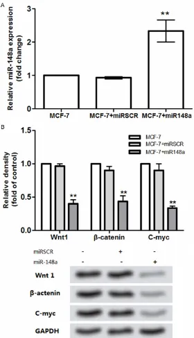
[image:3.629.104.528.79.240.2]All experiments were conducted independently for 3 times. Data are expressed as mean ± standard deviation (SD). Statistical analysis Figure 1. Expression of miR-148a in breast cancer MCF-7 cells. A: miR-148a mRNA level in MCF-7 cells was lower than that in the control. **: P<0.01, compared to the control (normal breast Hs578Bst cell); B: The relative miR-148a level is significantly increased by the overexpressed miR-148 vector transfection in MCF-7 cells. **: P<0.01, compared to the control cells (MCF-7 cell without transfection).
[image:3.629.101.295.313.459.2]for data was calculated using the graph prism 5.0 software (GraphPad Prism; San Diego, CA). Independent sample t-test was used to calcu-late the difference between two groups. The P<0.05 was considered as statistically signi-
ficant.
Results
Expression of miR-148a in breast cancer MCF-7 cells
The expression of miR-148a in MCF-7 cells was detected using the RT-PCR (Figure 1). The
results showed that miR-148a was significantly
down-regulated in MCF-7 cells compared to
the normal control cells (P<0.01; Figure 1A).
However, its expression was significantly
increased when cells were transfected with the overexpressed miR-148a vector (P<0.01; Figure 1B).
miR-148a overexpression decreased the MCF-7 cell viability
Compared to the control, cell viability was
sig-nificantly decreased by the overexpressed
miR-148a (Figure 2). No significant difference for
[image:4.629.102.527.82.466.2]the cell viability was observed between the con-trol group and the cells transfected with scram-ble miRNA.
patients with breast cancer and acts as a diag-nostic marker for breast cancer [16]. This study was aimed to investigate the possible role of miR-148a in breast cancer development and to reveal its potential mechanism. In agreement with previous data [16, 26], our results showed miR-148a overexpression induced MCF-7 cell
apoptosis
[image:5.629.101.381.79.566.2]We further analyzed the effects of miR-148a expression on breast cancer cell apoptosis (Figure 3). The results showed that the
apop-totic cells were significantly
increased by the overex-pressed miR-148a (about 6.13%) compared to the con-trol (about 2.66%) (P<0.001; Figure 3A and 3B), indicating that miR-148a expression was positively correlated to breast cancer cell apoptosis. Effects of miR-148a expres-sion on the Wnt/β-catenin signal pathway-related pro-tein expression
Since the results revealed that miR-148a expression was correlated to the breast cancer biological processes including cell viability and apoptosis, we therefore ana-lyzed the possible mecha-nism of its action (Figure 4). The results showed that the mRNA and protein levels for
Wnt1, β-catenin, and C-myc were significantly decreased
by the overexpressed miR-148a compared to the control (P<0.01; Figure 4B). However,
no significant difference for
the three proteins expression was found between the con-trol and the scramble miRNA group (Figure 4B).
Discussion
Due to the complicate molec-ular mechanism, increasing evidence has focused on the pathogenesis research for breast cancer in recent decades, including the differ-entially expressed genes, mi- RNAs and signal pathways [11, 25]. A recent evidence showed that miR-148a is down-regulated in serum from Figure 4. Influence of miR-148a expression on the Wnt/β-catenin signal
that miR-148a expression was lower than that in normal breast Hs578Bst cells (Figure 1), indicating the correlation between miR-148a expression and the pathogen of breast cancer. Accordingly, we investigated the effects of miR-148a expression on breast cell viability and apoptosis using the MCF-7 cells. Coincidence with the results presented in Figure 1, our results showed that MCF-7 cell viability was suppressed by the overexpressed miR-148a (Figure 2). Taylor et al said that miR-148a was
used to establish a cancer-specific signature
for ovarian cancer [27]. On the other hand, the results revealed that MCF-7 cell apoptosis was induced by the overexpressed miR-148a (Figure 3). Apoptosis plays a vital role in main-taining tissue homeostasis. Effects of miR-148a expression on breast cancer cell apopto-sis has been not been widely reported. However, studies have referred that the down-regulated miR-148a promotes colorectal cancer cell apoptosis by targeting Bcl-2 [28], similar results were also found in hepatocellular cancer and gastric cancer [29]. Hence, we speculated that miR-148a abnormal expression was correlated to breast cancer pathogen via involving in the cell viability and apoptosis processes.
Dysfunction of the cell apoptosis-related signal pathway has been widely considered as the piv-otal step in tumorigenesis [30]. Previous stud-ies revealed the pivotal roles of Wnt signal pathway in the tumorigenesis and the other bio-logical processes of cancers, including breast cancer [31]. Wissmann et al proved that WIF1, a component of Wnt signal pathway, was down-regulated in breast cancer [32]. Meanwhile, the activated Wnt1 and C-myc lead to the forma-tion and growth of breast tumors [33, 34]. In this study, the results showed that the Wnt1 and C-myc levels were suppressed by the over-expressed miR-148a (Figure 4). On the other
side, the role of β-catenin in breast cancer
remains controversial. Study said that the loss
of β-catenin resulted in the metastasis and
invasion of breast cancer [35], whereas Lin et
al said that the activation of β-catenin promot -ed the development of breast cancer [36]. Our
results revealed that β-catenin was highly
expressed in breast MCF-7 cells but was sup-pressed by the overexsup-pressed miR-148a. Taken together, we speculated that the overexpressed miR-148a may suppress the breast cancer
development via suppressing the activation of
Wnt/β-catenin signal pathway.
In conclusion, the data presented in our study reveals that the overexpressed miR-148a func-tions as a tumor suppressor for the develop-ment of breast cancer via involving in the cell proliferation and apoptosis processes and
inhibiting the activation of Wnt/β-catenin signal
pathway. This study may provide theoretical basis for the therapeutic target diagnosis of miR-148a in breast cancer. Further experimen-tal studies are still needed to investigate the deep molecular mechanism.
Disclosure of conflict of interest
None.
Address correspondence to: Dr. Dongmin Chang, De- partment of Surgical Oncology, The First Affiliated Hospital of Xi’an Jiaotong University, 277 Yanta West Road, Xi’an 710061, Shaanxi, China. E-mail: changdongmin5693@126.com
References
[1] Desantis CE, Lin CC, Mariotto AB, Siegel RL, Stein KD, Kramer JL, Rick A, Robbins AS and Ahmedin J. Cancer treatment and survivorship statistics, 2014. CA Cancer J Clin 2014; 64: 252-271.
[2] Wu S, Cai J, Wang H, Zhang H and Yang W. Association between 1p11-rs11249433 Polymorphism and Breast Cancer Suscepti- bility: Evidence from 15 Case-Control Studies. PLoS One 2013; 8: 800-802.
[3] Bourdeanu L and Luu T. Targeted Therapies in Breast Cancer: Implications for Advanced Oncology Practice. J Adv Pract Oncol 2014; 5: 246-260.
[4] Wright T and Mcgechan A. Breast cancer: new technologies for risk assessment and diagno-sis. Mol Diagn 2003; 7: 49-55.
[5] Nasongkla N. Biodegradable Polymeric Impla- nts as Drug Delivery Systems for Brain Cancer Therapy. IFMBE Proceedings 2009; 25/8: 11-14.
[6] Maurizio P, Marco M, Marco G, Carlotta Z, Jeff P, Pierluigi G and Stefano V. Next generation analysis of breast cancer genomes for preci-sion medicine. Cancer Lett 2013; 339: 1-7. [7] Early Breast Cancer Trialists’ Collaborative
Thürlimann B, van de Velde C, Pan H, Peto R, Davies C, Gray R. Aromatase inhibitors versus tamoxifen in early breast cancer: patient-level meta-analysis of the randomised trials. Lancet 2015; 386: 1341-52.
[8] Miller AB, Wall C, Baines CJ, Sun P, To T and Narod SA. Twenty five year follow-up for breast cancer incidence and mortality of the Canadian National Breast Screening Study: randomised screening trial. BMJ 2014; 348: 329-330. [9] Siegel R, Ma J, Zou Z, Jemal A. Cancer
statis-tics, 2014. CA Cancer J Clin 2014; 64: 9-29. [10] Ha M, Kim VN. Regulation of microRNA
biogen-esis. Nat Rev Mol Cell Biol 2014; 15: 605-610. [11] Iorio MV, Ferracin M, Liu CG, Veronese A,
Spizzo R, Sabbioni S, Magri E, Pedriali M, Fabbri M, Campiglio M, Ménard S, Palazzo JP, Rosenberg A, Musiani P, Volinia S, Nenci I, Calin GA, Querzoli P, Negrini M, Croce CM. MicroRNA gene expression deregulation in hu-man breast cancer. Cancer Res 2005; 65: 7065-7070.
[12] Li M, Julie TF and Weinberg RA. Tumour inva-sion and metastasis initiated by microRNA-10b in breast cancer. Nature 2007; 449: 682-688. [13] Gong J, Zhang JP, Li B, Zeng C, You K, Chen MX,
Yuan Y, Zhuang SM. MicroRNA-125b promotes apoptosis by regulating the expression of Mcl-1, Bcl-w and IL-6R. Oncogene 2012; 32: 3071-3079.
[14] Lombard A, Mooso B, Libertini S, Lim R, Costanzo N, Ghosh P and Mudryj M. Abstract 5239: MiR-148a promotes apoptosis in uro-thelial cell carcinoma of the bladder cells in part by targeting DNMT1. Cancer Res 2014; 74: 5239-5239.
[15] Naoya S, Yutaka N, Naohide O, Kazuhiro S, Naohiro U, Htoo ZO, Kazuyoshi Y, Kazuhiko A, Hiroki S and Wataru Y. Roles of miR-148a in gastric cancer invasion by regulating MMP7 expression. MiR-148a expression in gastric cancer tissue detected by in situ hybridization. See also Sakamoto, N., et al. (pages 236– 243 of this issue). Cancer Science 2014; 105: February cover-February cover. [16] Luo J, Zhao Q, Zhang W, Zhang Z, Gao J, Zhang
C, Li Y and Tian Y. A novel panel of microRNAs provides a sensitive and specific tool for the diagnosis of breast cancer. Mol Med Rep 2014; 10: 785-791.
[17] Joshi P, Waterhouse P, Kannan N, Narala S, Fang H, Digrappa M, Jackson H, Penninger J, Eaves C and Khokha R. RANK Signaling Amplifies WNT-Responsive Mammary Pro- genitors through R-SPONDIN1. Stem Cell Rep 2015; 9: 31-44.
[18] Montales MT, Melnyk SB, Simmen FA and Simmen RC. Maternal metabolic perturbations elicited by high-fat diet promote Wnt-1-induced mammary tumor risk in adult female offspring
via long-term effects on mammary and sys-temic phenotypes. Carcinogenesis 2014; 35: 2102-2112.
[19] Lien WH, Fuchs E. Wnt some lose some: tran-scriptional governance of stem cells by Wnt/β-catenin signaling. Genes Dev 2014; 28: 1517-1532.
[20] Fuentes RG, Toume K, Arai MA, Sadhu SK, Ahmed F and Ishibashi M. Scopadulciol, Isolated from Scoparia dulcis, Induces β-Catenin Degradation and Overcomes Tumor Necrosis Factor-Related Apoptosis Ligand Resistance in AGS Human Gastric Adenocar- cinoma Cells. J Nat Prod 2015; 78: 864-72. [21] Arendt LM, St LJ, Wronski A, Caballero S, Lyle
SR, Naber SP and Kuperwasser C. Human Breast Progenitor Cell Numbers Are Regulated by WNT and TBX3. PLoS One 2014; 9: e111442-e111442.
[22] Sreekumar A, Roarty KP and Rosen JM. The mammary stem cell hierarchy: a looking glass into heterogeneous breast cancer landscapes. Endocr Relat Cancer 2015; 22: T161-76. [23] Yogi K, Sridhar E, Goel N, Jalali R, Goel A,
Moiyadi A, Thorat R, Panwalkar P, Khire A, Dasgupta A, Shetty P and Shirsat NV. MiR-148a, a microRNA upregulated in the WNT subgroup tumors, inhibits invasion and tumori-genic potential of medulloblastoma cells by targeting Neuropilin 1. Oncoscience 2015; 2: 334-348.
[24] Arseculeratne SN, Atapattu DN, Kumarasiri R, Perera D, Ekanayake D and Rajapakse J. The use of MTT [3-(4, 5-dimethyl-2-thiazolyl)-2, 5-diphenyl-2H-tetrazolium bromide]-reduction as an indicator of the effects of strain-specific, polyclonal rabbit antisera on Candida albicans and C. krusei. Indian J Med Microbiol 2007; 25: 267-271.
[25] van de Vijver MJ, He YD, van’t Veer LJ, Dai H, Hart AA, Voskuil DW, Schreiber GJ, Peterse JL, Roberts C, Marton MJ, Parrish M, Atsma D, Witteveen A, Glas A, Delahaye L, van der Velde T, Bartelink H, Rodenhuis S, Rutgers ET, Friend SH, Bernards R. A gene-expression signature as a predictor of survival in breast cancer. N Engl J Med 2002; 347: 1999-2009.
[26] Blenkiron C, Goldstein LD, Thorne NP, Spiteri I, Chin SF, Dunning MJ, Barbosa-Morais NL, Teschendorff AE, Green AR and Ellis IO. MicroRNA expression profiling of human breast cancer identifies new markers of tumor sub-type. Genome Biol 2007; 8: 90-105.
[27] Taylor D and Gercel-Taylor C. MicroRNA signa-tures of tumor-derived exosomes as diagnostic biomarkers of ovarian cancer. Gynecol Oncol 2008; 110: 13-21.
[29] Fan KJ, Guo SL, Peng Z, Yang X, Ye H, Li ZH, Wang Y, Xu XL, Li J and Wang YL. miR-148a pro-moted cell proliferation by targeting p27 in gastric cancer cells. Int J Biol Sci 2011; 7: 567-574.
[30] Patra SK. Dissecting lipid raft facilitated cell signaling pathways in cancer. Biochim Biophys Acta 2008; 1785: 182-206.
[31] Howe LR and Brown AM. Wnt signaling and breast cancer. Cancer Biol Ther 2004; 3: 36-41.
[32] Christoph W, Peter Johannes W, Simone K, Stefan R, Robert S, Matthias W, Glen K, Jen-Chih H, Ferdinand H and Arndt H. WIF1, a com-ponent of the Wnt pathway, is down-regulated in prostate, breast, lung, and bladder cancer. J Pathol 2003; 201: 204–212.
[33] Li Y, Hively WP and Varmus HE. Use of MMTV-Wnt-1 transgenic mice for studying the genetic basis of breast cancer. Oncogene 2000; 19: 1002-1009.
[34] Hsieh TH, Tsai CF, Hsu CY, Kuo PL, Lee JN, Chai CY, Wang SC, Tsai EM. Phthalates induce prolif-eration and invasiveness of estrogen receptor-negative breast cancer through the AhR/ HDAC6/c-Myc signaling pathway. FASEB J 2012; 26: 778-787.
[35] Yoshida R, Kimura N, Harada Y, Ohuchi N. The loss of E-cadherin, α- and β-catenin expression is associated with metastasis and poor prog-nosis in invasive breast cancer. Int J Oncol 2001; 18: 513-520.