Original Article
Osthenol inhibits the viability of HCT116 cells through
suppressing PI3K/AKT signaling pathway
Biao Zhuang1, Zhijun Min1, Tangfeng Wang1, Peng Zhang2, Xiong Ni1, Huilong Qu1, Yuming Ding1
1Department of General Surgery, 2Scientific Innovation Center, Shanghai Pudong Hospital, Shanghai 201399,
People’s Republic of China
Received January 5, 2017; Accepted February 20, 2017; Epub June 1, 2017; Published June 15, 2017
Abstract: Colon cancer is one of the leading causes of cancer related deaths. The natural products show a great po-tential for anti-cancer use. The objective of the present study to explore the anti-tumor ability of osthenol in human colon cancer (HCT116) cells. The MTT assay revealed the antiproliferative activity of osthenol in a concentration de-pendent manner. And it also can promote HCT116 cells apoptosis in a concentration dede-pendent manner. Moreover
the expression of caspase-3, caspase-8 and caspase-9 in HCT116 were significant enhanced after treated with
osthenol. However the results also showed the osthenol was able to decrease HCT116 cells migration and arrest cell cycle in G1 phase. Furthermore the signal mechanism study demonstrated that the anti-tumor effect of osthe-nol was associated with PI3K/AKT signaling pathway. The current study strongly reveals that ostheosthe-nol inhibits colon cancer cells proliferation by inducing apoptosis mediated through cell cycle arrest and activation of caspases. These
findings may provide a promising method to treat the colon cancer.
Keywords: Colon cancer, apoptosis, osthenol, cell cycle, PI3K/AKT
Introduction
Colon cancer is the most common cancer in the world with an estimated 750,000 new cases diagnosed annually, and the severe cause of cancer death worldwide with an estimated 598,000 death annually [1]. More than 80% of colon cancer deaths occur in developing coun-tries. In China, colon cancer is the fourth most common cause of all cancer deaths and accounts for most of colon cancer deaths of the world [2]. Therefore, colon cancer
repre-sents a significant public health issues in devel -oping countries.
Surgery is the most common treatment approach for early stage colon cancer and have high rate of cure for patients with early stage colon cancer [3]. Unfortunately, more than 80% colon cancer cases are diagnosed in the inter-mediate or late stages with or unresectable advanced disease. The current life expectancy for patients with late stage colon cancer is only about a few months [4]. These late stage unre-sectable or metastatic colon cancers often carry a poor prognosis, and systemic therapy
with cytotoxic agents provides marginal benefit.
Because of the poor response of late stage colon cancer to systemic therapy, there has been a sense of nihilism for this disease for decades. The search for effective reagents with minimal adverse effects for the treatment of colon cancer remains the top priority of cancer research [3].
In recent decades, a number of studies have drawn attention to natural products extracted from Chinese medicinal herbs as anticancer agents in colon cancer therapy [5, 6]. Over 60% of the current anticancer drugs have their origin in one way or another from natural sources.
Nature continues to be the most prolific source
of biologically active and diverse chemotypes. Among natural products, natural coumarin rep-resent a structurally diverse group of organic compounds with potent bioactivity including antitumor effects [7]. However the antitumor mechanisms of these natural products are still not clear.
Osthenol inhibits colon cancer cells activity
3-kinase (PI3K)/AKT represent a critical path-way regulating the signaling of multiple biologi-cal processes such as apoptosis, metabolism, cell proliferation and cell growth [8]. PTEN acts as a tumor suppressor factor in AKT-mediated survival pathway via binding phosphatidyl-ino-sitol, 3, 4, 5 triphosphate (PIP3), the product of PI3K. Increase in PIP3 recruits AKT to the mem-brane, where it is activated by other kinases. Thus in the present study, we explored the anti-cancer effects of osthenol, isolated from
Angelica dahurica, in human colon cancer (HCT116) cells and its possible mechanism. Materials and methods
Materials
HPLC grade authentic standard of osthenol was purchased from Chengdu Congon Bio-tech Co., Ltd. (Sichuan, China), and dissolved in DMSO. Phospho-PTEN, PI3K, AKT, phospho-AKT, p38, phospho-p38, c-Jun, c-Fos, Bim, Bid, Puma, Bax, Bcl, Mcl-1, caspase-9, cleaved cas-pase-9, caspase-8, caspase-3, and cleaved caspase-3 anti-rabbit antibodies were pur-chased from Cell Signaling Technology (Danv-
ers, MA, USA). Loading control β-actin antibody
was purchased from CST. All other chemicals and solvents used were of the highest purity grade.
Cell culture
Human colon cancer HCT116 cell line was pur-chased from American Type Culture Collection
(ATCC, Rockville, MD). The normal colonic epi-thelial cells were obtained from Hongshun Biotechnology Company (Shanghai, China). The two cell lines were routinely cultured in RPMI 1640 medium supplemented with 10% fetal bovine serum (FBS; GIBCO/BRL, NY), 100 mg/l penicillin G and 100 U/ml streptomycin sulfate at 37°C in 5% CO2.
Cell viability assay
Cells were seeded at a 1×10⁴ density per well in 96-well plates and exposed to various
concen-trations of osthenol (0-200 μM) at different time points within 48 h. 10 μl 3-(4,5-dimethyl -thiazol-2-yl)-2,5-diphenyl tetrazolium bromide (5 mg/ml) (Sigma-Aldrich) were added to each well and cells were kept at 37°C for 4 h. The resulting formazan crystals were solubilized in 200 ml dimethyl sulfoxide (DMSO). The density of the solubilized formazan was read at 570 nm spectrophotometrically (bio-RAD, Hercules, CA, USA).
Invasion assays
Invasion assays were performed in 24-well Transwell chambers (Corning, Acton, MA, USA)
containing polycarbonate filters with 8-μm
pores coated with Matrigel (BD Biosciences,
Bedford, MA, USA). Briefly, HCT116 cells were allowed to grow to subconfluency and were
serum-starved for 48 h. After detachment with trypsin, the cells were washed with PBS, resus-pended in serum-free medium and 2×10⁴ cells were added to the upper chamber. Complete medium was added to the bottom chamber as a chemoattractant. Then, different
concentra-tions of osthenol (0, 25, 50, 100 μM) was
added to the inside of each insert and incubat-ed for 24 h in a cell culture incubator. Uninvadincubat-ed
cells on the upper surface of the filter were
mechanically removed with a cotton swab, and the invasive cells on the lower membrane
sur-face were fixed with methanol and stained with
0.1% crystal dark blue. The invading cells were counted and photographed under a microscope
at ×100 magnification. Five fields were counted per filter in each group and the experiment was
conducted in triplicate.
DAPI staining
HCT116 cells were treated with osthenol at the
final concentrations of 0, 25, 50 and 100 μM
[image:2.612.92.291.67.247.2]for 48 h, and then washed once in phosphate Figure 1. Osthenol inhibits HCT16 cells proliferation.
buffer saline (PBS) followed by fixation in cold
methanol: acetone (1:1) for 5 min. After wash-ing twice in PBS for 5 min, these cells were
stained with 4 μg/ml DAPI for 10 min at room
temperature and subsequently examined by
fluorescence microscopy (Eclipse E-800; Nikon, Tokyo, Japan). Apoptotic cells were identified by
chromatin condensation and nuclear fragment- ation.
Apoptosis assay
Annexin V-FITC/propidium iodide (PI) double-staining was per-formed with an Annexin V-FITC Kit (BDBioscience, USA). Human colon cancer (HCT116) cancer cells were treated with
differ-ent concdiffer-entrations (0, 25, 50 and 100 μM) of
osthenol for 48 h. The cells were trypsinized, rinsed twice with PBS, and resuspended in 1×binding buffer. The cells were labeled with
10 μl of FITC-conjugated annexin V and 10 μl of
propidium iodide. The cells were incubated for
20 min in dark at 37°C and then 450 μl of bind -ing buffer was added and the samples were
immediately analyzed with a flow cytometer
(Becton Dickinson, San Jose, CA). The annexin V-FITC-/PI- cell population was considered as normal, while the annexin V-FITC+/PI- and Annexin V-FITC+/PI+ cell populations were
con-sidered as indicators of early and late apoptotic cells, respectively.
Activity assay of caspases
Cells were treated with various concentrations
of osthenol (0-100 μM) for 48 h. For the activity
assay, the cell lysate was added into Protease Assay Buffer in 96-well plate. Reaction mix-tures with lysis buffer were used as negative controls. Cells treated with DMSO (0.1%) were treated as vehicle control. The reaction mix-tures were incubated for 1 h at 37°C. The AMC liberated from the substrates was measured
using spectrofluorometer of Victor 2 plate read -er (P-erkin Elm-er, Massachusetts, USA) with an excitation wave-length of 380 nm and an emis-sion wavelength of 430 nm.
Cell cycle analysis
Briefly, human colon cancer cells (1×10⁶) were seeded into each well of 6-well plates and incu-bated for 24 h for cell attachment and recovery. The cells were treated with different
concentra-tions (0, 25, 50 and 100 μM) of osthenol.
Untreated cells (control) were also incorporat-ed. After incubation for 24 h, the cells were
har-vested and fixed with ice-cold 70% ethanol (5
[image:3.612.94.520.73.327.2]mL) at -20°C for 2 h. Prior to analysis, the cells Figure 2. Osthenol promotes HCT116 cells apoptosis. A. DAPI staining showed the nucleus of HCT16 cells treated
Osthenol inhibits colon cancer cells activity
were washed with cold PBS and re-suspended
in 400 μl of PBS,20 μl PIand 20 μl RNase A. The DNA contents were recorded by a flow cytome -ter (Becton Dickinson, San Jose, CA) equipped with Cell Quest software.
Western blot analysis
Western blot assay was done as previously reported with slight modifications [9]. Bradford
assay (Bio-Rad) was used to determine the pro-tein content. After electrophoresing a total of 20-40 Ag of protein on 15% SDS-PAGE gels, it was transferred to nitrocellulose membranes. Membranes were blocked, incubated with pri-mary Abs at the suitable dose, and conse-quently incubated with primary antibody, washed and incubated with horseradish peroxi-dase conjugated secondary antibody (1:2500 dilution; Bio-Rad). Detection was performed using a chemiluminescent western detection kit (Cell Signaling Technology, Inc., Danvers, MA, USA).
Statistical analysis
Data are expressed as mean ± SD of three independent experiments. SPSS.13.0 software was used to perform statistical analysis. Differ- ences were analyzed using one-way analysis of variance (ANOVA) or two-way ANOVA. P<0.05
was considered statistically significant.
Results
Antiproliferative activity of osthenol in human colon cancer cells
To examine the inhibitory effect of osthenol on the proliferation of human colon cancer cells, MTT assay was conducted. HCT116 cells were treated with different concentrations (0, 12.5,
25, 50, 100 and 200 μM) of osthenol dissolved
in DMSO and the same volume of DMSO was used as a control. The results showed that osthenol exerted potent and dose-dependent as well as time dependent antiproliferative effects on HCT116 cancer cells after treatment for 24 and 48 h (Figure 1).
Effects of osthenol on the apoptosis of HCT116 cells
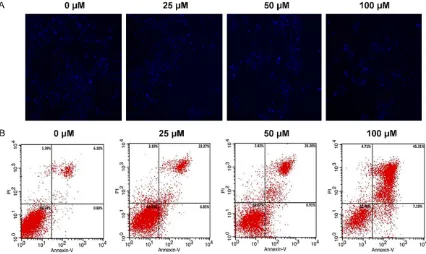
As shown in Figure 2A, DAPI staining showed the nucleus of HCT116 cells gradually reduced with the increased concentration of osthenol.
To further confirm the pro-apoptotic effect of osthenol, fluorescent Annexin V-FITC/PI double
staining was performed. HCT116 cells were treated with different concentration (0, 25, 50
and 100 μM) of osthenol for 48 h. Osthenol
induced both early and late apoptosis in a con-centration-dependent manner as compared to the untreated control cells (Figure 2B). When
[image:4.612.99.519.71.192.2]the cells were treated with 25, 50 and 100 μM
Figure 3. The expression of caspase-3, caspase-8 and caspase-9 in HCT116 cells treated with 0, 25, 50, 100 μM
osthenol respectively. *P<0.05, **P<0.01.
Figure 4. The number of invaded HCT116 cells
[image:4.612.91.287.250.395.2]for 48 h, the average proportion of Annexin V-staining positive cells (total apoptotic cells)
significantly increased from 7.06% in control to
30.12%, 33.11% and 52.5% respectively.
Osthenol induced caspase apoptosis in colon cancer cells
Osthenol induced a concentration-dependent activation of caspase-3, caspase-9, and cas-pase-8. As shown in Figure 3, the expression of caspase-3, caspase-9, and caspase-8 was increased with the increased concentration of
osthenol from 0 to 100 μM. And the expression
of caspase-3, caspase-9, and caspase-8 in
HCT 116 cells treated with 25, 50, 100 μM
osthenol were higher than HCT cells without osthenol treatments (P<0.05).
Osthenol inhibits invasion of HCT116
The effects of osthenol on HCT116 cells inva-sion were evaluated using Matrigel-coated transwell invasion assays. The results clearly
revealed that osthenol significantly inhibited
HCT116 cells invasion in a dose-dependent manner (Figure 4). The invaded HCT116 cells in
25 μM osthenol treated group were reduced
than HCT116 cells without osthenol treatment (P<0.05). Moreover the invaded HCT116 cells
in 50, 100 μM osthenol treated group were sig
-nificantly reduced than HCT116 cells without
osthenol treatment (P<0.01).
Osthenol induced G1 cell cycle arrest in HCT116 colon cancer cells
To determine the distribution of osthenol-treat-ed HCT116 cells in different phases of the cell cycle, DNA content in cells was detected by
[image:5.612.96.523.70.344.2]propidium iodide (PI) staining and flow cytome -try. The results showed that treatment with dif-ferent concentrations of osthenol for 48 h led to an increase in the population of cells in the G1 phase (apoptotic population) (P<0.01) (Figure 5). As compared to the control (Figure 5A), where 44.4% of the cells were in G1 phase,
25, 50 and 100 μM osthenol-treated cells
showed 50.9% (Figure 5B), 60.3% (Figure 5C) and 70.8% (Figure 5D) of the cells in the G1 phase (apoptotic phase) of the cell cycle.
Osthenol altered PI3K/AKT activation in HCT116 cells
To understand the effect of osthenol on PI3K/
AKT signaling cascade, we first investigated the
Figure 5. Effect of osthenol on the cell cycle arrest in human colon cancer (HCT116) cells. Cells were treated with
Osthenol inhibits colon cancer cells activity
expression level of phospho-PTEN, the phos-phorylated form of major negative regulator of the PI3K/Akt signaling pathway. Our results indicated that PTEN phosphorylation increased time-dependently following osthenol treatment in HCT116 cells (Figure 6). We also detected
that PI3K and AKT expression profiles were
both downregulated within 48 h after osthenol treatment in HCT116 cells. In addition, the phosphorylated form of AKT, which is required for its activation, was also found dephosphory-lated in HCT116 cells.
Discussion
Angelica dahurica, belonging to the family Apiaceae, is a perennial plant widely distribut-ed in China, Korea, Japan, and Russia. As a well-known herbal medicine, the roots of A. dahurica are commonly used for the treatment of headache, toothache, abscess, furunculosis, and acne. Up to date, more than 100 couma-rins have been obtained from A. dahurica, exhibiting notable and diverse pharmaceutical
properties, such as anti-tumor and, anti-inflam -matory, anti-oxidative, and acetylcholinester-ase inhibitory activities [10].
Apoptosis is a cellular suicide program that exterminates unwanted, faulty and potentially dangerous cells during the development and maintenance of cell homeostasis. Inducing apoptosis is a key tactic to eliminate cancer
cells without stimulating an inflammatory reac -tion. Regulation of apoptotic signaling
path-ways encompasses a complicated system con-sisting of several elements. Several conven-tional drugs are currently used in anticancer chemotherapy which are believed to induce cell apoptosis via activation of these elements [11, 12]. Therefore, the ability of cancer cells to induce the apoptotic program has been recog-nized as one of the major mechanisms which might serve for the development of novel approaches to treat cancer. Caspases are the vital machineries in the implementation of apoptosis [13, 14]. Usually, caspases associat-ed with apoptosis can be dividassociat-ed into the initia-tor caspases and the executioner caspases. Caspase-8 and -9 are the initiator caspases in the death receptor and the mitochondrial path-ways, respectively. Caspase-3, is the crucial executioner caspase in apoptosis pathway [15, 16]. Our results showed that osthenol induces apoptosis which is mediated through the acti-vation of caspase-3, -8 and -9. Procaspase-9, procaspase-8 and procaspase-3 decreased with the increased dose of osthenol, while as their cleaved form increased especially at high-er doses.
Dysregulation in the cell division and apoptosis are connected to the development of most can-cers. Many anticancer drugs function primarily to induce apoptosis in cancer cells and prevent tumor development [17, 18]. The morphological changes of apoptosis observed in most cell types initially start with a reduction in cell vol-ume and condensation of the nucleus [19]. In many cases extensive DNA damage age leads to activation of cell cycle check points and results in cell cycle arrest and apoptosis [20]. In the present study, we found that osthenol induced apoptosis in HCT116 colon cancer
cells as revealed by fluorescence microscopy
as well as Annexin V-FITC assay. When the cells
were treated with 25, 50 and 100 μM for 48 h,
the average proportion of Annexin V-staining
positive cells (total apoptotic cells) significantly
increased from 3.2% in control to 34.3%, 46.8% and 65.9% respectively. Further, we evaluated the effect of osthenol on cell cycle
phase distribution using flow cytometry. It was
observed that osthenol induced cell cycle arrest in the G1 phase.
[image:6.612.91.286.68.247.2]Various natural products have been reported to induce cell cycle arrest including the triter-penes, coumarins, lignans etc. Coumarins are highly multifunctional compounds and as a Figure 6. The expression of p-PTEN, AKT, p-AKT, PI3K
in HCT116 cells treated with 50 μM osthenol respec
result have promise as agents in the treatment of cancer because of their ability to block the
NF-κB activation, induce apoptosis, and pre -vent proliferation, invasion, metastasis and angiogenesis [21]. As far as osthenol is
con-cerned, we could not find reports of its antican -cer action or its effects on the cell cycle arrest. To the best of our knowledge, the current research work on this molecule has not been reported earlier and as such constitutes the
first such report.
In conclusion, we can summarize that osthenol exhibits anti-proliferative effects in HCT116 cancer cells by inducing apoptosis which is mediated by activation of caspase-3, cas-pase-8 and caspase-9. Consequently, the effect of the osthenol on survival and
stress-related pathway was described for the first time
in the literature and concluded that the altera-tions in the PI3K/AKT could lead to the apopto-sis and the alterations in these pathways might also cause by effects such as the induction of polyamine catabolism and related toxicity. Acknowledgements
This work was supported by the Pudong New Area health and Family Planning Commission of Shanghai (nos. PW2014A-28).
Disclosure of conflict of interest
None.
Address correspondence to: Biao Zhuang, Depart- ment of General Surgery, Shanghai Pudong Hospital, Shanghai 201399, People’s Republic of China. Tel: +86-18918355211; E-mail: zhuangb2005@163. com
References
[1] Siegel R, Naishadham D and Jemal A. Cancer statistics, 2013. CA Cancer J Clin 2013; 63: 11-30.
[2] Chen W, Zheng R, Zeng H, Zhang S and He J. Annual report on status of cancer in China, 2011. Chin J Cancer Res 2015; 27: 2-12. [3] Wang L, Yeung JH, Hu T, Lee WY, Lu L, Zhang L,
Shen J, Chan RL, Wu WK and Cho CH. Dihydrotanshinone induces p53-independent but ROS-dependent apoptosis in colon cancer cells. Life Sci 2013; 93: 344-351.
[4] Wang L, Hu T, Shen J, Zhang L, Chan RL, Lu L, Li M, Cho CH and Wu WK. Dihydrotanshinone I induced apoptosis and autophagy through cas-pase dependent pathway in colon cancer. Phytomedicine 2015; 22: 1079-1087.
[5] Chen J, Shi DY, Liu SL and Zhong L. Tanshinone IIA induces growth inhibition and apoptosis in gastric cancer in vitro and in vivo. Oncol Rep 2012; 27: 523.
[6] Li N, Fan LL, Sun GP, Wan XA, Wang ZG, Wu Q and Wang H. Paeonol inhibits tumor growth in gastric cancer in vitro and in vivo. World J Gastroenterol 2010; 16: 4483-4490.
[7] Petronelli A, Pannitteri G and Testa U. Triterpenoids as new promising anticancer drugs. Anticancer Drugs 2009; 20: 880-892. [8] Kim HA, Kim KJ, Seo KH, Lee HK and Im SY.
PTEN/MAPK pathways play a key role in plate-let-activating factor-induced experimental pul-monary tumor metastasis. FEBS Lett 2012; 586: 4296-4302.
[9] Zhang XD, Franco A, Myers K, Gray C, Nguyen T and Hersey P. Relation of TNF-related apopto-sis-inducing ligand (TRAIL) receptor and FLICE-inhibitory protein expression to TRAIL-induced apoptosis of melanoma. Cancer Res 1999; 59: 2747-2753.
[10] Yang WQ, Song YL, Zhu ZX, Su C, Zhang X,
Wang J, Shi SP and Tu PF. Anti-inflammatory
dimeric furanocoumarins from the roots of Angelica dahurica. Fitoterapia 2015; 105: 187-193.
[11] Earnshaw WC. Nuclear changes in apoptosis. Curr Opin Cell Biol 1995; 7: 337-343.
[12] Kundu T, Dey S, Roy M, Siddiqi M and Bhattacharya RK. Induction of apoptosis in hu-man leukemia cells by black tea and its
poly-phenol theaflavin. Cancer Lett 2005; 230:
111-121.
[13] Chiarugi P and Giannoni E. Anoikis: a neces-sary death program for anchorage-dependent cells. Biochem Pharmacol 2008; 76: 1352-1364.
[14] Kerr JF, Wyllie AH and Currie AR. Apoptosis: a basic biological phenomenon with wide-rang-ing implications in tissue kinetics. Br J Cancer 1972; 26: 239.
[15] Kantari C and Walczak H. Caspase-8 and bid: caught in the act between death receptors and mitochondria. Biochim Biophys Acta 2011; 1813: 558-563.
[16] Li J and Yuan J. Caspases in apoptosis and be-yond. Oncogene 2008; 27: 6194-6206. [17] Khoo BY, Chua SL and Balaram P. Apoptotic
ef-fects of chrysin in human cancer cell lines. Int J Mol Sci 2010; 11: 2188-2199.
[18] Li W, Wang J, Jiang HR, Xu XL, Zhang J, Liu ML and Zhai LY. Combined effects of cyclooxygen-ase-1 and cyclooxygenase-2 selective inhibi-tors on ovarian carcinoma in vivo. Int J Mol Sci 2011; 12: 668-681.
Osthenol inhibits colon cancer cells activity
of apoptosis by Garcinia mangostana (mango-steen) on SKBR3 human breast cancer cell line. J Ethnopharmacol 2004; 90: 161-166. [20] Pietenpol JA and Stewart ZA. Cell cycle
check-point signaling: cell cycle arrest versus apopto-sis. Toxicology 2002; 181: 475-481.