Original Article
Complement activation fragment C3a is a sensitive
biomarker for patients with recurrent miscarriage
Mei Chen, Li-Ting Wang, Peng-Sheng Zheng
Department of Reproductive Medicine, First Affiliated Hospital, Medical College of Xi’an Jiaotong University, Xi’an, People’s Republic of China
Received January 11, 2017; Accepted January 26, 2017; Epub April 1, 2017; Published April 15, 2017
Abstract: Purpose: This study aimed to investigate complement activation in recurrent miscarriage (RM) and to identify biomarkers for RM. Methods: A total of 108 female patients who were diagnosed with RM and 120 control female volunteers for reproductive health screening were included in this study. ELISA was used to measure plasma levels of the complement activation fragments C3a, C4a, C5a, and serum levels of anticardiolipin antibodies and
anti-β2 glycoprotein I. Nephelometric immunoassays were used to measure serum C3 and C4 levels. Results: All complement activation fragment (C3a, C4a and C5a) levels were significantly higher in RM patients than in controls
(P < 0.05). Only C3a levels were significantly higher in RM patients with normal C3 or C4 levels than in controls
(P < 0.05). C3a levels were significantly higher in RM patients with or without antiphospholipid antibodies than in
controls (P < 0.05). Conclusion: Complement activation is a common event that occurs in RM patients. Additionally, the complement activation fragment C3a is a sensitive biomarker for RM patients.
Keywords: Recurrent miscarriage, antiphospholipid syndrome, antiphospholipid antibodies, complements activa-tion, C3a
Introduction
Complement is one of the first lines of
de-fense in innate immunity and is important for cellular integrity, tissue homeostasis, and modifying the adaptive immune response [1]. Complement-mediated innate immune function not only recognizes and eliminates infectious agents, but also controls homeostatic process-es, such as clearance of cellular debris and apoptotic cells. Generally, there are three path-ways for complement activation: the classical pathway, the lectin pathway, and the alternative pathway. The classical pathway is primarily acti-vated by antigen-antibody complexes, and the other two pathways are activated by carbohy-drate groups and bacterial surfaces [2]. Activa- tion of the complement cascade leads to op- sonization of the target by C3b/C4b/C5b, fol-lowed by generation of the lytic membrane at-
tack complex, which releases pro-inflammatory
anaphylatoxins (C3a, C4a, and C5a) in blood to attract leukocytes. Therefore, the complement activation fragments C3a, C4a, and C5a in
blood may reflect the status of complement
activation in the whole body.
Recently, in recurrent miscarriage (RM) pa- tients with antiphospholipid syndrome (APS) had been found to be implicated with com- plement activation [3-6], Complement activa-tion is required for antiphospholipid antibo- dies (APLS)-induced fetal loss [3, 5, 7]. Fur- thermore, heparin prevents APLS-induced fe- tal loss by inhibiting complement activation [4, 6]. These studies suggest that complement activation is a critical event for APLS-induced fetal loss or RM.
RM is traditionally defined as three or more
Materials and methods
Patients
This prospective study was performed during July 2009 to December 2013. A total of 108 female patients (range, 20-36 years old) who were diagnosed with RM were included. We also included 120 control female volunteers for reproductive health screening who were recruit-ed from the outpatient clinic of the Department
of Reproductive Medicine at the First Affiliated
Hospital of Xi’an Jiaotong University Medical College.
RM was diagnosed according to the history of patients who had experienced three or more consecutive spontaneous miscarriages within 12 weeks of gestation [13]. All of the patients received standard evaluations consisting of thrombophilia screening, parental karyotypic evaluation, endocrine screening, and an ultra-sonographic examination for possible uterine anomalies. Women with any thrombophilic, en- docrine, karyotypic, or anatomical abnormali-ties were excluded from the patient group. All healthy female volunteers with at least one live birth and no history of pregnancy loss were enrolled as controls from the population who came for healthy screening. There was no
sig-nificant difference in age between the patient
and control groups (Table 1). None of the par-ticipants had complications associated with infection, malignancy, impaired circulation or tissue ischemia, a history of cigarette smok- ing and alcohol consumption, or exposure to any harmful substance in the recent 2 months prior to the date of blood collection. The study
h) was obtained for serum and plasma collec-tion on the 3rd day of the menstrual cycle to minimize hormonal effects, and after 3 months of their last miscarriage or delivery. For serum samples, venous blood was collected after 1500× g centrifugation for 10 min at room tem-perature, and samples were stored at -80°C until later analysis. For plasma samples, venous blood was collected in 3.8% sodium citrate (9:1) and centrifuged twice at 2000× g for 15 min at 4°C. Obtained plasma was stored at -80°C until later analysis. Repeated freezing and thawing of specimens was avoided, and plasma was stored before performing enzyme-linked immunoassay (ELISA) for complement activation within 1 year.
Detection of C3a, C4a, C5a, C3, and C4 levels
Plasma C3a, C4a, and C5a levels were mea-sured with an ELISA using monoclonal anti-
bodies specific to human C3a-desArg,
C4a-desArg, and C5a-C4a-desArg, with low detection limits of 0.007 ng/ml, 0.006 ng/ml, and 0.047 ng/ml, respectively. Immunoassay systems for C3a (human C3a BD OptEIATM ELISA), C4a (hu-
man C4a BD OptEIATM ELISA), and C5a (human
C5a BD OptEIATM ELISA II) were obtained from
BD Biosciences (San Jose, CA, USA). Comple- ment C3a, C4a, and C5a assays were per-formed with 1000× diluted plasma according to the manufacturer’s instructions. The plates were read at 450 nm by a Bio-Rad microplate reader with the wavelength correction set at 570 nm (iMarkTM Microplate Absorbance Read-
er; Bio-Rad Laboratories, Hercules, CA, USA). The amount of C3a, C4a, and C5a in each
[image:2.612.92.377.97.240.2]plasma sample was quantified by interpolation
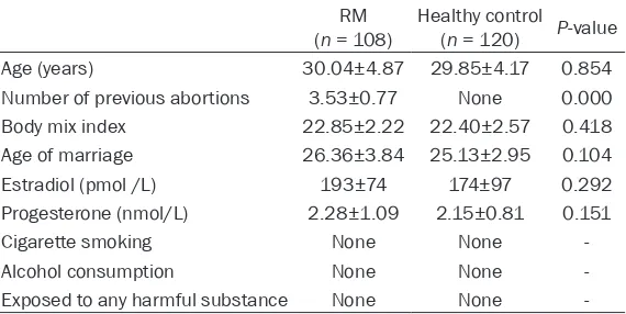
Table 1. Baseline characteristics of controls and recurrent miscar-riage (RM) patients
RM
(n = 108) Healthy control (n = 120) P-value
Age (years) 30.04±4.87 29.85±4.17 0.854
Number of previous abortions 3.53±0.77 None 0.000
Body mix index 22.85±2.22 22.40±2.57 0.418
Age of marriage 26.36±3.84 25.13±2.95 0.104
Estradiol (pmol /L) 193±74 174±97 0.292
Progesterone (nmol/L) 2.28±1.09 2.15±0.81 0.151
Cigarette smoking None None
-Alcohol consumption None None
-Exposed to any harmful substance None None
-The independent samples t-test was used for analysis.
was conducted in accord- ance with the Declaration of Helsinki and the Principles of Good Clinical Practice. The study procedures were ap- proved by the institutional re- view board and ethics com-mittee, and written informed consent was obtained from all participants.
Collection of serum and plas-ma samples
from individual standard curves composed of
purified human C3a, C4a, or C5a. The calculat
-ed inter- and intra-assay coefficients of varia -tion were all less than 10%. Serum C3 and C4 levels were determined with nephelometric immunoassays using goat anti-human C3 and goat anti-human C4 (Maike BioTeck, Sichuan, China), with normal ranges of 0.8-1.8 g/L and 0.2-0.4 g/L, respectively.
Anti-phospholipid antibody testing
Lupus anticoagulant (LA), anticardiolipin
anti-bodies (ACAs, IgG and IgM), and anti-β2 gly-coprotein I (anti-β2 GPI, IgG and IgM) were rec -ognized as APS-related autoantibodies in the revised international consensus statement for
definite APS. Serum levels of ACAs (IgG and IgM) and anti-β2 GPI (IgG and IgM) were
de-tected with an ELISA test kit (Euroimmun Me- dizinische Labordiagnostika AG, Lubeck, Ger- many). All of the procedures were performed according to the manufacturer’s instructions. The plates were read at 450 nm by a Bio- Rad microplate reader (Bio-Rad). The levels of
serum ACAs and anti-β2-GPI were quantified
with standard curves. The normal range of ACAs IgG and IgM is below 12 RU/ml (positive
sample ≥ 12 RU/ml), and the normal range of anti-β2-GPI IgG and IgM is below 20 RU/ml (positive sample ≥ 20 RU/L). LA was detected using a panel of two tests, the simplified dilute
Russell’s viper venom time test and an LA- sensitive test for activated partial thrombo-
plastin time (APTT). The simplified dilute
Rus-sell’s viper venom time test was carried out with a kit from Siemens (Siemens, Germany) using the CA-1500 coagulation Analyzer (Sys- mex Corporation, Japan). The LA-sensitive test for APTT was performed with a kit from Sys-
mex Corporation. LA was finally determined to
be present or absent based on mixing studies and phospholipid dependence. ACAs (IgG and
IgM), anti-β2 GPI (IgG and IgM), and LA needed
to be present in a high titer on two or more occasions at least 12 weeks apart.
Detection of D-dimer and fibrin (ogen) degra-dation product
A hypercoagulation state or thrombus forma-tion is recognized as a common event in RM. Fibrin (ogen) degradation product (FDP) and D-dimer are two classic markers of a hyper- coagulation state. After collection of plasma, D-dimer and FDP levels were immediately de- termined. D-dimer and FDP levels were
mea-sured by the Bead solidification method using
a blood coagulation analyzer (C2000-4; Stago, France). The normal range of D-dimer levels is 0-1.0 mg/L (positive sample > 1.00 mg/L) and the normal range of FDP levels is 0-5 mg/L (positive sample > 5.00 mg/L). All of the proce-dures were performed according to the manu-facturer’s instructions.
Statistical analysis
Statistical analysis was performed using SPSS software version 13.0 (SPSS Inc., Chicago, IL). The unpaired Student’s t-test, ANOVA, and chi-squared tests were used as appropriate. Con- tinuous variables are shown as the mean ± SD.
P < 0.05 was defined as statistically
signifi-cant. Results
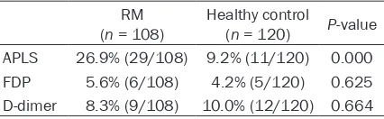
APLS, D-dimer, and FDP levels in RM patients and controls
LA/anti-β2-GPI and/or ACAs (IgG or IgM) were
found more frequently in RM patients (26.9%, 29/108) compared with controls (9.2%, 11/ 120, P < 0.001, Table 2). This finding suggest -ed that APLS were a trigger for a small part of RM. Only 26.9% RM patients suffered from APS in our study. Furthermore, there were no
significant differences in the levels of D-dimer
(P = 0.625) and FDP (P = 0.664) between RM patients and controls, suggesting that D-dimer and FDP could not be used as a monitoring tool for RM.
Complements and their activation fragments in RM patients
There was no significant difference in C4 levels
between RM patients (0.24±0.09 g/L) and con-Table 2. Positive rates of antiphospholipid
anti-bodies (APLS), Fibrin(ogen) degradation product (FDP) and D-dimer levels in controls and recur-rent miscarriage (RM) patients
RM
(n = 108) Healthy control (n = 120) P-value APLS 26.9% (29/108) 9.2% (11/120) 0.000
FDP 5.6% (6/108) 4.2% (5/120) 0.625
D-dimer 8.3% (9/108) 10.0% (12/120) 0.664
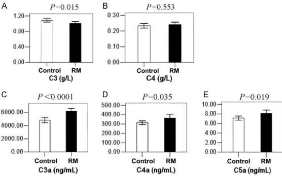
[image:3.612.91.304.120.186.2]trols (0.23±0.08 g/L, P = 0.553). However, C3
levels were significantly lower in RM patients
(1.01±0.24 g/L) than in controls (1.09±0.26 g/L, P = 0.015). These findings suggested that
C3, but not C4, was a sensitive indicator of immune reactions in RM patients. Moreover, C3a (6189±2126 ng/ml vs. 4815±2177 ng/
ml, P < 0.001), C4a (362±214 ng/ml vs. 314±
109 ng/ml, P = 0.035), and C5a (8.12±3.62
C3, C3a, C4a and C5a levels were compared between 120 controls and 72 RM patients with normal C4 levels (Figure 3 and Supplementary Table 3). There were no significant differences
[image:4.612.93.371.74.251.2]in C3, C4a, and C5a levels between the two groups. Only C3a was sensitive enough to dis-tinguish RM patients (6285±2047 ng/ml) with normal C4 levels from controls (4815±2177 ng/ml, P < 0.001). This finding indicated that
[image:4.612.93.372.334.534.2]Figure 1. Serum levels of complement C3, C4, C3a, C4a and C5a in recurrent miscarriage patients and controls. The levels of C3 (A), C4 (B), C3a (C), C4a (D) and C5a (E) in recurrent miscarriage patients and controls are shown as mean ± SD. The P-values are marked on the top of each bar. RM, recurrent miscarriage.
Figure 2. The comparison of C3, C3a, C4a and C5a between the healthy con-trols and recurrent miscarriage patients with normal C3 level. Comparisons of C3 (A), C3a (B), C4a (C) and C5a (D) levels between healthy controls and recurrent miscarriage patients with normal C3 level. P-values are marked on the top of the bar. RM, recurrent miscarriage.
ng/ml vs. 7.15±2.42 ng/ml,
P = 0.019) were significantly
elevated in RM patients com-pared with controls (Figure 1 and Supplementary Table 1).
Taken together, these findings
suggested that complement activation fragments (C3a, C4a, and C5a), but not com-plements (C3 and C4), were more sensitive for measur- ing complement activation in RM, although, C3 levels, but
not C4 levels, were signifi -cantly different between RM patients and controls.
C3a is a sensitive biomarker for RM patients with normal C3/C4 levels
To determine whether C3a, C4a, and C5a are sensitive enough to distinguish RM pa- tients from controls with nor-mal C3 levels, plasma levels of C3a, C4a, and C5a were compared between 120 con-trols and 89 RM patients with normal C3 levels (Figure 2 and Supplementary Table 2).
There were no significant dif -ferences in C3, C4a, and C5a levels between the two groups
(P > 0.05). However, C3a
lev-els were significantly higher
in RM patients with normal C3 levels (6289±2084 ng/ml) than in controls (4815±2177 ng/ml, P < 0.001). This finding
C3a was a sensitive biomarker for RM patients with normal C4 levels.
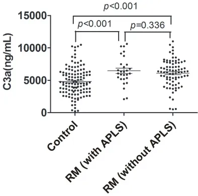
C3a is a sensitive biomarker not only for RM patients with APS, but also for RM patients without APS
RM patients with any one of the APLS (LA,
anti-β2-GP-1 IgG or IgM, ACAs IgG or IgM) may
ment activation was a common biomarker of RM patients, regardless of whether they had APLS.
Discussion
RM affects up to 1-5% of all pregnant couples and may be induced by multiple etiologies. To identify some common characteristics in most RM patients, RM patients with anatomical uter-ine abnormalities, endocruter-ine disorders, as well as chromosomal disorders in any of the par-ents, were excluded from this study. Therefore, in RM patients who were recruited in this study, the cause of RM was mainly attributed to AP- LS and unexplained etiology. Actually, only 29 of 108 (26.9%) RM patients in this study suf-fered from APS according to recommended APS guidelines by Jauniaux et al [14]. Comple- ment activation fragment C3a levels were much more elevated in RM patients with APS than those in controls, which is consistent with many previous studies [3]. Complement acti- vation is required in APS-induced fetal loss [3, 5, 15], and inhibiting complement activation through heparin treatment prevents APS-rela-
ted pregnancy loss [6]. These findings suggest
that complement activation is a critical pa- thophysiological process in APLS-induced fetal loss. Our results indicated that complement activation is a common event in RM patients
[image:5.612.93.371.74.263.2]with APS. Furthermore, for the first time, our
Figure 3. The comparison of C3, C3a, C4a and C5a between the healthy con-trols and recurrent miscarriage patients with normal C4 level. Comparisons of C3 (A), C3a (B), C4a (C) and C5a (D) levels between healthy controls and recurrent miscarriage patients with normal C4 level. P-values are marked on the top of each bar. RM, recurrent miscarriage.
Figure 4. The comparison of C3a among healthy con-trols, recurrent miscarriage patients with or without APLS. The level of C3a in the healthy controls and recurrent miscarriage patients with or without APLS are shown as above. P-values are marked on the top of each bar. RM, recurrent miscarriage.
be diagnosed as having APS. In the present study, C3a
levels were significantly
ele-vated in 29 RM patients with APLS (6499±2156 ng/ml) compared with 120 controls (4815±2177 ng/ml, P < 0.001), furthermore, C3a
lev-els were significantly higher
in 79 RM patients without APLS (6075±2118 ng/ml) than in the 120 controls (P < 0.001). There was no differ-ence in C3a levels between RM patients with and without APLS (P = 0.366) (Figure 4 and Supplementary Table 4). This result is in accordance with a previous study, which showed that complement was activated in RM patients with
[image:5.612.90.287.348.543.2]comple-study showed that complement activation frag-ment C3a levels were more highly elevated in RM patients without APS than those in con-
trols. This finding indicated that complement
activation is an event that not only occurs in RM patients with APS, but also occurs in patients without APS. Therefore, our study suggests that complement activation may be an important process in all cases of RM.
Complement components and their activated fragments have been evaluated as diagnostic and predictive markers for several diseases and outcomes of pregnancy [4, 12, 16-19]. In our study, we systemically compared C3 and C4, and the complement activation fragments C3a, C4a, and C5a in RM patients and con- trols. We found that C3a could distinguish all RM patients from controls, regardless of whe- ther RM patients suffered from APS, or whe- ther they had low or normal C3 or C4 levels. Therefore, the complement fragment C3a is the most sensitive marker to evaluate comple-ment activation in RM patients.
A hypercoagulation state or thrombus forma-tion has been recognized as a common event in RM. However, FDP and D-dimer, two classic markers of the hypercoagulation state, were not different between RM patients and controls
in our study. This finding suggests that these
markers cannot be used to monitor RM. C3a and C5a are two necessary complement ac- tivation components that are released from all complement activation pathways. Because C5a is rapidly degraded and has a short half-life in vitro, C5a is difficult to use as a monitor
-ing tool. For the first time, our study indicated
that C3a was the most sensitive indicator to evaluate complement activation in patients with RM. More studies are required in the fu- ture to determine if C3a is an ordinary and sen-sitive indicator that can used for monitoring development and progression of RM patients with and without APS.
In conclusion, our study shows that activation of complements is universally elevated in pa- tients with RM. Additionally, the complement activation fragment C3a is an effective and
sensitive biomarker in RM patients. This
find-ing provides a novel possibility to explore com-plement C3a as a clinical tool to evaluate the development and progression of RM.
Acknowledgements
This work was supported by the National Natural Science Foundation of China to Prof. Peng-Sheng Zheng for a general grant (No: 81270751).
Disclosure of conflict of interest
None.
Address correspondence to: Dr. Peng-Sheng Zheng,
Department of Reproductive Medicine, First
Affili-ated Hospital, Medical College of Xi’an Jiaotong University, Xi’an 710061, People’s Republic of China. Tel: +86-29-82657874; Fax: +86-29-8532- 4013; E-mail: [email protected]
References
[1] Ricklin D, Hajishengallis G, Yang K and Lam- bris JD. Complement: a key system for immune surveillance and homeostasis. Nat Immunol 2010; 11: 785-797.
[2] Nauta AJ, Roos A and Daha MR. A regulatory role for complement in innate immunity and autoimmunity. Int Arch Allergy Immunol 2004; 134: 310-323.
[3] Cohen D, Buurma A, Goemaere NN, Girardi G, le Cessie S, Scherjon S, Bloemenkamp KW, de Heer E, Bruijn JA and Bajema IM. Classical complement activation as a footprint for mu-rine and human antiphospholipid antibody-in-duced fetal loss. J Pathol 2011; 225: 502-511. [4] Sugiura-Ogasawara M, Nozawa K, Nakanishi
T, Hattori Y and Ozaki Y. Complement as a pre-dictor of further miscarriage in couples with recurrent miscarriages. Hum Reprod 2006; 21: 2711-2714.
[5] Holers VM, Girardi G, Mo L, Guthridge JM, Molina H, Pierangeli SS, Espinola R, Xiaowei LE, Mao D, Vialpando CG and Salmon JE. Complement C3 activation is required for an-tiphospholipid antibody-induced fetal loss. J Exp Med 2002; 195: 211-220.
[6] Girardi G, Redecha P and Salmon JE. Heparin prevents antiphospholipid antibody-induced fetal loss by inhibiting complement activation. Nat Med 2004; 10: 1222-1226.
[7] Salmon JE, Girardi G and Holers VM. Activation of complement mediates antiphospholipid an-tibody-induced pregnancy loss. Lupus 2003; 12: 535-538.
[8] Stirrat GM. Recurrent miscarriage. II: clinical associations, causes, and management. Lan- cet 1990; 336: 728-733.
Grudzinskas JG and Hustin J. The Euro-Team Early Pregnancy (ETEP) protocol for recurrent miscarriage. Hum Reprod 1995; 10: 1516-1520.
[10] Coulam CB, Clark DA, Beer AE, Kutteh WH, Silver R, Kwak J and Stephenson M. Current clinical options for diagnosis and treatment of recurrent spontaneous abortion. Clinical guidelines recommendation committee for di-agnosis and treatment of recurrent spontane-ous abortion. Am J Reprod Immunol 1997; 38: 57-74.
[11] Bricker L and Farquharson RG. Types of preg-nancy loss in recurrent miscarriage: implica-tions for research and clinical practice. Hum Reprod 2002; 17: 1345-1350.
[12] Micheloud D, Sarmiento E, Teijeiro R, Jensen J, Rodriguez Molina JJ, Fernandez-Cruz E and Carbone J. Hypocomplementemia in the ab-sence of autoantibodies in women with recur-rent pregnancy loss. Allergol Immunopathol (Madr) 2007; 35: 90-94.
[13] Practice Committee of American Society for
Reproductive Medicine. Definitions of infertility
and recurrent pregnancy loss: a committee opinion. Fertil Steril 2013; 99: 63.
[14] Jauniaux E, Farquharson RG, Christiansen OB and Exalto N. Evidence-based guidelines for the investigation and medical treatment of re-current miscarriage. Hum Reprod 2006; 21: 2216-2222.
[15] Caucheteux SM, Kanellopoulos-Langevin C and Ojcius DM. At the innate frontiers between mother and fetus: linking abortion with com-plement activation. Immunity 2003; 18: 169-172.
[16] Oku K, Atsumi T, Bohgaki M, Amengual O, Kataoka H, Horita T, Yasuda S and Koike T. Complement activation in patients with prima-ry antiphospholipid syndrome. Ann Rheum Dis 2009; 68: 1030-1035.
[17] Mankee A, Petri M and Magder LS. Lupus anti-coagulant, disease activity and low
comple-ment in the first trimester are predictive of
pregnancy loss. Lupus Sci Med 2015; 2: e000095.
[18] Madhukaran SP, Alhamlan FS, Kale K, Vatish M, Madan T and Kishore U. Role of collectins and complement protein C1q in pregnancy and parturition. Immunobiology 2016; 221: 1273-1288.
Supplementary Table 1. C3, C4, C3a, C4a, and C5a levels in controls and recurrent miscarriage (RM) patients
RM
(n = 108) Healthy control (n = 120) P-value
C3 (g/L) 1.01±0.24 1.09±0.26 0.015
C4 (g/L) 0.24±0.09 0.23±0.08 0.553
C3a (ng/ml) 6189±2126 4815±2177 0.000
C4a (ng/ml) 362±214 314±109 0.035
C5a (ng/ml) 8.12±3.62 7.15±2.42 0.019
The independent samples t-test was used for analysis.
Supplementary Table 2. C3, C3a, C4a, and C5a levels in controls and recurrent miscarriage (RM) patients with normal C3 levels
RM with normal
C3 (n = 89) Healthy control (n = 120) P-value
C3 (g/L) 1.07±0.22 1.09±0.26 0.626
C3a (ng/ml) 6289±2084 4815±2177 0.000
C4a (ng/ml) 350±205 314±109 0.132
C5a (ng/ml) 7.96±3.59 7.15±2.42 0.066
The independent samples t-test was used for analysis.
Supplementary Table 3. C3, C3a, C4a, and C5a levels in controls and recurrent miscarriage (RM) patients with normal C4 levels
RM with normal
C4 (n = 72) Healthy control (n = 120) P-value
C3 (g/L) 1.07±0.25 1.09±0.26 0.607
C3a (ng/ml) 6285±2047 4815±2177 0.000
C4a (ng/ml) 338±220 314±109 0.385
C5a (ng/ml) 8.00±3.90 7.15±2.42 0.096
The independent samples t-test was used for analysis.
Supplementary Table 4. C3a levels in controls and in recurrent miscarriage (RM) patients with or without antiphospholipid antibodies (APLS)
RM with
APLS (n = 29) APLS (RM without n = 79) Healthy control (n = 120) C3a (ng/ml) 6499±2156 6075±2118 4815±2177
One way ANOVA was used for analysis. RM patients with APLS ver-sus controls: P < 0.001. RM patients without APLS versus controls:
P < 0.001. RM patients with APLS versus RM patients without APLS: