Original Article
Enhanced expression of stem cell markers and drug
resistance in sphere-forming non-small cell lung
cancer cells
Feng-Feng Sun1*, Yong-He Hu2*, Lv-Ping Xiong1, Xiao-Yun Tu1, Ji-Hua Zhao1, Sheng-Song Chen1, Juan Song1,
Xiao-Qun Ye1
1Department of Respiratory Diseases, The Second Affiliated Hospital of Nanchang University, Nanchang 330006, China; 2The General Hospital of Chengdu Military Region, Chengdu 610083, Sichuan Province, China. *Equal con -tributors.
Received March 16, 2015; Accepted May 17, 2015; Epub June 1, 2015; Published June 15, 2015
Abstract: There is growing evidence suggesting that cancer stem cells (CSCs) are playing critical roles in tumor pro-gression, metastasis and drug resistance. However, the role of CSCs in non-small cell lung cancer (NSCLC) remains elusive. In this study, we enriched for stem-like cells from tumor spheres derived from NSCLC cell line A549 cultured in serum-free medium. Our results showed that sphere-derived cells expressed various stem cell markers such as CD44, CD133, Sox2 and Oct4. Compared with the corresponding cells in monolayer cultures, sphere-derived cells showed marked morphologic changes and increased expression of the stem cell markers CD133. Furthermore, we found that sphere-derived cells exhibited increased proliferation, cell-cycle progression as well as drug-resistant properties as compared to A549 adherent cells. Consistently, expression of several drug resistance proteins,
includ-ing lung resistance-related protein (LRP), glutathion-S-transferase-π (GST-π) and multidrug resistance proteins-1 (MRP1) were all significantly enhanced in sphere-derived cells. These results indicate the enrichment of CSCs in
sphere cultures and support their role in regulating drug resistance in NSCLC.
Keywords: Cancer stem cell, multidrug resistance, CD133, MRP1, LRP, GST-π
Introduction
Lung cancer is the leading cause of cancer-related mortality in the world with 1.3 million deaths worldwide annually [1, 2]. In spite of cur-rent developments in multidisciplinary treat-ment strategies and an expanding panel of che-motherapy agents, the chemoresistance of lung cancer remains a major unresolved clinical and scientific problem. Data from the research -es reveals that chemor-esistance to carboplatin and cisplatin was documented in 68% and 63% in non-small cell lung cancer (NSCLC), respec-tively [3], and often recurs with a chemoresis-tance phenotype shortly in approximately 80% of small cell lung cancer (SCLC) cases [4]. Although some patients have a good initial response, all lung cancers will eventually devel-op resistance to the chemotherapeutic agents to which they are exposed [5]. Taking into
account the chemoresistance is a ringleader to the tumor relapse after chemotherapeutic ther-apy, refined investigation on the mechanisms of chemoresistance is urgently desired in lung cancer to improve survival rate.
colorectal and ovarian carcinoma [13, 14]. According the reports, the mechanisms respon-sible for the multidrug resistance in these CSCs was involved in increasing efflux of drug, enhancing repair/increasing tolerance to DNA damage, high antiapoptotic potential, decreas-ing permeability and enzymatic deactivation. In addition, some molecular characterizations associated with drug resistance, such as over expression ABCA2 and ABCG2 of the ATP-binding cassette (ABC) transporters, and heat shock proteins (Hsps), were also revealed in side population or stem-like cells derived from lung cancer by researchers over the few years [15-17]. However, Apart from these important results, the actual mechanisms underlying the chemoresistance of lung cancer stem or initia-tion cells are not fully understood thus far. Due to the central role performed by CSCs in the recurrence of cancer after chemotherapy, Understanding the mechanisms of drug resis-tance in this lung cancer subpopulation may improve the results of treatment.
In this study, after the isolation and identifica -tion of clusters of lung stem-like cells growing as floating tumorospheres from the A549 lung cancer cell line, some of these features, such as the capability of cell proliferation and resist apoptosis, the half-maximal inhibitory concen-tration (IC50) study to first-line chemotherapy agents (cisplatin, DDP; and gemcitabine, GEM), the Cell Cycle and the expression of multidrug resistance-associated protein 1 (MRP1) mRNA, Lung resistance-related protein (LRP) mRNA and glutathion-S-transferase-π (GST-π) protein were examined. Our results indicate that, com-paring to A549 lung cancer cell line, lung can-cer stem cells (LCSCs) have more capability of anti-apoptotic and drug resistance. Selective targeting of LCSCs could help to improve the development of novel therapeutics for lung cancer.
Materials and methods
Cells lines and tumorsphere culture
A549 human lung carcinoma cells (ATCC) were grown in Dulbecco’s modification of eagle’s medium (DMEM, Hyclone, USA) supplemented with 10% fetal bovine serum (FBS, Gibco, USA), in a 37°C humidified atmosphere with 5% CO2. To generate suspended and stem-like sphere
growing cells, adherent cells were dissociated into a single-cell and seeded in 6-well plates at 1 × 10³ cells/well with DMEM supplemented
with 10% fetal FBS for 2 weeks. Thereafter, holoclones [18] (50-200 cells) were isolated using cloning cylinders (Corning, USA) and cul-tured with serum-free stem cell medium con-taining DMEM/F12 (Gibco), B27 (1×, Gibco), recombinant human epidermal growth factor (rhEGF, 20 ng/ml; Sigma, USA), basic fibroblast growth factor (bFGF, 20 ng/ml; Upstate, USA), and insulin (4 U/l; Sigma) for another 2-3 weeks. The medium is referred to as ‘Cancer Stem Cell medium’ (CSM). After primary tumor spheres reached approximately 100-200 cells/sphere, the spheres were dissociated and single cells were cultured for another 1-2 weeks with CSM until form secondary spheres. The secondary spheres were collected before initiating the characterization experiments.
Detection of the CD133 expression
Cells of A549 cell line and A549 spheres expressing CD133 antigen were identified by direct immunofluorescent staining using CD133/1 (AC133) antibody (Miltenyi Biotec, Auburn, CA, Germany) directly conjugated with phycoerythrin (PE). Cells were trypsinized by 0.25% trypsin and rinsed in 0.01% phosphate-buffered saline (PBS pH 7.4), at least 500,000 cells were first treated with FcR blocking reagent (Miltenyi Biotec, Auburn, CA) and then incubated in the dark at 4°C for 10 minutes with CD133/1 fluorescent-labelled monoclonal antibody. After washing steps, the expression of CD133 was evaluated by flow cytometry (Beckman, USA).
Cell proliferation analysis
the same time established a well of only medi-um without cells as blank control. The experi-ment was repeated 3 times.
Viability study
Cells dissociated from the A549 cell line and A549 secondary spheres were injected to a 96-well plate at a cell density of 1 × 10⁴ cells/
well and incubated overnight. On the next day, old medium was removed and fresh medium supplemented with various concentrations (1.5-24 μg/ml) of DDP (Sigma, St. Louis, MO, USA) and (80-400 μg/ml) of GEM (Sigma,). Control cells received fresh medium without anticancer drugs. Three wells were prepared for each group. After 24 h of incubation at 37°C in 5% CO2, Cell viability was determined by the MTT cell viability/cytotoxicity assay kit (Bey- otime) according to the manufacturer’s instruc-tions. Absorbance was then measured with an Enzyme-linked immunosorbent (U-Quat) at 570
nm. The IC50 value, defined as the drug con -centration required to reduce cell survival to 50% as determined by the relative absorbance of MTT, was assessed by probit regression analysis using SPSS11.5 statistical software. Cell cycle and apoptosis analysis
Cells dissociated from the A549 cell line, A549 secondary spheres were injected into 6-well plates at the cell density of 106 cells/well
cul-tured in serum-free DMEM. The cells were treated with IC50 concentrations of chemo-therapeutic drugs (DDP and GEM) for 48 h. Then, cells were collected for cell cycle and apoptosis analyses. Control A549 cell line and control A549 spheres were not treatment by drugs.
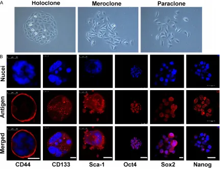
For cell cycle analyses, the collected cells fixed in 75% cold ethanol for 1 day at -20°C. Later, the cells were washed in cold PBS and incubat-Figure 1. Isolation and identification of LCSCs. A. Three types of colonies formed by A549 cells. (400×). B. Immu -nostaining of stem cell markers in secondary spheres. Nuclei were stained using DAPI (blue). LCM, scale bar = 25
[image:3.612.92.523.73.405.2]ed with PI (50 μg/ml) containing RNase at 37°C for 30 min in darkness. Cell cycle was analyzed by flow cytometry (Beckman, USA).
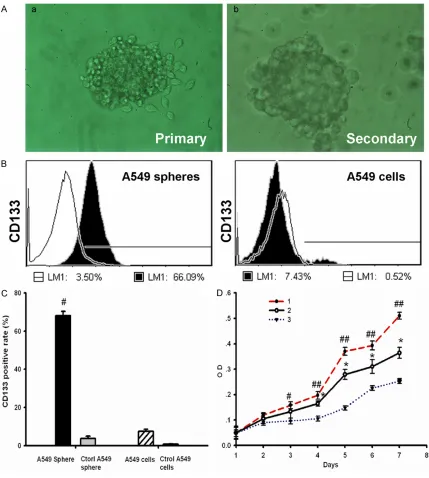
For apoptosis analyses, the collected cells were washed with the collected culture medium, and suspended in PBS. Then, the cells were incu-Figure 2. A549 cancer spheres have high express CD133 and more self renewal capability. A. The morphology of tumor spheres. a. Primary tumor spheres formed in the CSM medium. b. Secondary spheres derived from single parental cells of primary tumor spheres under microscopy. a and b. 200×. B. Flow cytometry analysis of CD133 in
A549 cancer spheres (left) and A549 cells lines (right). Cells were labeled with fluorescent anti-CD133 antibody,
which are shown as black areas. The white area on each box represents the corresponding negative control label-ing, and the line denotes a positive gate. Numbers are the percentage of positive cells. Data are representative of three independent experiments. C. Detection of CD133 positive rate among four groups cells. The results are
expressed as the mean ± SD of three experiments (n = 3). #P<0.05, compared to A549 cells. D. Growth curves 1.
[image:4.612.91.520.69.547.2]bated with the Human Annexin V-FITC Apoptosis Kit (Bender MedSystems, Vienna, Austria) according to the manufacturer’s instructions before analysis by flow cytometry (Beckman) analysis.
Western-blot analysis
Protein extracts of A549 cells line, A549 sec-ondary spheres and HBE were resolved by 12% SDS-PAGE and transferred on PVDF (Millipore, USA) membranes using ECL Semi-dry Blotters (TE70PWR, USA) electro transfer system. After blocking, the PVDF membranes were washed three times with TBST at room temperature and incubated with primary antibodies for GST-π (1:1500, Booster, China), MRP1 (1:1000, Abcam, UK) and LRP (1:1000, Abcam, UK) at 4°C overnight. After extensive washing, mem-branes were incubated with secondary peroxi-dase-labelled goat anti-rabbit IgG (Santa Cruz, USA) for 1 h. After washing four times for 15 min with TBST at room temperature once more, the bands were detected by a Chemilumi- nescence detection kit (ECL) (Beyotime Biotech, Jiangsu, China). The films were scanned and quantitation was carried out with Optiquant software (GBOX/CHEM, USA).
Immunofluorescence staining
Immunofluorescence staining was used for detecting the expression of some stem cell-related markers in tumor spheres, GST-π in tumor spheres and A549 cells. The cells were fixed in 4% paraformaldehyde-PBS for 15 min and permeabilized in 0.2% Triton X-100 in PBS for 30 min at room temperature, and incubated with 3% BSA for 30 min at room temperature to block nonspecific binding. Then, cells were incubated with primary antibodies against, CD133 (mouse monoclonal IgG1, Abcam, Great Britain), stem cell antigen-1 (Sca-1, goat poly-clonal, R&D, USA), CD44s (mouse monoclonal IgG1, Neo Markers, USA), Oct4 (mouse mono
-clonal IgG1, Abcam), Nanog (mouse monoclo -nal IgG1, Abcam), Sox2 (sex determining region Y-box 2, Novus Biologicals, USA), GST-π (1:200, Boster). The appropriate secondary antibodies (TRITC red goat anti-rabbit, Cy3 red donkey anti-goat and Cy3 red rabbit anti- mouse; Molecular Probes, USA) were used. The cells were counterstained with 4, 6-diamidino-2-phe-nylindole (DAPI, Sigma) to visualize cell nuclei and examined under a LCM (Leica, Germany). The mean fluorescence intensity of GST-π in cells was measured using the Leica Confocal Software (Image-Pro Plus 10.0).
Statistical analysis
All experiments were repeated at least three times and representative results are present-ed. Where applicable, quantitative data were presented as means ± SD. P < 0.05 was con-sidered significant. Tumor sphere formation and growth curves were analyzed by ANOVA with a SPSS11.5 statistical software.
Results
Generation of A549 tumor spheres from A549
cell line
A549 cells formed three morphologically differ-ent colonies (Figure 1A): holoclone, meroclone and paraclone by using single-cell cloning cul-ture. Then, the holoclones were dissociated and directly incubation with CSM. Cells started to lose their characteristic epithelial morpholo-gy within 48-72 hours. Some adherent cells typically lost a rhomboidal epithelial shape and became floating cells or cell clusters. The small spheres/well each containing 5-10 cells were observed after 5-6 days. In 2 weeks, the diam-eter of these Primary spheres increased by 10- to 30-fold (Figure 2A). Many adherent non-sphere forming cells could be seen in the bot-tom of the wells. Single cell suspension pre-pared from primary tumor spheres was exam-Figure 3. Measurement of the sensitivity of A549 cancer spheres and A549 cells line to anticancer drugs (DDP and
GEM). A. The lethal doses (IC50) of DDP (left) and GEM (right) in A549 cancer spheres and A549 cells line, The data
shown represent the mean ± SD of three independent experiments(n = 3). #P<0.05, compared to A549 cells line.
B. Graphs shows flow cytometry of the apoptotic induced by DDP and GEM. The cells were stained with annexin
V-FITC and Propidium Iodideed. a. A549 cells line; b. A549 cancer spheres; c. A549 cells line/DDP; d. A549
can-cer spheres/DDP; e. A549 cells line/GEM; f. A549 cancan-cer spheres/GEM. C. Histograms represent the percents of Apoptotic of DDP and GEM in A549 cancer spheres and A549 cells line. The data shown represent the mean ± SD
of three independent experiments (n = 3), #P < 0.05, compared with A549 spheres/DDP; ★P < 0.05, compared
nificantly increased than A549 cells line in DMEM with 10% FBS on 3 days (Figure 2D, sphere, 0.16 ± 0.01, cells line, 0.09 ± 0.01; n = 3, P < 0.05). Meanwhile, Optical density of dissociated A549 tumor spheres in CSM was also signifi -cantly increased than A549 cells line in DMEM with 10% FBS on 4 days (Figure 1D, sphere, 0.20 ± 0.02; cells line, 0.11 ± 0.01; n = 3,
[image:7.612.90.356.98.190.2]P < 0.05). The doubling time of A549 tumor spheres cells in DMEM with 10% FBS was 46.71h com-pared to 53.96 h in CSM. However,
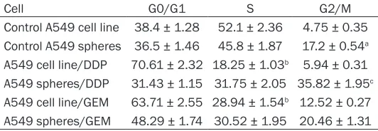
Table 1. Cell cycle distribution of A549 cells line and A549 tumor spheres cells in each groups (%)
Cell G0/G1 S G2/M
Control A549 cell line 38.4 ± 1.28 52.1 ± 2.36 4.75 ± 0.35 Control A549 spheres 36.5 ± 1.46 45.8 ± 1.87 17.2 ± 0.54a
A549 cell line/DDP 70.61 ± 2.32 18.25 ± 1.03b 5.94 ± 0.31
A549 spheres/DDP 31.43 ± 1.15 31.75 ± 2.05 35.82 ± 1.95c
A549 cell line/GEM 63.71 ± 2.55 28.94 ± 1.54b 12.52 ± 0.27
A549 spheres/GEM 48.29 ± 1.74 30.52 ± 1.95 20.46 ± 1.31
aP < 0.05, comparing with control A549 cell line with significant high propor -tion in the G2/M phase. bP < 0.05, comparing with control A549 cell line with significant decrease in S phase; cP < 0.05, comparing with control A549 spheres with significant increase in G2/M phase.
ined for the capacity to form secondary spheres by single parental cells in fresh CSM. The result shows that secondary spheres (Figure 2A) were formed in the 75-ml flask seeded with cells from Primary tumor spheres. The tumor spheres could be passaged every 2-3 weeks for many generations in fresh CSM.
Expression of stem cell-related markers mark
-er in A549 tumor sph-eres
In order to investigate the expression of CD133 (one of the widely accepted CSCs marker) in A549 cancer sphere-growing cells, the single cell suspensions of A549 cells and A549 tumor spheres were analyzed by flow cytometry. Our results showed that the fractions of CD133+
expressing cells in A549 tumor spheres was significantly high than A549 cells line (Figure 2B and 2C, sphere, 66 ± 1.23%; sphere control, 3.5 ± 1.12%; cell line, 10.2 ± 0.83%; cell line control, 1.79 ± 0.56%; n = 3, P < 0.05). These results indicate a good enrichment of CD133+
subpopulations in the A549 tumor spheres. In addition, fluorescent immunostaining revealed that some stem cell-related markers, such as CD133, Sca-1 (A normal bronchioalveolar stem cell or LCSCs marker), CD44s (A stem cell marker of some epithelium-derived tumors, such as breast CSCs), Oct4, Sox2 and Nanog [Embryonic stem cell (ES)/induced pluripotent stem cell (iPS) or CSCs markers] were positive in these secondary tumor spheres (Figure 1B).
Proliferation capability of A549 tumor spheres
cells
To evaluate A549 tumor spheres proliferation capability,We examined the growth of A549 tumor spheres cells and A549 cells line by the MTT assay. Optical density of dissociated A549 tumor spheres in DMEM with 10% FBS was
sig-A549 cells line more quiescent in DMEM with 10% FBS, These cells proliferated with a dou-bling time of 75.6 h (Figure 2D).
A549 tumor spheres express more drug resis -tant capability
Viability study revealed that the growth and inhibition of DDP or GEM to A549 cells line and A549 tumor spheres cells. The results of IC50 shown that A549 tumor spheres cells have sig-nificantly high drug resistant than A549 cells line (Figure 3A). DDP inhibited the growth of A549 tumor spheres cells and A549 cells line with an IC50 of 18.25 ± 0.66 mg/L and 3.8 ± 0.36 mg/L respectively. GEM inhibited the gro-wth of A549 tumor spheres cells and A549 cells line with an IC50 of 280.38 ± 8.2 mg/L and 189.47 ± 5.65 mg/L respectively. Mean- while, under the pressure of DDP or GEM, The apoptotic percentages of A549 tumor spheres cells and A549 cells line had increase com-pared with controls, and A549 cells line was more significant increasing the apoptotic per -centages than A549 tumor spheres in DDP or GEM (Figure 3B and 3C, sphere control, 7.16 ± 1.12%; cell line control, 11.83 ± 1.56%; sph- ere/DDP, 14.64 ± 1.23%; cell line/DDP, 72.16 ± 5.63%; sphere/GEM, 34.38 ± 2.94%; cell line/DDP, 77.32 ± 6.73%; n = 3, P < 0.05). It al- so suggested that A549 tumor spheres cells have stronger drug resistant than A549 cells line.
Cell cycle distribution of A549 cells line and A549 tumor spheres on effects of chemother
-apeutic drugs
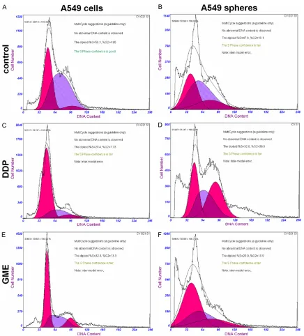
Figure 4. Effect of the IC50 concentration of Cisplatin (DDP) and Gemcitabine (GEM) on the cell cycle profile of A549 cells line and A549 spheres. A. A549 cells line; B. A549 cells line/DDP; C. A549 cells line/GEM; D. A549 cancer spheres; E. A549 cancer spheres/DDP; F. A549 cancer spheres/GEM. This experiment was repeated three separate times, and similar results were obtained. The representative flow cytometry pattern is shown.
results. We investigated whether the cell cycle distribution was related to “stemness” or “drug resistant” of cells. As shown in Table 1 and
Figure 4, cell cycle analysis revealed that the number of A549 spheres cells in the G0/G1 phase and S phase similar to A549 cells line cells. However, an increase in the number of A549 spheres cells in the G2/M phase was apparent. Both DDP and GME can block cells
treated groups (A549 spheres/DDP and A549 spheres/GME) had no significant changes in the G0/G1 phase and S phase were seen as compared to the control group. In addition, the percentages of G2/M phase significant increas -ing in A549 spheres/DDP group and a little accumulation in spheres/GME group compared their control group.
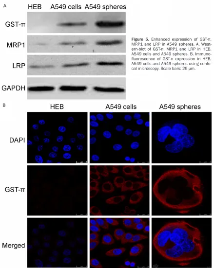
Upregulation of drug resistant proteins GST-π, MRP1 and LRP in A549 tumor spheres
[image:9.612.88.527.68.619.2]One possible mechanism for chemoresistance in A549 tumor spheres is enhanced expression of drug resistant proteins. To test this possibili-ty, we use western-blot to examine expression of several drug resistant proteins, including
Figure 5. Enhanced expression of GST-π,
MRP1 and LRP in A549 spheres. A.
West-ern-blot of GST-π, MRP1 and LRP in HEB,
A549 cells and A549 spheres. B.
Immuno-fluorescence of GST-π expression in HEB,
GST-π, MRP1 and LRP, in HEB, A549 cells and A549 tumor spheres. As expected, expression of all three proteins was enhanced in A549 tumor spheres with the most dramatic change occur for GST-π (Figure 5A). We further validat-ed this result using Immunofluorescence stain -ing of GST-π in these cells. As shown in Figure 5C, in line with western-blot, robust expression of GST-π expression is observed in A549 cells and tumor spheres but not HEB cells. Moreover, GST-π expression is significantly enhanced in A549 tumor spheres shown higher than that of A549 cells line (average signal intensity: sphere, 35.4 ± 2.32; cell line, 27.76 ± 2.24; n = 20, P < 0.05).
Discussion
CSCs are thought to play multiple roles in tumorigenesis. Previous studies selected CSCs, which can grow as spheres in the serum-free medium, from many human lung cancer cell lines and tumors by magnetic activated cell sorting (MACS) or fluorescence-activated cell sorter (FACS) methods depend on some stem cell markers [15, 16, 19-22]. As some studies have demonstrated that cell lines derived from prostate carcinoma and glioma can form mor-phologically heterogeneous colonies in vitro, due to the intrinsic stem cell hierarchies of their parental cells. The holoclones consist of CSCs, while the meroclone and paraclone show non-CSCs properties [18, 23-26]. Therefore, in this current study, we chose A549 holoclones cells cultured with serum-free medium in the pres-ence of EGF and FGF2 for obtaining tumor spheres. Some LCSC markers (such as CD133, Sca-1 and CD34), many embryonic stem (ES)- and induced pluripotent stem (iPS) - related core transcription factors (such as Sox2, Oct4 and Nanog) are overexpressed in these small subset of stem cell-like cells.
More interestingly, it has previously been shown that the expression of these markers and fac-tors is not only related to the stemness but also to drug resistance. For example, CD133 cur-rently serves as a useful marker for the isola-tion of CSCs in various tumors, including lung cancer [16, 27-29]. In this study, our results show that A549 spheres considerably higher expression of CD133 than A549 cell line. It con-sistent with the previous reports that CD133+
fractions varied from 1.4% in A549 cells,
whereas 31.7% in A549-derived tumor spheres and nearly 100% positive in H82 (Small-cell lung cancer lines) side population cells [19, 30]. Moreover, this molecule also performs an important role in sustaining the resistance to chemo- or radiotherapy of CSCs in various tumors [31-33], such as NSCLC and glioma cells. Possible mechanisms may involve unreg-ulated expressing of multiple drug resistance transporters, detoxifying enzymes, activated DNA repair machinery and resistance to apop-tosis. Additionally, while Nanog, Sox2 and Oct4 are key transcription factors in maintaining ES cell pluripotency, they also influence CSCs’ resistance property. Knock down of Sox2 in gli-oma stem-like cells resulted in loss of the acti-vation of ATP-binding cassette (ABC) transport-ers [34-36]. These resistance characteristics inhibitions are also accompanied by the down-regulation of Oct4 and Nanog in LCSCs and prostate cancer stem cell [37-39]. In addition, there is some evidence to indicate that other factors, such as CD44s and Sca-1, can also influence radiation resistance [40, 41]. Thus, it is possible that LCSCs, which commonly over-express either the surface markers or tran-scriptional factors mentioned previously, may also possess some resistance phenotype. In this study, the results suggest that A549 spheres cells are exhibited more extensive pro-liferative potential and significantly resistant to the two tested chemotherapeutic agents. It was high IC50 and low apoptosis rat of A549 spheres cells to DDP or GEM than A549 cells, which is coincidence with the literatures [14, 16].
treat-ment with DDP or GEM. This data in line with recently findings that normal and malignant stem-like cells have an extended G2 cell cycle phase, the phase of the cell cycle associated with DNA repair, which is associated with apop-totic resistance. Targeting G2 checkpoint pro -teins releases these cells from the G2-block and makes them more prone to apoptosis [46]. This novel finding suggests that G2 phase con -tribute to drug resistance of A549 spheres and might be an attractive target for CSCs therapy. Proposed mechanisms of multidrug resistance (MDR) in CSCs include enhanced expression of multidrug resistance transporters, anti-apop-totic factors, or increased levels of DNA dam-age repair proteins [47, 48]. In our research, there are illustrated a significant increase in MRP1 (ABCC1), LPR and GST-π expression in A549 spheres cells. MRP1 belongs to ATP-binding cassette (ABC) proteins, involved in export pumps that hamper the accumulation of anionic conjugates and oxidized glutathione in cytoplasm, and therefore play a critical role in reduces the formation of platinum-DNA adducts, detoxification and development of platinum-based drug resistance [49]. A series of studies has reported that MRP1 up-regula-tion in CD133 positive glioblastoma stem-like cells and SP lung cancer cells [15, 48, 49]. Likewise, LPR, another important MDR-associated protein, vaults act by effuxing drugs from the nucleus and/or the sequestration of drugs into exocytotic vesicles [50, 51]. Recently study demonstrate that it can high expression of those genes in leukemic stem cell [52], but not involve in A549 CSCs so far. In addition, GST-π, the cytosolic detoxifcation protein, is generally accepted that the MRP1 is act in syn-ergy with GST-π to confer resistance to the che -motherapeutic agents, especially platinum drugs [53-55]. Expression of GST-π varies between different CSCs, being high in human fetal liver hematopoietic stem cells [56], but low in human bone marrow mesenchymal stem cells and prostate CSCs [57, 58]. In A549 spheres, we presumed the overexpression of a GST-π in concert with MRP1 and/or LRP would give optimal resistance to chemotherapeutic agents. The available evidence, presented above, suggests that stem-like cell populations in A549 spheres could play an important role in drug resistance, possibly through activation of cell membrane drug effux transporters, and
altered expression of detoxifying enzymes. Moreover, the concomitance increase of the genes and protein, MRP1, LPR and GST-π, sug -gested that lung CSCs was not the solo factor to maintain resist apoptosis and to pump the anti-tumor drugs out of cells.
In conclusion, the present work suggests that 549 spheres, which derived from 549 cells line directly, has enriched cancer stem-like cells and elevated capability of proliferation, anti-apoptotic and drug resistance than 549 cells. In addition, the combined working of MRP1, LPR and GST-π might responsible for the fact that lung CSCs may be spared by traditional chemotherapy and be rapidly recurrence. These observations have potentially important implications for future therapeutic approaches that target the lung CSCs and correlated drug resistance mechanisms.
Acknowledgements
This work is supported by National Natural Science Foundation of China (Grant numbers: 81160027) and Natural Science Foundation of Jiang Xi Province (Grant numbers: 20114BA-B205001).
Disclosure of conflict of interest
None.
Address correspondence to: Dr. Xiao-Qun Ye, Depart-
ment of Respiratory Diseases, The Second Affiliated
Hospital of Nanchang University, Nanchang 3300- 06, China. E-mail: [email protected]
References
[1] Eramo A, Haas T and Maria RD. Lung cancer
stem cells: tools and targets to fight lung can -cer. Oncogene 2010 29: 4625-4635.
[2] Spiro SG, Tanner NT, Silvestri GA, Janes SM,
Lim E, Vansteenkiste JF and Pirker R. Lung cancer: progress in diagnosis, staging and therapy. Respirology 2010; 15: 44-50.
[3] d’Amato TA, Landreneau RJ, McKenna RJ, Santos RS and Parker RJ. Prevalence of in vitro extreme chemotherapy resistance in resected nonsmall-cell lung cancer. Ann Thorac Surg 2006; 81: 440-446; discussion 446-447. [4] Kohmo S, Kijima T, Otani Y, Mori M, Minami T,
Takahashi R, Nagatomo I, Takeda Y, Kida H,
Goya S, Yoshida M, Kumagai T, Tachibana I,
tet-raspanin CD9 mediates chemoresistance in small cell lung cancer. Cancer Res 2010; 70: 8025-8035.
[5] Chang A. Chemotherapy, chemoresistance and the changing treatment landscape for NSCLC. Lung Cancer 2011; 71: 3-10.
[6] Visvader JE and Lindeman GJ. Cancer stem
cells in solid tumours: accumulating evidence and unresolved questions. Nat Rev Cancer 2008; 8: 755-768.
[7] Salmaggi A, Boiardi A, Gelati M, Russo A,
Calatozzolo C, Ciusani E, Sciacca FL, Ottolina
A, Parati EA, La Porta C, Alessandri G, Marras C, Croci D and De Rossi M.
Glioblastoma-derived tumorospheres identify a population of tumor stem-like cells with angiogenic potential and enhanced multidrug resistance
pheno-type. Glia 2006; 54: 850-860.
[8] Fuchs D, Daniel V, Sadeghi M, Opelz G and
Naujokat C. Salinomycin overcomes ABC trans-porter-mediated multidrug and apoptosis
re-sistance in human leukemia stem cell-like
KG-1a cells. Biochem Biophys Res Commun 2010; 394: 1098-1104.
[9] Li R, Wu R, Zhao L, Wu M, Yang L and Zou H. P-glycoprotein antibody functionalized carbon nanotube overcomes the multidrug resistance of human leukemia cells. ACS Nano 2010; 4: 1399-1408.
[10] Xu XL, Xing BC, Han HB, Zhao W, Hu MH, Xu ZL,
Li JY, Xie Y, Gu J, Wang Y and Zhang ZQ. The
properties of tumor-initiating cells from a he-patocellular carcinoma patient’s primary and recurrent tumor. Carcinogenesis 2010; 31: 167-174.
[11] Liu T, Xu F, Du X, Lai D, Zhao Y, Huang Q, Jiang L, Huang W, Cheng W and Liu Z. Establishment and characterization of multi-drug resistant, prostate carcinoma-initiating stem-like cells from human prostate cancer cell lines 22RV1. Mol Cell Biochem 2010; 340: 265-273. [12] Zheng X, Cui D, Xu S, Brabant G and Derwahl
M. Doxorubicin fails to eradicate cancer stem cells derived from anaplastic thyroid carcino-ma cells: characterization of resistant cells. Int J Oncol 2010; 37: 307-315.
[13] Saigusa S, Tanaka K, Toiyama Y, Yokoe T, Okugawa Y, Kawamoto A, Yasuda H, Morimoto Y, Fujikawa H, Inoue Y, Miki C and Kusunoki M. Immunohistochemical features of CD133 ex-pression: association with resistance to chemoradiotherapy in rectal cancer. Oncol Rep 2010; 24: 345-350.
[14] Hu L, McArthur C and Jaffe RB. Ovarian cancer stem-like side-population cells are tumouri-genic and chemoresistant. Br J Cancer 2010; 102: 1276-1283.
[15] Ho MM, Ng AV, Lam S and Hung JY. Side popu-lation in human lung cancer cell lines and
tu-mors is enriched with stem-like cancer cells. Cancer Res 2007; 67: 4827-4833.
[16] Bertolini G, Roz L, Perego P, Tortoreto M, Fontanella E, Gatti L, Pratesi G, Fabbri A,
Andriani F, Tinelli S, Roz E, Caserini R, Lo Vullo S, Camerini T, Mariani L, Delia D, Calabro E,
Pastorino U and Sozzi G. Highly tumorigenic
lung cancer CD133+ cells display stem-like features and are spared by cisplatin treat-ment. Proc Natl Acad Sci U S A 2009; 106: 16281-16286.
[17] Hsu HS, Lin JH, Huang WC, Hsu TW, Su K, Chiou SH, Tsai YT and Hung SC. Chemoresis- tance of lung cancer stemlike cells depends on activation of Hsp27. Cancer 2011; 117: 1516-1528.
[18] Locke M, Heywood M, Fawell S and Mackenzie IC. Retention of intrinsic stem cell hierarchies in carcinoma-derived cell lines. Cancer Res 2005; 65: 8944-8950.
[19] Salcido CD, Larochelle A, Taylor BJ, Dunbar CE and Varticovski L. Molecular characterisation of side population cells with cancer stem cell-like characteristics in small-cell lung cancer. Br J Cancer 2010; 102: 1636-1644.
[20] Lei H, Zhai B, Yin S, Gygi S and Reed R.
Evidence that a consensus element found in naturally intronless mRNAs promotes mRNA export. Nucleic Acids Res 2013; 41: 2517-2525.
[21] Wang P, Yin S, Zhang Z, Xin D, Hu L, Kong X and Hurst LD. Evidence for common short natural trans sense-antisense pairing between
tran-scripts from protein coding genes. Genome
Biol 2008; 9: R169.
[22] Yin S, Yang J, Lin B, Deng W, Zhang Y, Yi X, Shi
Y, Tao Y, Cai J, Wu CI, Zhao G, Hurst LD, Zhang
J, Hu L and Kong X. Exome sequencing
identi-fies frequent mutation of MLL2 in non-small
cell lung carcinoma from Chinese patients. Sci Rep 2014; 4: 6036.
[23] Li H, Chen X, Calhoun-Davis T, Claypool K and
Tang DG. PC3 human prostate carcinoma cell
holoclones contain self-renewing tumor-initiat-ing cells. Cancer Res 2008; 68: 1820-1825. [24] Zhou ZH, Ping YF, Yu SC, Yi L, Yao XH, Chen JH,
Cui YH and Bian XW. A novel approach to the
identification and enrichment of cancer stem
cells from a cultured human glioma cell line. Cancer Lett 2009; 281: 92-99.
[25] Yin S, Deng W, Hu L and Kong X. The impact of nucleosome positioning on the organization of replication origins in eukaryotes. Biochem Biophys Res Commun 2009; 385: 363-368. [26] Yin S, Deng W, Zheng H, Zhang Z, Hu L and
Biochem Biophys Res Commun 2009; 383: 378-382.
[27] Yu SC, Ping YF, Yi L, Zhou ZH, Chen JH, Yao XH,
Gao L, Wang JM and Bian XW. Isolation and
characterization of cancer stem cells from a human glioblastoma cell line U87. Cancer Lett 2008; 265: 124-134.
[28] Miki J, Furusato B, Li HZ, Gu YP, Takahashi H, Egawa S, Sesterhenn IA, McLeod DG, Srivastava S and Rhim JS. Identification of pu -tative stem cell markers, CD133 and CXCR4, in hTERT-immortalized primary nonmalignant and malignant tumor-derived human prostate epithelial cell lines and in prostate cancer specimens. Cancer Res 2007; 67: 3153-3161. [29] Yin S, Wang P, Deng W, Zheng H, Hu L, Hurst
LD and Kong X. Dosage compensation on the active X chromosome minimizes transcription-al noise of X-linked genes in mammtranscription-als.
Genome Biol 2009; 10: R74.
[30] Levina V, Marrangoni A, Wang TT, Parikh S, Su
YY, Herberman R, Lokshin A and Gorelik E.
Elimination of Human Lung Cancer Stem Cells through Targeting of the Stem Cell Factor-c-kit Autocrine Signaling Loop. Cancer Res 2010; 70: 338-346.
[31] Angelastro JM and Lame MW. Overexpression of CD133 promotes drug resistance in C6 glio-ma cells. Mol Cancer Res 2010; 8: 1105-1115. [32] Salnikov AV, Gladkich J, Moldenhauer G, Volm
M, Mattern J and Herr I. CD133 is indicative for a resistance phenotype but does not represent a prognostic marker for survival of non-small cell lung cancer patients. Int J Cancer 2010; 126: 950-958.
[33] Shervington A and Lu C. Expression of multi-drug resistance genes in normal and cancer stem cells. Cancer Invest 2008; 26: 535-542. [34] Jeon HM, Sohn YW, Oh SY, Kim SH, Beck S,
Kim S and Kim H. ID4 imparts chemoresis-tance and cancer stemness to glioma cells by derepressing miR-9*-mediated suppression of Sox2. Cancer Res 2011; 71: 3410-3421. [35] Jia X, Li X, Xu Y, Zhang S, Mou W, Liu Y, Lv D, Liu
CH, Tan X, Xiang R and Li N. Sox2 promotes tumorigenesis and increases the anti-apoptot-ic property of human prostate cancer cell. J Mol Cell Biol 2011; 3: 230-238.
[36] Deng WJ, Nie S, Dai J, Wu JR and Zeng R. Proteome, phosphoproteome, and hydroxypro-teome of liver mitochondria in diabetic rats at early pathogenic stages. Mol Cell Proteomics 2010; 9: 100-116.
[37] Chen YC, Hsu HS, Chen YW, Tsai TH, How CK, Wang CY, Hung SC, Chang YL, Tsai ML, Lee YY, Ku HH and Chiou SH. Oct-4 expression main-tained cancer stem-like properties in lung can-cer-derived CD133-positive cells. PLoS One 2008; 3: e2637.
[38] Jeter CR, Liu B, Liu X, Chen X, Liu C, Calhoun-Davis T, Repass J, Zaehres H, Shen JJ and Tang
DG. NANOG promotes cancer stem cell charac -teristics and prostate cancer resistance to an-drogen deprivation. Oncogene 2011; 30: 3833-3845.
[39] Chan CT, Pang YL, Deng W, Babu IR, Dyavaiah M, Begley TJ and Dedon PC. Reprogramming of
tRNA modifications controls the oxidative
stress response by codon-biased translation of proteins. Nat Commun 2012; 3: 937.
[40] Yin H and Glass J. The phenotypic radiation re -sistance of CD44+/CD24(-or low) breast can-cer cells is mediated through the enhanced activation of ATM signaling. PLoS One 2011; 6: e24080.
[41] Chen MS, Woodward WA, Behbod F, Peddibhotla S, Alfaro MP, Buchholz TA and Rosen JM. Wnt/beta-catenin mediates radia-tion resistance of Sca1+ progenitors in an im-mortalized mammary gland cell line. J Cell Sci 2007; 120: 468-477.
[42] Gangemi R, Paleari L, Orengo AM, Cesario A,
Chessa L, Ferrini S and Russo P. Cancer Stem Cells: A New Paradigm for Understanding
Tumor Growth and Progression and Drug
Resistance. Curr Med Chem 2009; 16: 1688-1703.
[43] Zito G, Richiusa P, Bommarito A, Carissimi E,
Russo L, Coppola A, Zerilli M, Rodolico V,
Criscimanna A, Amato M, Pizzolanti G, Galluzzo A and Giordano C. In vitro identification and
characterization of CD133(pos) cancer stem-like cells in anaplastic thyroid carcinoma cell lines. PLoS One 2008; 3: e3544.
[44] Baas P, Belderbos JS and van den Heuvel M. Chemoradiation therapy in nonsmall cell lung cancer. Curr Opin Oncol 2010;
[45] Honoki K. Do stem-like cells play a role in drug resistance of sarcomas? Expert Rev Anticancer Ther 2010; 10: 261-270.
[46] Harper LJ, Costea DE, Gammon L, Fazil B,
Biddle A and Mackenzie IC. Normal and malig-nant epithelial cells with stem-like properties
have an extended G2 cell cycle phase that is
associated with apoptotic resistance. BMC Cancer 2010; 10: 166.
[47] Sarkar B, Dosch J and Simeone DM. Cancer Stem Cells: A New Theory Regarding a Timeless Disease. Chem Rev 2009; 109: 3200-3208. [48] Jin F, Zhao L, Guo YJ, Zhao WJ, Zhang H, Wang
HT, Shao T, Zhang SL, Wei YJ, Feng J, Jiang XB
and Zhao HY. Influence of Etoposide on
anti-apoptotic and multidrug resistance-associated protein genes in CD133 positive U251 glio-blastoma stem-like cells. Brain Res 2010; 1336: 103-111.
[49] Jin F, Zhao L, Zhao HY, Guo SG, Feng J, Jiang
Paradoxical expression of anti-apoptotic and MRP genes on cancer stem-like cell isolated from TJ905 glioblastoma multiforme cell line. Cancer Invest 2008; 26: 338-343.
[50] Bartkowiak D, Stempfhuber M, Wiegel T and Bottke D. Radiation- and chemoinduced multi-drug resistance in colon carcinoma cells. Strahlenther Onkol 2009; 185: 815-820. [51] Kitazono M, Sumizawa T, Takebayashi Y, Chen
ZS, Furukawa T, Nagayama S, Tani A, Takao S, Aikou T and Akiyama S. Multidrug resistance and the lung resistance-related protein in hu-man colon carcinoma SW-620 cells. J Natl Cancer Inst 1999; 91: 1647-1653.
[52] de Figueiredo-Pontes LL, Pintao MC, Oliveira
LC, Dalmazzo LF, Jacomo RH, Garcia AB,
Falcao RP and Rego EM. Determination of P-glycoprotein, MDR-related protein 1, breast cancer resistance protein, and lung-resistance protein expression in leukemic stem cells of acute myeloid leukemia. Cytometry B Clin Cytom 2008; 74: 163-168.
[53] Morrow CS, Smitherman PK, Diah SK, Schneider E and Townsend AJ. Coordinated
ac-tion of glutathione S-transferases (GSTs) and
multidrug resistance protein 1 (MRP1) in
anti-neoplastic drug detoxification. Mechanism of GST A1-1- and MRP1-associated resistance to
chlorambucil in MCF7 breast carcinoma cells. J Biol Chem 1998; 273: 20114-20120.
[54] Laborde E. Glutathione transferases as media -tors of signaling pathways involved in cell pro-liferation and cell death. Cell Death Differ 2010; 17: 1373-1380.
[55] Mellor HR and Callaghan R. Resistance to che-motherapy in cancer: A complex and integrat-ed cellular response. Pharmacology 2008; 81: 275-300.
[56] Shao J, Stapleton PL, Lin YS and Gallagher EP.
Cytochrome p450 and glutathione s-transfer-ase mRNA expression in human fetal liver he-matopoietic stem cells. Drug Metab Dispos 2007; 35: 168-175.
[57] Kumanov A, Hayrabedyan S, Karaivanov M and Todorova K. Basal cell subpopulation as puta-tive human prostate carcinoma stem cells. Folia Histochem Cytobiol 2007; 45: 75-80. [58] Allameh A, Esmaeli S, Kazemnejad S and