Original Article
Partial hepatectomy promotes implanted mouse
hepatic tumor growth by activating hedgehog signaling
Junbin Zhang1,4*, Li Wang1,4*, Hui Li1,4*, Jing Zhou3, Zhiying Feng3, Yichun Xu5, Xiaolong Chen1,4, Huilin Liu2,4,
Hai Jin6, Jianxu Yang7, Yang Yang1,4, Guihua Chen1,4, Genshu Wang1,4
Departments of 1Hepatic Surgery, 2Gastroenterology, 3Pathology, The Third Affiliated Hospital of Sun Yat-sen University, Guangzhou, Guangdong Province, PR China; 4Guangdong Provincial Key Laboratory of Liver Disease Research, Guangzhou, Guangdong Province, PR China; 5Department of Orthopedics, The Third Affiliated Hospital of Sun Yat-sen University, Lingnan Hospital, Guangzhou, Guangdong Province, PR China; 6Department of Medical Ultrasonics, Guangzhou First People’s Hospital, The Second Affiliated Hospital of South China University of Technology, Guangzhou, Guangdong Province, PR China; 7Department of Intensive Care Unit, Henan Provincial People’s Hospital, Zhengzhou, Henan Province, PR China. *Equal contributors.
Received February 25, 2018; Accepted March 25, 2018; Epub May 1, 2018; Published May 15, 2018
Abstract: Objective: To investigate the role hedgehog signaling (Hh) in the growth of implanted hepatic tumors after partial hepatectomy (PH) in mice. Methods: H22 cells were implanted to the scapula of 2 BALB/c (nu/nu) nude mice and tumor developed in 2 weeks. 40 nude mice were randomized into 4 groups: non-hepatectomy group (Sham operation group), 30% hepatectomy group, 70% hepatectomy group, and 70% hepatectomy with cyclopamine (Hh inhibitor). The hepatectomy model of nude mice was established. After hepatectomy, the tumor tissues incised from the scapula were implanted to the rest of the livers of the 4 groups. After 2 weeks, the tumor formation rates and the volumes of the implanted tumors were compared. Hh related proteins and downstream cytokine VEGF were tested by Western blot and Immunohistochemistry. All the data were analyzed to explore the role of Hh in the growth
of tumor after PH. Results: The volumes of the implanted tumors after liver resection were significantly higher in the
70% PH group than those in 0% and 30% PH groups; meanwhile, we also found that expression of the Hh ligand Indian Hh, its downstream transcription factor protein Gli-1, and its target VEGF were remarkably increased after PH, especially in the 70% PH group. Additionally, applying the Hh inhibitor cyclopamine to mice that underwent 70%
PH significantly inhibited the growth of implanted tumors. Conclusions: The Hh signaling pathway was activated
after PH and promoted liver regeneration. The growth of implanted hepatic tumors was also accelerated after PH via paracrine signaling.
Keywords: Hepatocellular carcinoma, hedgehog signaling pathway, partial hepatectomy, tumor growth, Balb/c nu, Hcc model
Introduction
Worldwide, hepatocellular carcinoma (HCC) is the fourth most common malignant disease and the third leading cause of cancer-related deaths. Currently, a multidisciplinary treatment is the optimal management strategy for HCC [1], and radical hepatectomy is the treatment of choice for early and intermediate-stage liver cancer with sufficiently reserved hepatic func-tion [2]. However, tumors recur within 5 years of surgery in over 80% of patients that undergo radical hepatectomy, which seriously affects long-term survival [3]. Recurrence following ra- dical hepatectomy is closely related to
multi-centric growth and micro hepatic tumor throm-bus [4].
foci. Additionally, the activation of coagulation factor chains following hepatectomy, postoper-ative temporary local or systemic immune sup-pression, and the activation of epithelial pro-genitor cells and other bone marrow-derived hepatic progenitor cells may also can promote tumor recurrence and metastasis [5, 6].
The hedgehog (Hh) signaling pathway is an important intercellular communication system in animal development. It promotes cell prolif-eration and differentiation in a tissue-specific manner during embryogenesis and determines the formation of embryonic patterns. The Hh signaling pathway regulates the self-renewal and proliferation of stem cells in many tissues and organs and maintains the normal morphol-ogies and functions of tissues and organs. Hh pathway activation promotes the differentia-tion and proliferadifferentia-tion of tissue stem cells to repair damaged tissues and organs. However, abnormal Hh pathway activation in adult tis-sues and mature organs can lead to the occur-rence of multiple tumors such as liver cancer, gastric cancer, colon cancer, and lung cancer [7].
The Hh signaling pathway consists of Hh ligand, two transmembrane protein receptors (Patched [Ptch] and Smoothened [Smo]), and the down-stream transcription factor Gli. In mammals, there are three distinct Hh ligands: Sonic (Shh), Indian (Ihh), and Desert (Dhh) Hedgehog. Ptch and Smo are transmembrane proteins located on the cell membrane, and Ptch is the Hh re- ceptor that binds to the three Hh ligands. When Hh ligand is absent, Ptch inhibits Smo activity, which leads to the inhibition of downstream gene expression. When the Hh ligand binds to Ptch, the inhibitory effect on Smo is relieved, which activates the downstream transcription-al regulator Gli, which can induce target gene expression including cyclin, c-myc, and vascular endothelial cell growth factor (VEGF) [8]. Hh signaling is closely related to the develop-ment and metastasis of liver cancer. In HCC tis-sues, one study found the rate of positive Shh expression was 60% (69/115), and the rates of positive Gli and Smo expression exceeded 50% [9]. Hh pathway suppression can inhibit the pro-liferation of some HCC cell lines, promote their apoptosis, and remarkably affect the metasta-sis and invasion capabilities of tumor cells
[9-11]; it can also significantly inhibit the growth and metastasis of HCC xenografts in nude mice [12].
The Hh pathway is activated during liver regen-eration and repair after partial hepatectomy (PH) or severe liver injury. In our studies [13, 14] as well as in studies performed by Ochoa et al. [15] and Cai et al. [16], the Hh pathway was activated following 70% PH in rats and mice, and strongly promoted liver regeneration. While the Hh pathway can be activated by PH, and thus promotes liver regeneration, whether it facilitates liver cancer recurrence and metasta-sis following resection remains unclear. This study investigated the role and mechanism of Hh signaling in the growth of implanted tumors in nude mice after PH.
Materials and methods
HCC cell line and experimental animals and grouping
The procedures for all animal experiments were approved by the Institutional Animal Care and Use Committee at The Third Affiliated Hospital of Sun Yat-sen University. The H22 mouse hep-atoma cell line was used in this study to intra-peritoneally inoculate 4-week-old male C57BL/ 6J mice. Also, 4-week-old male BALB/c nude mice weighing 20-25 g were used to prepare models of subscapular implanted tumors and models of hepatic implanted tumors. Mice were housed in plastic cages under a 12:12 light: dark cycle with free access to food and water. Cell lines and mice were purchased from the Animal Experimental Center of the East Campus of Sun Yat-sen University. All experiments were carried out at the Experimental Animal Center of the North Campus of Sun Yat-sen University.
Reagents
Establishing mouse models of implanted liver tumors and grouping
Culturing H22 mouse hepatoma cells: H22
cells were cultured in ascites. H22 cells (2×10⁶)
and PBS were prepared into a 1 ml cell suspen-sion at a ratio of 1:1, and then intraperitoneally injected into C57BL/6J mice. After one week, the ascites was collected and centrifuged. After repeated rinsing with PBS, a purified mouse H22 hepatoma cell suspension was obtained.
Establishing mouse models with subscapular
implanted tumors: All mice operations were
conducted in the morning (9:00-12:00 am). Animal surgeries were performed using midline laparotomy after the mice were anaesthetized by intraperitoneal injection of 1.5% pentobarbi-tal (30 mg/kg). H22 cells (2×10⁶) and 0.2 ml of
PBS were prepared into a cell suspension, which was injected into the subscapular region of nude mice. After two weeks, tumors were vis-ible in the scapular region and were pathologi-cally confirmed to be HCC tissues by two pathologists from the Third Affiliated Hospital of Sun Yat-sen University.
Establishing the 0%, 30%, and 70% PH mod
-els: A left lateral lobe hepatectomy was per-formed to establish the 30% PH model, and combined middle lobe and left lateral lobe hep-atectomies were performed to establish the 70% PH model [17].
Establishing mouse models of implanted liver
tumors: The implanted tumor tissue in the
sub-scapular region of nude mice was cut into tis-sue blocks, each sized 1 mm3. After being wa-
shed in a sterile PBS solution, the tissue blocks were implanted into the right liver lobe.
Grouping: Forty BALB/c nude mice were divided
into four groups using a random number table: group A (Sham operation group): the tumor tis-sue blocks were implanted into the right liver lobe, but neither liver resection nor ligature were performed; group B (30% PH group): tu- mor tissue blocks were implanted following 30% PH; group C (70% PH group): tumor tissue blocks were implanted following 70% PH; and group D (70% PH + cyclopamine group): tumor tissue blocks were implanted following 70% PH, and then the Hh inhibitor cyclopamine (30 mg/ kg/d) was intraperitoneally injected on a daily basis for 14 days.
Specimen collection and analysis
Mice in each group were sacrificed on day 14 after the operation, then the implanted tumors and para-tumor liver tissues were collected. The tumor formation rate, tumor volumes (V=L ×W2×0.52) were measured, and the character
of the implanted tumors were pathologically confirmed (by two pathologists from the Third Affiliated Hospital of Sun Yat-sen University). Thereafter, a part of each sample was frozen with liquid nitrogen, then stored at -80°C. The left tissues were fixed with 4% paraformalde-hyde, then manufactured into paraffin sections. The expression of the Hh pathway ligand Ihh, downstream protein Gli-1, target gene VEGF, and the proliferation marker proliferating cell nuclear antigen (PCNA) in the implanted tumors and para-tumor samples were detected using Western blotting and Immunohistochemical staining.
Immunohistological staining and Western blot
-ting analysis
extensity score and intensity score (0, 1, 2, 3, 4, 6, 9). Scores < 4 were defined as low expres-sion; scores ≥ 4 were assigned as high expre- ssion.
Western blotting using whole cell lysates from implanted tumor and para-tumor liver tissue were performed as previously described [18]. The protein concentration was quantified using the BCA Protein Assay (Kaiji, Jiangsu, China). Protein lysates (30 μg/sample) were subjected to 12% sodium dodecyl sulfate polyacrylamide gel electrophoresis and then transferred to nitrocellulose membranes (Bio-Rad, Hercules, CA). After transferring, membranes were block- ed with 5% nonfat-milk for 1 hour at room tem-perature and then incubated using the follow-ing antibodies at 4°C for overnight: Ihh (ab- 39634, 1:1000, Abcam, USA), Gli-1 (ab151796, 1:1000, Abcam, USA), PCNA (ab29, 1:1000,
Abcam, USA), and VEGF (ab32152, 1:1000, Abcam, USA), and β-actin (#4970, 1:1000, Cell Signal Technology, USA). Then the membranes were washed using (Tris Buffered Saline Tween, TBST) followed by incubation with an anti-rabbit or anti-mouse IgG antibody (#7074, 1:5000, Cell Signal Technology, USA) for 1 hour at room temperature. The specific bands were detected using Chemidoc™ MP Imaging System after visualizing with Enhanced Chemiluminescence, ECL, (Advansta, USA). The protein expressions were calculated by using β-actin (#4970, 1:1000, Cell Signal Technology, USA) antibody as a control.
Statistical analysis
All data were analyzed using GraphPad Prism 7.0 software (IBM, Armonk, NY, USA), and the density of western blotting bands were
[image:4.612.86.522.73.400.2]lyzed by Image J software 1.8.0. The tumor for-mation rates were compared by X2 test. The
rest of the data were presented as mean ± SD. and were analyzed by either a One-way ANOVA or a Student’s-t test. A value of P < 0.05 was considered to be statistically significant.
Results
Mouse models of implanted liver tumors after PH were successfully established
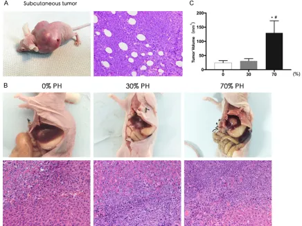
Implanted tumors were visible two weeks after inoculating H22 hepatoma cells into the scapu-lar region. After hematoxylin and eosin staining, it was pathologically confirmed to be HCC. Microscopy of the tumor tissue revealed that the cells had large and deeply stained nuclei, and some nuclei were lobulated (Figure 1A). Two weeks after inoculating the tumor tissue blocks, the liver mass in each group was har-vested. The mass was white, with an irregular shape and clear boundaries. Pathology reveal- ed that there was a clear boundary between the liver and tumor tissue; the nuclei of the tumor cells were large and deeply stained, and some were multinucleated or lobulated. The pathological morphologies were the same as those of the scapular implant tumor tissue, so the mass was confirmed as HCC. In group C, the implanted tumor tissue had more necrosis (Figure 1B). The tumor formation rates of groups A, B and C were 90%, 90% and 80%, respectively, showing no significant difference (P=0.075).
PH promoted the growth of implanted liver tumors
The implanted tumors were harvested 2 weeks after their establishment in nude mice. The vol-umes of the implanted tumors in groups A, B,
and C were 24.0±8.1 mm3, 30.8±8.3 mm3, and
129.9±42.6 mm3, respectively, showing
signifi-cant differences (P < 0.01). However, the im- planted tumor volumes were not significantly different between groups A and B (P > 0.05). Furthermore, group C had significantly larger tumor volumes than groups A and B (A vs C and B vs C, P < 0.05) (Figure 1C). It was found that the volumes of implanted tumors in group C were larger than those in groups A and B. Thus, larger resections significantly promoted the growth of implanted liver tumors in nude mice.
Enhanced proliferation of liver cells after PH
Western blotting revealed significantly different levels of PCNA expression in the para-tumor liver tissues among the three groups (P < 0.01); PCNA expression was significantly higher in group B and group C (P < 0.05) and highest in group C (P < 0.05) (Figure 2A). Immunohisto- chemical staining showed that PCNA was expressed in the nucleus, and there was a sig-nificant difference in the number of PCNA-positive cells among the three groups (P < 0.01); the number of PCNA-positive cells was significantly higher in group C than in groups A or B (P < 0.05), and significantly higher in group B than group A (P < 0.05) (Figure 2B, 2G). These results showed that PCNA expression was increased in para-tumor liver tissues fol-lowing PH, especially in the 70% PH group, sug-gesting the proliferation capability of liver tis-sue cells was enhanced after PH. Furthermore, this capability was associated with the size of the removed liver tissue: liver cell proliferation was stronger in mice with larger resection sizes.
Activated Hh signaling in liver tissues after PH
Western blotting revealed that the level of Ihh protein expression in para-tumor liver tissues
Figure 2. PH enhanced the proliferation of liver cells and activated the Hh signaling. PCNA, Ihh and Gli-1 expression in para-tumor liver tissue in mice after partial hepatectomy increased, but Ihh and Gli-1 expression was not shown in the implanted tumor. All the protein levels were detected by western blotting and the band density quantitative ana-lyzed. All the immunohistochemical staining positive cells were quantitative anaana-lyzed. All the data are expressed as mean ± S.D. (n=10). A. PCNA protein level in Group B and C was higher than that in Group A. Data were analyzed by One-way ANOVA. (#P < 0.05 for B vs A; *P < 0.05 for C & B vs A; &P < 0.05 for C vs B). B, G. The Immunohistochemical
staining of PCNA in para-tumor liver tissue (×200). Data were analyzed by One-way ANOVA. (#P < 0.05 for B vs A; *P
< 0.05 for C vs A; &P < 0.05 for C vs B). C. Ihh protein level in para-tumor liver tissue and implanted tumor. Data were
analyzed by One-way ANOVA. (*P < 0.05 for C vs A, &P < 0.05 for C vs B). D, G. The Immunohistochemical staining of
Ihh in para-tumor liver tissue (×200). Data were analyzed by One-way ANOVA. (*P < 0.05 for C vs A, &P < 0.05 for C vs
B). E. Gli-1 protein level in para-tumor liver tissue and implanted tumor. Data were analyzed by One-way ANOVA. (*P
significantly differed among the three groups (P
[image:7.612.91.522.74.530.2]< 0.01); Ihh was significantly higher in groups B and C than in group A (group C than group B (PP < 0.05), and higher in < 0.05) (Figure 2C).
Figure 3. Suppressing Hh signaling inhibited the growth of implanted tumors after PH. Cyclopamine inhibited the growth of hepatic implanted tumor, and the expression of Ihh, Gli-1 and PCNA in para-tumor liver tissue in mice after 70% partial hepatectomy. All the protein levels were detected by western blotting and were quantitative analyzed of the band density. All the immunohistochemical staining positive cells were quantitative analyzed. All the data are expressed as mean ± S.D. (n=10). A, E. Naked eye and microscopic morphology of hepatic implanted tumor in mice after partial hepatectomy and volume comparison of the hepatic implanted tumors in mice of C and D group. Data were analyzed by student’s-t test. (*P < 0.05 for C vs D). B. Ihh protein level in para-tumor liver tissue of C
and D. Data were analyzed by student’s-t test. (*P < 0.05 for C vs D). B, E. Immunohistochemical staining of Ihh in
para-tumor liver tissue of C and D group. Data were analyzed using Student’s-t test. (*P < 0.05 for C vs D). C. Gli-1
protein level in para-tumor liver tissue of C and D. Data were analyzed using Student’s-t test. (*P < 0.05 for C vs D).
C, E. Immunohistochemical staining of Gli-1 in para-tumor liver tissue of C and D group. Data were analyzed Using Student’s-t test. (*P < 0.05 for C vs D). D. PCNA protein level in para-tumor liver tissue of C and D. Data were
ana-lyzed using Student’s-t test. (*P < 0.05 for C vs D). D, E. Immunohistochemical staining of PCNA in para-tumor liver
However, no obvious Ihh expression was detect-ed in the implantdetect-ed tumor tissue in each group (Figure 2C). Immunohistochemical staining sho- wed that Ihh was expressed on the cell mem-brane, and the number of Ihh-positive cells in para-tumor liver tissues was significantly differ-ent among the three groups (P < 0.05); addi-tionally, it was significantly higher in groups B and C than in group A (P < 0.05), and signifi-cantly higher in group C than group B (P < 0.05) (Figure 2D, 2G).
Western blotting revealed that the level of Gli-1 protein expression in the para-tumor liver tis-sues significantly differed among the three groups (P < 0.01); it was significantly higher in groups B and C than group A (P < 0.05), where-as there wwhere-as no significant difference between groups C and B (P > 0.05) (Figure 2F). Addi- tionally, no obvious Gli-1 protein expression was detected in the implanted tumor tissue in any group (Figure 2F). Immunohistochemical staining showed that Gli-1 was mainly expressed in the nucleus, although some cytoplasms had a positive signal. The number of Gli-1-positive cells in para-tumor liver tissues was significant-ly different among the three groups (P < 0.05); additionally, it was significantly higher in groups B and C than in group A (P < 0.05), and signifi-cantly higher in group C than in group B (P < 0.05) (Figure 2E, 2G).
Therefore, the expression of the Hh pathway ligand Ihh and its downstream transcription factor Gli-1 were remarkably increased after PH, especially in the 70% PH group. Hh signal-ing was activated in liver tissues after PH, and the degree of activation was related to the size of PH. However, Hh signaling was not activated in the implanted tumors themselves.
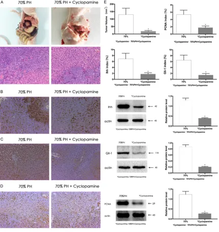
Suppressing Hh signaling inhibited the growth of implanted tumors after PH
Hh signaling was blocked by cyclopamine in group D, and the tumor formation rate was 90%, which was not significantly different from group C (80%; P > 0.05); additionally, the vol-ume of implanted tumors was 18.0±5.4 mm3 in
group D, which was significantly smaller than that of group C (129.9±42.6 mm3; P < 0.05)
(Figure 3A, 3E). Therefore, Hh suppression inhi- bited the growth of implanted tumors after PH. Western blotting and immunohistochemical staining showed that the expression of Ihh and Gli-1 were lower in group D than in group C (Figure 3A, 3E). Therefore, cyclopamine inhibit-ed the expression of Hh signaling components in liver tissues after PH. Western blotting and immunohistochemical staining also showed that PCNA expression was significantly lower in group D than group C (Figure 3D, 3E) (P < 0.05), suggesting that suppressing Hh signaling might inhibit liver regeneration after PH.
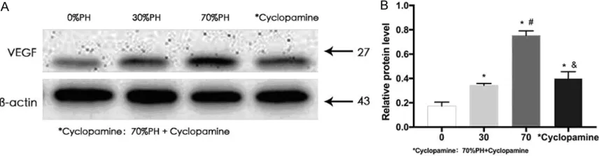
Hh signaling promoted tumor growth by in
-creasing VEGF expression in para-tumor
tis-sues
Hh signaling promotes tumor growth by activat-ing downstream VEGF and other target genes [8]. Therefore, we detected VEGF protein ex- pression in para-tumor liver tissues after PH. Western blotting showed that VEGF expression significantly differed among the four groups (P
[image:8.612.92.520.73.185.2]cantly inhibited in group D (P < 0.05). Therefore, Hh pathway activation promoted tumor growth by increasing VEGF expression in para-tumor liver tissues (Figure 4).
Discussion
The growth rate of metastatic lesions can be 8 times that of normal liver tissues after PH. Additionally, PH of different proportions may also have different effects on liver tumor growth [18]. After 70% PH, the number and vol-ume of residual liver tumors were significantly higher than after 30% PH [19]. After partial li- ver transplantation, grafts can regenerate to restore the normal liver mass; however, after an HCC patient receives a partial liver trans-plantation, the recurrence rate of HCC is higher than that of patients who receive a full liver transplantation [20]. In this study, the growth rate of implanted tumors in the liver of mice was higher in the 70% PH group than those in the 30% PH group or 0% PH group, suggesting major PH promoted the growth of implanted liver tumors, further indicating that hepatecto-my promotes tumor recurrence.
Recent studies have suggested that liver re- generation following liver resection, especially after major hepatectomy or partial liver trans-plantation, may promote tumor recurrence and metastasis, although the exact mechanism is not fully understood. When the apoptosis rate of tumor cells is balanced with their prolifera-tion rate, micrometastases are dormant. After partial liver resection (especially after major hepatectomy), some liver regeneration-related signaling pathways and growth factors, such as HGF, EGF, b-FGF, VEGF, TGF-β, and MMPS, can activate dormant micrometastases, promoting tumor recurrence and metastasis.
Hh signaling is markedly activated during liver regeneration after PH. Abnormal Hh activation can cause the development of a variety of tumors, including HCC [7]; however, few studies have explored whether Hh signaling, which motes liver regeneration after PH, can also pro-mote tumor recurrence and metastasis after HCC resection.
In this study, the growth rate of implanted tumors after resection was significantly higher in the 70% PH group than in the 0% and 30% PH groups; meanwhile, we also found that the
expression of Hh ligand Ihh and its downstream transcription factor Gli-1 were remarkably in- creased after PH, especially in the 70% PH group. Additionally, application of the Hh inhibi-tor cyclopamine in mice after 70% PH signifi-cantly inhibited the growth of implanted tumors. Therefore, the Hh signaling pathway that pro-motes liver regeneration after hepatectomy may also promote the growth of implanted liver tumors.
downstream cytokines warrants further experi- ments.
Currently, three categories of Hh pathway inhib-itors are under development: a) Smo inhibinhib-itors including cyclopamine, vitamin D3, vismodegib, SANT, BMS-833923, LDE225, and LEQ506, among which, the role of vismodegib in rectal cancer and ovarian cancer has been studied in phase II clinical trials [23]; SANT has been manufactured by several drug companies as a promising anti-cancer reagent [15]; b) The Shh ligand antagonists, i.e. robotnikinin, which bind to the N-terminal fragments of Shh, blocking translation of Hh target genes [21]. c) Gli inhibi-tors, including GANT58, GANT61, and HPI-1, -2, -3 and -4, which block Gli-mediated gene trans-lation. Both GANT58 and GANT61 can inhibit the growth of implanted prostate cancer [25]. Additionally, both itraconazole (a commonly used antifungal drug that can act on Smo) and arsenic trioxide (an anti-cancer drug that inhib-its Gli) can block Hh signaling. In some clinical trials, itraconazole has been used to treat basal cell carcinoma [26]. Our study revealed that Hh signaling promotes liver regeneration after hep-atectomy and also promotes the growth of liver implanted tumors, suggesting Hh signaling may be a new target for the prevention and treat-ment of HCC recurrences and metastases. Some commonly used drugs (e.g. vitamin D3 and itraconazole) have been shown to exert anti-cancer effects by blocking Hh signaling [27, 28]. Therefore, the prevention and treat-ment of HCC recurrence by blocking Hh signal-ing is worthy of further study. Our findsignal-ings may thus provide new evidence for the prevention and treatment of HCC recurrence following hepatectomy.
In summary, Hh signaling, which is activated after PH and promotes liver regeneration, can promote the growth of implanted hepatic tu- mors. The possible mechanism may be that Hh signaling promotes the growth of liver tumors by promoting VEGF expression through a Hh ligand-dependent paracrine pathway. However, further research is warranted regarding Hh sig-naling-related genes and their target genes in HCC.
Acknowledgements
This work was supported by the National Na- tural Science Foundation of China 81372243,
81170422, 81570593, 81370575. Key Scien- tific and Technological Projects of Guangdong Province, 2014B020228003, 2014B03030- 1041. Natural Science Foundation of Guang- dong Province, 2015A030312013. Science and Technology Planning Project of Guang- zhou, 201400000001-3, 201508020262, 2014J4100128. Science and Technology Pla- nning Project of Guangdong Province, 2017- A020215178.
Disclosure of conflict of interest
None.
Address correspondence to: Genshu Wang, Depart-
ment of Hepatic Surgery, The Third Affiliated Hospital
of Sun Yat-sen University, Guangzhou, Guangdong Province, PR China; Guangdong Provincial Key Laboratory of Liver Disease Research, Guangzhou, Guangdong Province, PR China. E-mail: wanggshu@ mail.sysu.edu.cn
References
[1] Siegel R, Ma J, Zou Z, Jemal A. Cancer statis-tics, 2014. CA Cancer J Clin 2014; 64: 104-17. [2] Tang CW, Zhu M, Feng WM, Bao Y, Zheng YY. Chinese herbal medicine, Jianpi Ligan decoc-tion, improves prognosis of unresectable hepa-tocellular carcinoma after transarterial chemo-embolization: a retrospective study. Drug Des Dev Ther 2016; 10: 2461-2466.
[3] Imamura H, Matsuyama Y, Tanaka E, Ohkubo T, Hasegawa K, Miyagawa S, Sugawara Y, Minagawa M, Takayama T, Kawasaki S, Makuu-chi M. Risk factors contributing to early and late phase intrahepatic recurrence of hepato-cellular carcinoma after hepatectomy. J Hepa-tol 2003; 38: 200-207.
[4] Cheng HY, Wang X, Chen D, Xu AM, Jia YC. The value and limitation of transcatheter arterial chemoembolization in preventing recurrence of resected hepatocellular carcinoma. World J Gastroentero 2005; 11: 3644-3646.
[5] Paschos KA, Bird NC. Liver regeneration and its impact on post-hepatectomy metastatic tu-mour recurrence. Anticancer Res 2010; 30: 2161-2170.
[6] Gandhi CR, Chaillet JR, Nalesnik MA, Kumar S, Dangi A, Demetris AJ, Ferrell R, Wu T, Divanovic S, Stankeiwicz T, Shaffer B, Stolz DB, Harvey
SA, Wang J, Starzl TE. Liver-specific deletion of
[7] Omenetti A, Diehl AM. The adventures of sonic hedgehog in development and repair. II. Sonic
hedgehog and liver development, inflamma -tion, and cancer. Am J Physiol Gastrl 2008; 294: G595-598.
[8] Scales SJ, de Sauvage FJ. Mechanisms of Hedgehog pathway activation in cancer and implications for therapy. Trends Pharmacol Sci 2009; 30: 303-312.
[9] Huang S, He J, Zhang X, Bian Y, Yang L, Xie G, Zhang K, Tang W, Stelter AA, Wang Q, Zhang H, Xie J. Activation of the hedgehog pathway in human hepatocellular carcinomas. Carcino-genesis 2006; 27: 1334-1340.
[10] Patil MA, Zhang J, Ho C, Cheung ST, Fan ST, Chen X. Hedgehog signaling in human hepato-cellular carcinoma. Cancer Biol Ther 2006; 5: 111-117.
[11] Cheng WT, Xu K, Tian DY, Zhang ZG, Liu LJ, Chen Y. Role of Hedgehog signaling pathway in proliferation and invasiveness of hepatocellu-lar carcinoma cells. Int J Oncol 2009; 34: 829-836.
[12] Xu Y, Chenna V, Hu C, Sun HX, Khan M, Bai H, Yang XR, Zhu QF, Sun YF, Maitra A, Fan J, An-ders RA. Polymeric nanoparticle-encapsulated hedgehog pathway inhibitor HPI-1 (NanoHHI) inhibits systemic metastases in an orthotopic model of human hepatocellular carcinoma. Clin Cancer Res 2012; 18: 1291-1302. [13] Hai J, Hui-Ling L, Xuejun L, Zhiying F, Bing W,
Jianxu Y, Genshu W, Bin W. Expression of hedgehog signaling molecules in rat livers af-ter partial hepatectomy. Chin J Patho 2012; 28: 90-93.
[14] Yang JX, Jin H, Liu HL, et al. Effect of ischemia and reperfusion on expression of Hedgehog signaling in liver after rat partial hepatectomy. Chin J Exp Surg 2012; 29: 671-673.
[15] Ochoa B, Syn WK, Delgado I, Karaca GF, Jung Y, Wang J, Zubiaga AM, Fresnedo O, Omenetti A, Zdanowicz M, Choi SS, Diehl AM. Hedgehog signaling is critical for normal liver regenera-tion after partial hepatectomy in mice. Hepa-tology 2010; 51: 1712-1723.
[16] Cai Y, Zheng H, Gong W, Che Y, Jiang B. The role of hedgehog signaling pathway in liver re-generation. Hepatogastroenterol 2011; 58: 2071-2076.
[17] Stocker E, Pfeifer U. [On the manner of prolif-eration of the liver parenchyma after partial hepatectomy. Autoradiography studies using 3H-thymidine]. Naturwissenschaften 1965; 52: 663.
[18] Elias D, De Baere T, Roche A, Mducreux, Leclere J, Lasser P. During liver regeneration following right portal embolization the growth rate of liver metastases is more rapid than that of the liver parenchyma. Br J Surg 1999; 86: 784-788.
[19] Sowa JP, Best J, Benko T, Bockhorn M, Gu Y, Niehues EM, Bucchi A, Benedetto-Castro EM, Gerken G, Rauen U, Schlaak JF. Extent of liver resection modulates the activation of tran-scription factors and the production of cyto-kines involved in liver regeneration. World J Gastroenterol 2008; 14: 7093-7100.
[20] Lo CM, Fan ST, Liu CL, Chan SC, Ng IO, Wong J. Living donor versus deceased donor liver transplantation for early irresectable hepato-cellular carcinoma. Br J Surg 2007; 94: 78-86. [21] Kudchadkar R, Lewis K, Gonzalez R. Advances
in the treatment of Basal cell carcinoma: Hedgehog inhibitors. Semin Oncol 2012; 39: 139-144.
[22] Wang X, Venugopal C, Manoranjan B, McFar-lane N, O’Farrell E, Nolte S, Gunnarsson T, Hol-lenberg R, Kwiecien J, Northcott P, Taylor MD, Hawkins C, Singh SK. Sonic hedgehog regu-lates Bmi1 in human medulloblastoma brain tumor-initiating cells. Oncogene 2012; 31: 187-199.
[23] Kaye SB, Fehrenbacher L, Holloway R, Amit A, Karlan B, Slomovitz B, Sabbatini P, Fu L, Yauch RL, Chang I, Reddy JC. A phase II, randomized, placebo-controlled study of vismodegib as maintenance therapy in patients with ovarian cancer in second or third complete remission. Clin Cancer Res 2012; 18: 6509-6518. [24] Saqui-Salces M, Merchant JL. Hedgehog
sig-naling and gastrointestinal cancer. Biochim Biophys Acta 2010; 1803: 786-795.
[25] Chung MK, Kim HJ, Lee YS, Han ME, Yoon S, Baek SY, Kim BS, Kim JB, Oh SO. Hedgehog signaling regulates proliferation of prostate cancer cells via stathmin1. Clin Exp Med 2010; 10: 51-57.
[26] Tian H, Callahan CA, DuPree KJ, Darbonne WC, Ahn CP, Scales SJ, de Sauvage FJ. Hedgehog signaling is restricted to the stromal compart-ment during pancreatic carcinogenesis. Proc Natl Acad Sci U S A 2009; 106: 4254-4259. [27] Xin M. Hedgehog inhibitors: a patent review
(2013 - present). Expert Opin Ther Pat 2015; 25: 549-565.