Original Article
Differential expression of serum microRNAs in
glucocorticoid-resistant patients with ulcerative colitis
Juan Luo1*, Yijie Wang3*, Danfeng Lan1, Junkun Niu1, Jiarong Miao1, Xiangqian Dong1, Gang Yang1, Fengrui
Zhang1, Yu Cao4, Kunhua Wang2, Yinglei Miao1
Departments of 1Gastroenterology, 2General Surgery, The First Affiliated Hospital of Kunming Medical University, Yunnan Institute of digestive disease, Kunming , China; 3Department of Critical Care Medicine, the First Affiliated Hospital of Kunming Medical University, Kunming, China; 4Department of Cardiac Surgery, The First People’s Hospital of Yunnan Province, Kunming, China. *Equal contributors.
Received December 18, 2017; Accepted January 12, 2017; Epub February 1, 2018; Published February 15, 2018
Abstract: Inflammatory bowel disease (IBD) includes ulcerative colitis (UC) and Crohn’s disease (CD). Glucocorti -coids (GCs) are the most effective treatment for moderate to severe active UC. However, one-third of patients are not sensitive to GCs (i.e., they are GC resistant). The mechanism of GC resistance in IBD is unknown, and it remains unclear how to predict resistance in IBD patients. This study aimed to explore the possible correlation between miR -NA expression and variability in GC-resistant and GC-sensitive patients with ulcerative colitis. A comparative serum microRNA analysis in GC-resistant and GC-sensitive patients with ulcerative colitis was conducted by microarray. Differential microRNA expression was further validated in serum samples by quantitative real-time PCR. We found that downregulated microRNAs had a significant correlation with several signal transduction pathways (the PI3K-Akt and MAPK signaling pathways) and target genes (HSP90B1, MAPK13, MAPK9, PIK3AP1 and TLR4) related to GC resistance. Eight downregulated microRNAs were chosen for further validation in 76 serum samples. The results showed that miR-16-2-3p, miR-30e-3p, miR-32-5p, miR-642a-5p, miR-150-5p, and miR-224-5p were significantly downregulated in the GC-resistant group. Receiver operating characteristic analysis showed that the area under the curves (AUCs) for those microRNAs were 0.94, 0.93, 0.85, 0.87, 0.92, and 0.99, with specificities of 97.30%, 89.20%, 59.50%, 73.00%, 97.30%, and 97.30% and sensitivities of 74.40%, 84.60%, 97.40%, 92.30%, 66.70%, and 89.70%, respectively. Our study provides preliminary evidence for the pathogenic mechanism of GC resistance and shows that serum microRNAs might serve as biomarkers for GC resistance in IBD.
Keywords: Ulcerative colitis, glucocorticoid resistance, molecular mechanism, MicroRNA
Introduction
Inflammatory bowel disease (IBD) is a term used to describe chronic and non-specific inflammatory disorders of the intestine, includ -ing ulcerative colitis (UC) and Crohn’s disease (CD). IBD is difficult to cure, and the symptoms recur often. The incidence of this disease in China and other Eastern countries is increasing every year [1].
Despite newer therapies with highly effective biological agents, glucocorticoids (GCs) are still the most effective treatment for inducing rapid remission in moderate to severe active UC. In clinical settings, one-third of patients are not sensitive to GCs (i.e., they are GC-resistant). Studies have reported that GC therapy is
hetero-com-plex may contribute to alteration of GC respon-siveness [2]. The present study confirmed that several kinases that facilitate GR phosphoryla-tion, including the c-Jun amino terminal kinase (JNK), serine/tyrosine kinase (MAPKs p38), and extracellular signal regulated protein kinase (ERK), are closely related to GC resistance [3]. In addition, the proportion of GR (GR-α/GR-β) plays an important role in sensitivity to GCs. Extensive evidence shows that the activities of histone deacetylase 2 (HDAC2) and mitogen-activated protein kinase phosphatase 1 are attenuated in GC-resistant patients [4]. Furthermore, the role of immune cells, such as T helper 17 (Th17) cells and regulatory T (Treg) cells, and cytokines, such as IL-2, IL-10, IL-17, and TNF-α, in GC resistance suggest they have functional significance [5].
MicroRNAs (miRNAs) are small (~22 nucleo-tides) non-coding RNA molecules that bind to and downregulate expression of target mRNAs by degrading and/or blocking translation. Additionally, they serve as fine-tuning regulators of diverse biological processes, including the development and function of the immune sys-tem, apoptosis, metabolism, and inflammation. Emerging data indicates that miRNAs are asso-ciated with drug metabolism and drug sensitivi-ty. Several research studies have shown that miRNAs are involved in the modulation of the GC response in multiple diseases, such as hematologic neoplasms and airway hyperre-sponsiveness. Zhao JJ et al. found that the miR-221-222/PUMA/BAK/BAX pathway can abro -gate dexamethasone resistance in multiple myeloma [6]. It was reported that miR-103 is upregulated in GC-sensitive leukemia cells treated by the hormone. Additionally, miR-103 expression in GC-resistant cells facilitates GC-induced apoptosis, marking miR-103 as a potential molecule for therapeutic intervention in ALL [7]. Therefore, the causal relationship between the function of GCs and the expression of miRNAs is perplexing. Another study indicat-ed that miR-19a could be a potential molecular biomarker for predicting and monitoring resis-tance to FOLFOX chemotherapy regimens in advanced colorectal cancer patients [8]. However, the possible correlation between the miRNA expression and variability in GC-resistant and GC-sensitive patients in IBD has not yet been evaluated.
Therefore, in this study, we systematically per-formed comparative serum miRNA analyses in
9 GC-resistant patients and 9 GC-sensitive patients with UC using a PCR-microRNA micro -array. After analysis with a series of bioinfor-matic methods, several potential signaling pathways and targets of GC responses were identified. Differential microRNA expression was then further validated in 76 samples by quantitative real-time PCR. Potential biomark-ers were determined by receiver operating characteristic (ROC) curve analysis.
Patients and methods
Ethical considerations
The study and protocol were reviewed and approved by the Bioethics Committee of the First Affiliated Hospital of Kunming Medical University. The study was carried out in accor -dance with the approved guidelines. Written informed consent was obtained from all patients and their legal guardians. All proce-dures in this study were performed in compli-ance with the Helsinki Declaration and national laws.
Subjects and samples
were randomly divided into three groups and 9 GC-sensitive patients who were similarly divid-ed into 3 groups. The samples were screendivid-ed for disease-associated miRNAs by PCR-microRNA array analysis. Approximately 76 samples obtained from 37 GC-resistant patients and 39 GC-sensitive patients were used to ensure the validity of the results by quantitative reverse-transcription polymerase chain reaction (qRT-PCR).
RNA extraction and miRNA PCR array
Total RNA was extracted from serum samples using a TRIzol-based (Invitrogen, Carlsbad, CA, USA) isolation kit (Shanghai Kangcheng Biotechnology Company, Shanghai, China) according to the manufacturer’s protocol. The concentration and purity of RNA was analyzed with a NanoDrop-1000 (Thermo Scientific, Waltham, MA, USA). Then, approximately 20-25 ng RNA was reverse-transcribed into cDNA using a MicroRNA Reverse Transcription Kit and RT Primer Pools (Exiqon, Denmark) accord-ing to the manufacturer’s protocol. qRT-PCR was conducted on an ABI PRISM7900 system (Applied Biosystems, Foster, CA, USA) with a microRNA PCR Panel (V3.M) (Exiqon, Denmark) that detected 752 human microRNAs using a miRCURY LNA TM Universal RT microRNA PCR system and Ready-to-use human panel I+II (Exiqon, Kangcheng, China). Three small RNA (U6snRNA, SNORD38B, and SNORD49A) and two microRNA (miR-103a-3p and miR-423-5p) reference genes were included in the panel. Fold changes in miRNA expression between the two groups were calculated using the 2-ΔCT method.
Computational analysis
The data obtained from the microRNA PCR panel were analyzed with the GenEx qPCR anal-ysis software (www.exiqon.com/mirna-pcr-analysis) by KangChen Bio-tech, Shanghai, PR China. Gene ontology (GO) analysis (http:// www.geneontology.org) and pathway analyses (pathway identifiers used in KEGG) were per -formed to describe the roles of the differentially expressed miRNAs. Furthermore, Microcosm (http://www.ebi.ac.uk/enright-srv/microcosm/ht- docs/targets/v5/), Miranda (http://www.micr- orna.org/microrna/home.do) and Targetscan (http://www.targetscan.org/vert_60/) were us- ed to analyze target genes associated with glu-cocorticoid resistance for the selected miRNAs.
miRNA and mRNA networks were drawn using Cytoscape.
Quantitative real-time PCR analysis
The differentially altered miRNAs in the serum of 37 GC-resistant patients and 39 GC-sensitive individuals were further validated by qRT-PCR. Total RNA was extracted from freshly frozen serum using TRIzol reagent (Qiagen, Hilden, Germany). The complementary DNA was syn-thesized using SYBR PrimeScript RT reagent kits (TaKaRa, Dalian, China) according to the manufacturer’s instructions. Quantitative real-time PCR was performed on an ABI prism 7900 HT sequence detector (Applied Biosystems, Foster City, CA, USA) using SYBR green method -ology. Briefly, in a 20 µl reaction volume, 1 µl of cDNA was added to 10 µl of SYBR green Master mix (Darmstadt, Germany) and 0.3 μmol/L of each primer. The specific primers of miR-16-2-3p (#HmiRQP0228), miR-30e-3p (#Hmi-RQP0399), 32-5p (#HmiRQP0404), miR-425-5p (#HmiRQP0495), miR-642a-5p (#Hm-iRQP0752), 150-5p (#HmiRQP0210), miR-224-5p (#HmiRQP0343), miR-486-3p (#Hmi-RQP0522) and RNU6 (#HmiRQP9001) were purchased from GeneCopoeia (Guangzhou, China). All PCR reactions were conducted iden-tically according to the following sequence: 95°C for 10 min; 95°C for 15 sec and 60°C for 30 sec for 40 cycles; 65°C for 5 sec; and 95°C. The fluorescence signal was normalized to unify the internal reference level, and the threshold cycle (CT) was set in the exponential amplifica -tion phase of the PCR. The relative expression levels of miRNAs were calculated based on the number of PCR cycles for miRNAs. The compar-ative 2-ΔCT method was used to calculate the relative expression level of each target gene with RNU6 as the internal control. Each sample was run in triplicate.
Statistics
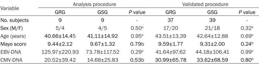
Table 1. Characteristics of the entire patient cohort
Variable Analysis procedure Validated procedure
GRG GSG P value GRG GSG P value
No. subjects 9 9 - 37 39
-Sex (M/F) 5/4 4/5 0.50a 17/20 21/18 0.32a
Age (years) 40.66±14.45 41.11±14.92 0.95b 43.51±13.39 42.64±12.88 0.69b
Mayo score 9.44±2.12 9.67±1.32 0.79b 9.59±1.77 9.31±2.00 0.24b
EBV-DNA 125.97±220.93 73.78±117.52 0.29b 41.64±97.62 44.18±106.41 0.99b
CMV-DNA 20.52±39.42 14.68±25.83 0.53b 30.99±65.78 33.62±68.59 0.80b
GRG: glucocorticoids resistant group, GSG: glucocorticoids sensitive group, M: male, F: female. Age, Mayo score, EBV-DNA
and CMV-DNA were presented as mean ± standard deviation (_x ± s), EBV-DNA and CMV-DNA were E+2. aPearson Chi-Square; bIndependent sample-t test.
[image:4.612.93.519.246.653.2]Results
Clinical parameters in UC patients
As shown in Table 1, there were no significant differences among the patient groups with
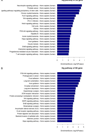
ed to upregulated transcripts, with the “neuro-trophin signaling pathway” as the most repre-sented pathway Figure 3A. With respect to downregulated transcripts, there were 69 path-ways represented, and the most highly enriched was the “PI3K-Akt signaling pathway”. In
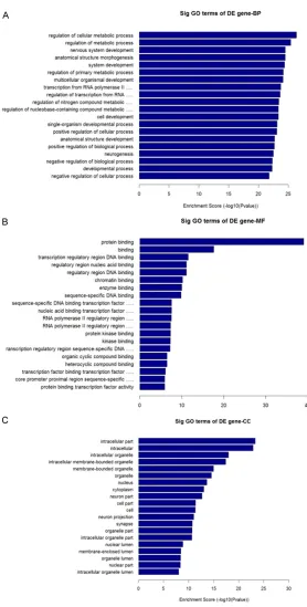
par-Figure 2. GO analysis of downregulated transcripts. A. Biological Process, BP. B. Cellular Component, CC. C. Molecular Function, MF.
respect to sex, age, Mayo score, EBV-DNA, and CMV-DNA (P>0.05).
Profiling of serum miRNAs by
microarrays
As shown in Figure 1A-C, 128 miRNAs were identified as dif -ferentially expressed between two groups with a fold change >2. Of these miRNAs, 50 were upregulated, and 78 were do- wnregulated. We observed that only 16 miRNAs were sig-nificantly downregulated (P< 0.05; 4.58-fold on average) in the GC-resistant group com-pared to the GC-sensitive group. The 50 upregulated miRNAs were not significantly different between the two gr- oups (P>0.05). Hierarchical clustering analysis indicated that there were 128 differen-tially expressed miRNAs. GO analysis and pathway
analysis
[image:5.612.91.369.65.614.2]correspond-ticular, the “PI3K-Akt signaling pathway” and “MAPK signaling pathway”, which were associ-ated with downregulassoci-ated transcripts, were shown to be involved in GC mechanisms of action Figure 3B.
miRNA target prediction
Based on the results of the miRNA PCR arrays and pathway analysis, we carefully selected 8 downregulated miRNAs (miR-16-2-3p, miR- 30e-3p, miR-32-5p, miR-425-5p, miR-642a-5p, miR-150-5p, miR-224-5p and miR-486-3p) wh- ose fold changes were >2.0 and P<0.05 for
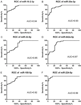
fur-confidence interval [CI] 0.858-0.974, P< 0.0001) and 0.99 (95% confidence interval [CI] 0.968-1.00, P<0.0001), with specifici-ties of 97.30%, 89.20%, 59.50%, 73.00%, 97.30%, and 97.30% and sensitivities of 74.40%, 84.60%, 97.40%, 92.30%, 66.70%, and 89.70%, respectively.
Discussion
In this study, we demonstrate distinct differ-ences in miRNA expression profile from the sera from GC-resistant and GC-sensitive patients with UC in Yunnan, China. We chose Figure 3. The result of pathway analysis. A. Pathways associated with
upregu-lated transcripts. B. Pathways associated with downreguupregu-lated transcripts.
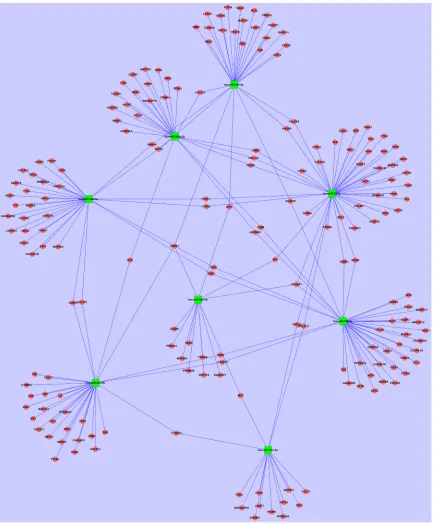
[image:6.612.88.372.77.516.2]ther analysis. The predicted target genes of these objec-tive miRNAs are shown in
Figure 4. The analysis found target genes related to the mechanisms of action of GCs, including HSP90B1, MAPK13, MAPK9, PIK3AP1, and TLR4. Validation of selected serum
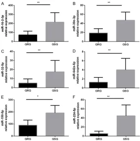
miRNAs
As shown in Figure 5, the expr- ession levels of miR-16-2-3p, 30e-3p, 32-5p, miR-642a-5p, miR-150-5p, and miR-224-5p were significantly lower in GC-resistant patients than in GC-sensitive patients (P<0.05). The expression lev-els of 425-5p and miR-486-3p were not statistically different.
Receiver operating character-istic (ROC) curve analysis
patients in Yunnan, China, for the following rea-sons: (1) Growing evidence has indicated that different miRNAs are downregulated in differ-ent IBD populations, presumably owing to dif -ferent environmental and genetic influences; (2) Yunnan is located in the southwest plateau
[image:7.612.91.525.69.594.2]the population in this area, several novel mech-anisms or potential biomarkers of GC resis-tance may be found.
We found 6 miRNAs (miR-16-2-3p, miR-30e-3p, miR-32-5p, miR-642a-5p, miR-150-5p, and miR-224-5p) that were significantly downregu -lated in GC-resistant patients. Among these miRNAs, miR-150-5p can sensitize the resp- onse to GC therapy in MM1S multiple my- eloma cells by evoking GR-specific effects through indirect mRNA regulation of GR- interacting transcription factors, hormone receptors, and GR chaperones [10]. Another study indicated that miR-150 downregulation may contribute to pertuzumab resistance in ovarian cancer via the PI3K-Akt pathway [11]. Another study found that miR-642 increased cisplatin sensitivity in eight bladder cell lines [12]. Additionally, miR-224 was observed to promote the sensitivity of osteosarcoma cells to cisplatin [13]. Meanwhile, miR-224-5p was shown to function as an oncogene and induce platinum resistance in ovarian papillary serous
plays an important role in regulating GC effects. A previous study showed that steroid resis-tance in COPD is associated with impaired molecular chaperone Hsp90 expression by pro-inflammatory lymphocytes [16]. A C-terminal HSP90 inhibitor restored GC sensitivity in a mouse allograft model of Cushing’s disease [3]. Our study found that 642a-5p and miR-150-5p had the same target gene, HSP90B1. A recent study also found that HSP90 has the potential to assess the activity and prognosis of UC [17].
Many prior studies have shown that p38 MAPK and JNK, as mitogen-activated protein kinase (MAPK) family members, are key enzymes for GR phosphorylation and affect GC sensitivity. We found that all of the downregulated miRNAs could affect expression of various MAPK sub-type target genes. For example, miR-150-5p clearly regulated the target genes for MAPK13 (p38 MAPK) and MAPK9 (JNK2).
Although there have been few studies investi-gating IBD, one found that p38 MAPK phos
-Figure 5. Further validation of miRNAs that are significantly downregulated in the serum. The miRNA levels were determined in the serum by qRT-PCR. GRG: GC-resistant group (n=37); GSG: GC-sensitive group (n=39). miRNA expression was normalized to baseline levels in each sample. *P<0.05, **P<0.01 versus the other groups. The data were expressed as the means ± SD.
carcinoma, which was at least partially promoted by down-regulating PRKCD [14]. In ad- dition, a study examining the drug sensitivity of Hsp90 in-hibitors on human cancer cell lines showed that miR-16-2*, miR-30e*, miR-32 and 12 oth-ers significantly modulated miRNA and gene expression to affect drug responses [15]. These results show that down-regulated miRNAs in our study might be closely related to GC resistance in IBD.
In addition, our analysis of tar-get genes and the pathways of downregulated miRNAs in- cluded important genes relat-ed to GC-resistance, such as HSP90B1 (HSP90), MAPK13 (p38 MAPK), MAPK9 (JNK2), PIK3AP1 (PI-3Kδ), and TLR4. We also found several path-ways related to the mecha-nism of GC resistance, such as the PI3K-Akt and MAPK sig-naling pathways.
[image:8.612.90.373.71.356.2]phorylation and membrane desmoglein-2 expression were reduced in colonic epithelial cells of GC-refractory patients [5]. The p38 MAPK-mediated synergism between IL-10 and GCs improved desmosome straightness and contributed to the recovery of intestinal epithe-lium and reduced contact between luminal antigens and lamina propria in UC. Another investigation showed that GCs regulated barri-er function by increasing the activity of MAPK phosphatase-1 (MKP-1) and changing claudin expression. Barrier augmentation might poten -tially contribute to the therapeutic efficacy of GCs in IBD [18]. While an increasing number of studies concentrate on the blood and respira-tory systems, these studies could provide a theoretical basis for understanding the mecha-nisms of GC resistance in IBD. For example,
curred because the inhibition reduced the expression of impaired HDAC [21]. Despite these advancements, there have been no stud-ies on GC resistance in IBD.
[image:9.612.90.373.72.438.2]In addition, we detected an interesting target gene, TLR4, associated with miR-642a-5p. A study reported that miR-642a possibly binds to a genetic variation of rs11536889 that contrib-utes to translational regulation of TLR4 [22]. Recently, research indicated that TLR4, which is a cell surface TLR, plays an essential role in the development of autoimmune diseases and offers multiple therapeutic targets. Kong et al. [23] found that GC resistance for TAK1 activation was associated with TLR4 engage-ment and might be an important contributor to GC resistance in inflammatory disorders.
Figure 6. Receiver operating characteristics curve evaluation of serum mi-croRNAs for predicting GC resistance in IBD: miR-16-2-3p (A), miR-30e-3p (B), miR-32-5p (C), miR-642a-5p(D), miR-150-5p (E) and miR-224-5p (F). AUC: area under the curve.
p38 MAPK inhibition could reverse GC insensitivity of pe- ripheral blood mononuclear cells in patients with COPD by preventing phosphorylation of GR at serine 211 [19]. Sp- ecific gram-negative bacteria trigger TAK1/MAPK activation and induce GC resistance in asthma. Transforming growth factor-β-associated kinase-1 (TAK1) inhibition also restored cellular sensitivity to GCs [20]. This finding suggests that th-ere is a strong relationship between the microbiome and GC resistance. Similarly, intes-tinal microflora abnormalities exist in patients with IBD, which might also be a cause of GC-resistance.
Although no research has shown that TLR4 is directly associated with GC resistance in IBD, it is well known that TLR4 initiates a variety of immune responses by detecting microbial products. A meta-analysis suggested that CMV-positive IBD patients have nearly double the risk of steroid resistance compared with CMV-negative IBD patients, indicating that CMV infection is a probable cause of steroid-resis-tant IBD [24]. Another study showed that ben-eficial bacteria, such as Lactobacillus rhamno
-sus and Bifidobacterium breve, have a similar effect on GCs, mainly by inducing regulatory T cells to secrete more IL-10 and by increasing CD4 T cell Foxp3 transcription. Additionally, th- ese beneficial bacteria regulate the expression of Toll-like receptors (TLRs) and Nod-like recep-tors (NLRs), cytokines, and T cell transcription factors [25]. In dextran sulfate sodium-induced murine colitis, the anti-inflammatory action se-ems to operate via affecting the TLR4-mediated p38 mitogen-activated protein kinase pathway [26]. Thus, TLR4 might be an important factor causing GC resistance in IBD.
Finally, using ROC curve analysis, we found that miR-16-2-3p, miR-30e-3p, miR-32-5p, miR-642a-5p, miR-150-5p, and miR-224-5p displayed high specificity and sensitivity for diagnosis of GC resistance in IBD.
In conclusion, we demonstrate that miRNAs are downregulated in serum samples from GC-resistant patients with UC. These results, analyzed using bioinformatic methods and ROC curves, may provide a framework for under-standing the mechanism of GC resistance and potential biomarkers for GC resistance. Ho-wever, owing to the small sample size and lack of studies similar to our work, a well-designed, large-scale study with more samples is urgently needed to support our conclusions. Meanwhile, further research could reveal specific roles for miRNAs in GC resistance and could determine whether miRNAs can be developed as biomark-ers and/or therapeutic targets for GC resis-tance in IBD.
Acknowledgements
This work was supported by the Nation- al Natural Science Foundation of China (81260074, 81160055); Applied Basic Res-earch Key Projects of Yunnan Province (2016FA033); Social Development of Science and Technology Projects of Yunnan Province
(2013CA021); Foundation of Yunnan Institute of Digestive Disease (2014NS123, 2016- NS002, 2017NS004); Kunming Engineering Research Center of Digestive Disease (2015-3-A-02243).
Disclosure of conflict of interest
None.
Address correspondence to: Yinglei Miao, Depar- tment of Gastroenterology, The First Affiliated Hospital of Kunming Medical University, Yunnan Institute of Digestive Disease, 295 Xichang Road, Kunming 650032, China. E-mail: [email protected]
References
[1] Ye Y, Pang Z, Chen W, Ju S, Zhou C. The epide-miology and risk factors of inflammatory bowel disease. Int J Clin Exp Med 2015; 8: 22529-22542.
[2] Scheschowitsch K, Leite JA, Assreuy J. New in-sights in glucocorticoid receptor signaling-more than just a ligand-binding receptor. Front Endocrinol (Lausanne) 2017; 8: 16.
[3] Lorén V, Cabré E, Ojanguren I, Domènech E, Pedrosa E, García-Jaraquemada A, Mañosa M, Manyé J. Interleukin-10 enhances the intesti-nal epithelial barrier in the presence of cortico-steroids through p38 MAPK activity in Caco-2 monolayers: a possible mechanism for steroid responsiveness in ulcerative colitis. PLoS One 2015; 10: e0130921.
[4] Jiang Z, Zhu L. Update on molecular mecha -nisms of corticosteroid resistance in chronic obstructive pulmonary disease. Pulm Pharma-col Ther 2016; 37: 1-8.
[5] Vazquez-Tello A, Halwani R, Hamid Q, Al-Muh -sen S. Glucocorticoid receptor-beta up-regula-tion and steroid resistance inducup-regula-tion by il-17 and il-23 cytokine stimulation in peripheral mononuclear cells. J Clin Immunol 2013; 33: 466-78.
[6] Zhao JJ, Chu ZB, Hu Y, Lin J, Wang Z, Jiang M, Chen M, Wang X, Kang Y, Zhou Y, Ni Chonghaile T, Johncilla ME, Tai YT, Cheng JQ, Letai A, Mun-shi NC, Anderson KC, Carrasco RD. Targeting the miR-221-222/PUMA/BAK/BAX pathway abrogates dexamethasone resistance in multi-ple myeloma. Cancer Res 2015; 75: 4384-4397.
[7] Kfir-Erenfeld S, Haggiag N, Biton M, Stepensky P, Assayag-Asherie N, Yefenof E. MiR-103 in-hibits proliferation and sensitizes hemopoietic tumor cells for glucocorticoid-induced apopto-sis. Oncotarget 2017; 8: 472-489.
-cer cases. Asian Pac J Can-cer Prev 2013; 14: 7421-7426.
[9] Niu J, Miao J, Tang Y, Nan Q, Liu Y, Yang G, Dong X, Huang Q, Xia S, Wang K, Miao Y. Iden -tification of environmental factors associated with inflammatory bowel disease in a south -western highland region of China: a nested case-control study. PLoS One 2016; 11: e0153524.
[10] Palagani A, Op de Beeck K, Naulaerts S, Did -dens J, Sekhar Chirumamilla C, Van Camp G, Laukens K, Heyninck K, Gerlo S, Mestdagh P, Vandesompele J, Berghe WV. Ectopic microR -NA-150-5p transcription sensitizes glucocorti-coid therapy response in MM1S multiple my-eloma cells but fails to overcome hormone therapy resistance in MM1R cells. PLoS One 2014; 9: e113842.
[11] Wuerkenbieke D, Wang J, Li Y, Ma C. MiR-NA-150 downregulation promotes pertuzumab resistance in ovarian cancer cells via AKT acti-vation. Arch Gynecol Obstet 2015; 292: 1109-1116.
[12] Nordentoft I, Birkenkamp-Demtroder K, Ager -bæk M, Theodorescu D, Ostenfeld MS, Hart -mann A, Borre M, Ørntoft TF, Dyrskjøt L. MiR -NAs associated with chemo-sensitivity in cell lines and in advanced bladder cancer. BMC Med Genomics 2012; 5: 40.
[13] Geng S, Gu L, Ju F, Zhang H, Wang Y, Tang H, Bi Z, Yang C. MicroRNA-224 promotes the sensi-tivity of osteosarcoma cells to cisplatin by tar-geting Rac1. J Cell Mol Med 2016; 20: 1611-1619.
[14] Zhao H, Bi T, Qu Z, Jiang J, Cui S, Wang Y. Ex -pression of miR-224-5p is associated with the original cisplatin resistance of ovarian papil-lary serous carcinoma. Oncol Rep 2014; 32: 1003-1012.
[15] Yang DS. Novel prediction of anticancer drug chemosensitivity in cancer cell lines: evidence of moderation by microRNA expressions. Conf Proc IEEE Eng Med Biol Soc 2014; 2014: 4780-4786.
[16] Hodge G, Roscioli E, Jersmann H, Tran HB, Holmes M, Reynolds PN, Hodge S. Steroid re -sistance in COPD is associated with impaired molecular chaperone Hsp90 expression by pro-inflammatory lymphocytes. Respir Res 2016; 17: 135.
[17] Abou El Azm AR, Yousef M, Kobtan A, Awad A, Elkassas G, Elfert A. Colonic mucosal expres-sion of heat-shock proteins may have a poten-tial prognostic value in ulcerative colitis. Arab J Gastroenterol 2015; 16: 20-24.
[18] Fischer A, Gluth M, Weege F, Pape UF, Wieden -mann B, Baumgart DC, Theuring F. Glucocorti -coids regulate barrier function and claudin ex-pression in intestinal epithelial cells via MKP-1. Am J Physiol Gastrointest Liver Physiol 2014; 306: G218-228.
[19] Khorasani N, Baker J, Johnson M, Chung KF, Bhavsar PK. Reversal of corticosteroid insensi -tivity by p38 MAPK inhibition in peripheral blood mononuclear cells from COPD. Int J Chron Obstruct Pulmon Dis 2015; 10: 283-291.
[20] Goleva E, Jackson LP, Harris JK, Robertson CE, Sutherland ER, Hall CF, Good JT Jr, Gelfand EW, Martin RJ, Leung DY. The effects of airway mi-crobiome on corticosteroid responsiveness in asthma. Am J Respir Crit Care Med 2013; 188: 1193-1201.
[21] Matsumura Y. Inflammatory cellular pheno -types and molecular mechanisms of glucocor-ticoid resistance in patients with bronchial asthma. Antiinflamm Antiallergy Agents Med Chem 2013; 12: 189-200.
[22] Sato K, Yoshimura A, Kaneko T, Ukai T, Ozaki Y, Nakamura H, Li X, Matsumura H, Hara Y, Ogata Y. A single nucleotide polymorphism in 3’-un -translated region contributes to the regulation of Toll-like receptor 4 translation. J Biol Chem 2012; 287: 25163-25172.
[23] Kong F, Laryea G, Liu Z, Bhattacharyya S. Transforming growth factor-β-activated kinase 1 resistance limits glucocorticoid responsive-ness to Toll-like receptor 4-mediated inflamma -tion. Immunology 2015; 145: 136-149. [24] Wu XW, Wu L, Ji HZ, Wang FY. Relationship be
-tween cytomegalovirus infection and steroid resistance in inflammatory bowel disease: a meta-analysis. Dig Dis Sci 2015; 60: 3203-3208.
[25] Sagar S, Morgan ME, Chen S, Vos AP, Garssen J, van Bergenhenegouwen J, Boon L, Georgiou NA, Kraneveld AD, Folkerts G. Bifidobacterium breve and Lactobacillus rhamnosus treatment is as effective as budesonide at reducing in-flammation in a murine model for chronic asth -ma. Respir Res 2014; 15: 46.