Original Article
MEG3 regulates the invasion and migration of lung
cancer via targeting miR-219a and through
MAPK pathway
Chengyan Jin, Jindong Li, Donglei Shi, Peiyan Hua, Guangxin Zhang, Bin Wang
Department of Thoracic Surgery, The Second Hospital of Jilin University, Changchun 130041, China
Received March 26, 2017; Accepted May 24, 2017; Epub June 1, 2017; Published June 15, 2017
Abstract: Objective: To investigate the expression of MEG3 in lung cancer and the role of MEG3 in the invasion and migration of lung cancer cells and its mechanism. Methods: The expression of MEG3 in lung cancer and adjacent lung cancer cells was detected by qPCR. Transwell invasion assay was used to detect the invasion ability of lung cancer cells after silencing MEG3. The migration ability of lung cancer cells after silencing MEG3 was detected by
scratch test. The expression of miR-219a was detected by flow cytometry. The expression of miR-219a in lung can -cer and adjacent tissues and different lung can-cer cells was detected by qPCR. Transwell invasion assay was used to detect the effect of miR-219a on the invasion of lung cancer cells after silencing MEG3. The effect of miR-219a on the tumor size and volume of lung cancer was detected by subcutaneous tumorigenesis of MEG3 in nude mice. The effect of miR-219a on the tumor size and volume of lung cancer cells was detected by scintigraphy. Western blotting was used to detect the expression of Notch pathway protein after silencing MEG3. The morphological changes of microcapsule microtubules after silencing MEG3 were detected by phalloidin staining. Results: The expression of
MEG3 was significantly increased in lung cancer tissues compared with adjacent tissues; The expression of MEG3 was the highest in lung cancer cell A549; MEG3 could specifically bind to 3’UTR of miR-219a; The expression of 219a was significantly decreased in lung cancer tissues compared with adjacent tissues; Inhibition of
miR-219a could promote the invasion and migration of lung cancer cells after silencing MEG3; The tumor volume and
weight of MEG3-siRNA + miR-219a-inhibitor group were significantly increased compared with MEG3-siRNA group. The expression of Notch pathway protein was down-regulated after silencing MEG3. The number of microfilament
microtubules decreased and the number of pseudopods decreased after silencing MEG3. Conclusion: MEG3 plays an important role in the development of lung cancer. MEG3 can regulate the invasion and migration ability of miR-219a through Notch signaling pathway.
Keywords: MEG3, lung cancer, MiR-219a, transwell
Introduction
In recent years, the incidence of lung cancer and mortality rate shows a sharp upward trend,
lung cancer ranks first in the respiratory malig -nancy, the mortality rate accounts for the third rate of malignant tumor mortality [1]. Early
diag-nosis is difficult due to the lack of specificity in
the early stage and diagnosis is more than in the late, 5-year survival rate is low [2]. Therefore,
finding effective early diagnosis, targeted ther -apy and prognostic markers is a hot topic for current research. The new molecular targeting therapy strategy of lung cancer has been grad-ually promoted to the direction of diagnosis and treatment of lung cancer with the deepen-ing of molecular biology research.
The role of meg3 in invasion and migration of lung cancer cells and its mechanism
MicroRNA (miRNAs) is an endogenous single-stranded small molecule RNA that regulates protein expression by inhibiting or degrading messenger RNA [9]. MiRNAs plays a unique role in the diagnosis, treatment and prognosis of tumors [10]. MiR-219a is relatively less
stud-ied in the field of miRNA, it’s reported that the expression of specific in brain tissue, but also
in the brain glioma has a certain role in the inhi-bition of cancer [11]. Recent investigations have shown that miR-219a expression in lung cancer, breast cancer and other malignant tumors, may be involved in the process of tumor invasion and migration [12]. Mitogen activated protein kinase (MAPK) signal trans-duction pathway is also known as extracellular signal-regulated kinase (ERK) cascade pathway [13]. The structure of MAPK family members is highly conservative, and plays an important role in cell proliferation and differentiation, and plays a different role in the occurrence and development of different tumors. This pathway is a signal transduction pathway will stimulate signal transmitted to the nucleus to the cell surface and internal mediated cell response in most a major pathway, such as growth, differ-entiation, division and apoptosis are regulated by MAPK signal transduction pathway [14]. This investigation intends to analyze the expression of MEG3 in lung cancer, and further investigate the interaction between MEG3 and miR-219a, and investigate the role and mechanism of MAPK pathway in the invasion and metastasis of lung cancer.
Materials and methods
Samples collection
50 cases of lung cancer patients and 50 cases of adjacent tissues were collected admitted to our hospital from March 2015 to May 2016. All patients had no chemotherapy or radiotherapy before operation, and they were postoperative pathological staging by two sub-high above the pathologist jointly read the tumor tissue in vitro then put into RNA preservation solution quickly.
Cell lines
Human cells PGCI3, H1299, 95D, A549 were purchased from Shanghai Zhongshan cell bank and they were cultured and passaged in culture medium containing 10% fetal bovine serum in 5% CO2 incubator at 37°C. Fetal bovine serum, RPMI 1640 medium were purchased from
Hyclone Corporation (Hyclone, Logan, UT).
Transwell Chamber was purchased from Mil- lipore (Millipore, Billerica, MA), Matrigel was purchased from Bio-Rad (Bio-Rad, Madrid, Spain). Lipofectamine 2000 and miR-219a-in-hibitor were purchased from (Gnenpharma Co., Shanghai, China). Trierol was purchased from
Ambion (Ambion Inc., Austin, TX, USA), reverse
transcription kit (FSQ-101) was purchased from Japan TOYOBO Corporation (TOYOBO, FSQ-101, Japan), PCR kit was purchased from Sigma
(KapaBiosystems Inc., Boston, US). The lucifer -ase activity assay kit was purch-ased from Promega Corporation (Promega Biotech Co., Beijing, China). The luciferase reporter vector was synthesized by Promega Corporation (Promega Biotech Co., Beijing, China).
Quantitative real-time polymerase chain reac-tion
Total RNA from tumor tissues and cell lines were extracted using the Trizol reagent (In-
vitrogen, Carlsbad, CA, USA) according to the manufacturer’s instructions. Total RNA was
eluted with RNase-free water and stored at -80°C. qRT-PCR was performed by using SYBR-green PCR Master Mix in a Fast Real-time PCR 7500 System (Applied Biosystems). The RT-PCR primers were purchased from GeneCopoeia
(SanDiego, California, USA). GAPDH was used
as the internal control of the mRNA or miRNA, respectively. Fold change of MEG3 or miR-219 was calculated by the equation 2-ΔΔCt.
Cell transfection
Mimics/inhibitors specific for miR-219 and
short hairpin RNA (shRNA)/scramble fragments targeting MEG3 were designed and purchased
from Invitrogen (USA). Cells were seeded in
24-well plates at 1×10⁵ cells per well. Mimics/
inhibitors or shRNA/scramble fragments was transfected into cells with lipofectamine 3000
(Invitrogen) following the manufacturer’s
ins-tructions for 24 h, respectively. After transfec-tion, the cells were allowed to recover by incu-bating them for 4 h at 37°C. The experiment was replicated thrice for data calculations.
Transwell invasion assays
The two transwell invasion chambers with Ma- trigel (1 mg/ml) (Becton-Dickinson, New Jersey,
USA) were used in assay invasion assays of cells in vitro. Firstly, 200 μl serum-free medium
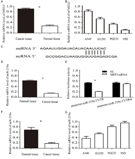
Figure 1. A. The mRNA expression of MEG3 were detected by qPCR in lung cancer tissue and normal tissue. B. The mRNA expression of MEG3 were detected by qPCR in different lung cancer cells. C, D. MEG3 binds to miR-219a. E. Luciferase report gene detects miR-219a the direct target of MEG3-siRNA. F. The mRNA expression of miR-219a were detected by qPCR in pancreatic lung cancer tissue and normal tissue. G. The mRNA expression of miR-219a were detected by qPCR in different pancreatic cancer cells. Error bars represent standard error. *P < 0.05.
upper chamber, and the lower chamber contained 0.6 ml medium containing 20% FBS. After incuba-tion at 37°C for 24 h, non-invading cells on the upper membranes were removed with a cotton swab. The migrated or invaded cells were fixed in
95% ethanol, stained with hematoxylin. The cell numbers were counted by ImageJ software and photographed under an inverted microscope on
10 random fields in each well. Each experiment
The role of meg3 in invasion and migration of lung cancer cells and its mechanism
Wound-healing assays
Wound-healing assay was performed to evalu-ate the migration revalu-ate of cells transfected with MEG3-siRNA/MEG3 scramble/miR-219 inhibi-tor. To accomplish this, 1.5×10⁶ cells/well
were seeded in 6-well plates and cultured
overnight until the cells reached 90% conflu -ence. A straight scratch was created by a ster-ile pipette tip. The destroyed cells were rinsed off with PBS 3 times gently and cultured in medium for another 24 h. Cell migration was observed and imaged at 0 h, 24, 48, 72 h with a digital camera (Leica DFC300FX).
Luciferase activity assays
The luciferase reporter vector was co-trans-fected with A549 cells with MEG3-siRNA. The transfected pRL-TK was used as standard internal control. The cells were harvested after transfection for 36 h. The luciferase ac-
tivity of A549 cells was detected by Promega’s
luciferase activity assay kit. Calculate relative
luciferase activity = firefly luciferase activity
value/bloody luciferase activity value.
Lung cancer xenografts
Nude mice are selected from 4 to 6 weeks old. The lung cancer siRNA cells and MEG3-siRNA + miR-219a-inhibitor cells were cultured in logarithmic phase. Adjusted to cell concen-tration of 2×10⁸/ml. Take 0.1 ml cell
suspen-sion injection in each nude mice left forelimb axillary subcutaneous, a total of 10. Within 4 weeks after injection, the survival, weight, and survival status of the mice were monitored, and the size and weight of the tumor in the immediately-dead mice were measured.
Statistical analysis
The results are presented as the mean ± the standard error of the mean of 3 replicates. Differences between means were analyzed
using Student’s t test. The difference was con
-sidered statistically significant at P < 0.05. Results
Expression of MEG3 mRNA in lung cancer tis-sues, adjacent tissues and lung cancer cells
The results (Figure 1A) of qPCR showed that the expression of MEG3 mRNA in lung cancer
tissues was significantly higher than that in
adjacent tissues [(0.86±0.05) vs (0.22±0.03), P < 0.05], the difference was statistically
sig-nificant; The expression level of MEG3 in A549
cells was the highest in different lung cancer cells (Figure 1B). We selected A549 for further experimental cell lines combined with the above results and consider MEG3 in lung can-cer play a role in cancan-cer.
Luciferase reporter gene detection of MEG3 and miR-219a relationship
We use the bioinformatics predictor to make clear that MEG3 may interact directly with miR-219a to clarify the case of miRNAs asso- ciated with MEG3. MEG3-siRNA and miR-219a were co-transfected into lung cancer cell A549
to confirm whether MEG3 binds to miR-219a 3’UTR. The luciferase reporter gene results showed that MEG3-siRNA significantly inhibited
luciferase activity in miR-219a (Figure 1C-E).
The results show that MEG3-siRNA can specifi
-cally bind to the 3’UTR of miR-219a.
Expression of miR-219a mRNA in lung cancer tissue, adjacent tissues and lung cancer cells
The results of qPCR showed that the expres-sion of miR-219a mRNA in lung cancer tissues
was significantly lower than that in adjacent tis -sues [(0.78±0.03) vs (0.29±0.01), (P < 0.05)] (Figure 1F), the difference was statistically
sig-nificant; The expression level of MEG3 was the
lowest in A549 cells in different lung cancer cells (Figure 1G).
Effect of MEG3-siRNA on invasion and migra-tion of human lung cancer cell line A549
The ability of cells to penetrate through Matrigel
can reflect the ability of cells to invade. The
results of Transwell showed that the number of cells in Matrigel matrix was (179.2±7.4) (Figure 2A), which was significantly higher than that of
MEG3-siRNA group (44.8±2.9), the difference
was statistically significant (P < 0.01). It indi -cated that MEG3-siRNA can inhibit the invasion of human lung cancer cell A549. The results of scratches showed that the mobility of
MEG3-siRNA group was significantly decreased at 24
h, 48 h and 72 h compared with NC group [24 h (34.2±2.3)% vs (18.6±1.3)%, P < 0.05 and 48 h (62.8±4.8)% vs (39.9±3.1)%, P < 0.05; 72 h (86.4±7.8)% vs (52.1±5.0)%, P < 0.01] (Figure 2B), the difference was statistically significant.
Figure 2. A. Effect of MEG3-siRNA on the invasion ability of A549 cells were detected by Transwell matrigel invasion assays. B. Effect of MEG3-siRNA on the A549 cells migration ability were detected by wound healing assays. Error bars represent standard error. *P < 0.05.
migration ability of human lung cancer cell A549.
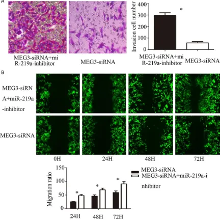
Effects of miR-219a on the invasion and migration ability of human lung cancer cell line A549 after silencing MEG3
The results of Transwell showed that the num-ber of cells in Matrigel matrix was (63.4±5.3),
which was significantly less than that of
MEG3-siRNA + miR-219a-inhibitor group (187.2±6.9) (Figure 3A), the difference was statistically
sig-nificant (P < 0.01). The results indicated that
inhibition of miR-219a could promote the inva-sion of human lung cancer cell line A549 after silencing MEG3. The results of scratches sho- wed that the mobility of MEG3-siRNA +
miR-219a-inhibitor group was significantly higher
than that of MEG3-siRNA group at 24 h, 48 h and 72 h [24 h (29.2±2.4)% vs (12.1±1.3)%, P < 0.05; 48 h (62.4±4.9)% vs (38.2±2.8)%, P < 0.05; 72 h (89.5±9.1)% vs (53.4±3.8)%, P < 0.01] (Figure 3B), the difference was
statisti-cally significant. It indicated that inhibition of
miR-219a could promote the migration of hu- man lung cancer cell line A549 after silencing MEG3.
Effect of miR-219a on tumor growth after silencing MEG3 showed by the experiment of subcutaneous tumor formation in nude mice
The role of meg3 in invasion and migration of lung cancer cells and its mechanism
Figure 3. A. Effect of miR-219a on the invasion ability of A549 cells were detected after silencing MEG3 by Transwell matrigel invasion assays. B. Effect of miR-219a on the A549 cells migration ability were detected after silencing MEG3 by wound healing assays. Error bars represent standard error. *P < 0.05.
weeks. The autopsy showed that all the tumors in the left armpit were growing, and the tumors were gray white, solid, round or oval, with
nodu-lar protrusions on the surface and fish in the profile. The tumor formation rate was 100%.
The tumor growth in nude mice (Figure 4A): the
tumor size of MEG3-siRNA group was signifi -cantly higher than that of MEG3-siRNA group compared with MEG3-siRNA group. The tumor weight and volume of contrast (Figure 4B, 4C): MEG3-siRNA miR-219a-inhibitor group the
tumor volume and weight were significantly
higher compared with MEG3-siRNA group
[vol-ume (2.99 + 0.35) cm3 vs (0.29 + 0.11) cm3, P
< 0.05; weight (3.12 + 0.34) g vs (0.36 + 0.07) g, P < 0.05].
Changes in cytoskeleton morphology and ROCK family protein levels
The role of meg3 in invasion and migration of lung cancer cells and its mechanism
[(81.26%±5.23%) vs (19.62±1.62), P < 0.05; (78.28±8.15) vs (11.31±2.61) g, P < 0.05]. It was detected by Phalloidin staining (Figure 4E, 4F) that microfilament microtubule cytoskele -ton in MEG3 silencing pseudopodia increased intracellular reduction, burr like structure,
which can influence the movement and migra -tion of cells.
Discussion
Lung cancer is the most common malignant tumor of the respiratory system, its rapid devel-opment of the disease, recurrence rate and mortality is high, the prognosis is very poor, which has become a serious threat to human health and life in China and even malignant dis-ease [2]. So the prognosis of lung cancer patients and early intervention is very impor-tant. DNA abnormal methylation can lead to microRNA and other tumor suppressor gene inactivation and oncogene activation in the occurrence and development of tumor [15]. Therefore, miR-219a may be associated with lung cancer progression and prognosis.
It was reported that only about 2% of the genome sequence was translated into protein, most of which were transcribed as non-coding RNA with the deepening of research and the understanding of genomics deepened [16]. Non-coding RNA is divided into lncRNA and small non-coding RNA according to the se- quence of the transcribed gene. LncRNA is a class of RNA molecules of about 200 nucleo-tides in length, and there is no relatively com-plete open reading frame that was once con-sidered to have no biological function. However, recent researches have shown that lncRNA is closely related to a variety of diseases, such as some malignant tumors, cardiovascular dis-ease, neurodegenerative diseases, autoim-mune diseases [17].
MEG3 is located on chromosome 14q32.3, about 1.6 kb, and is initially found to have a class of lncRNA with anti-cancer effect. Re- searches have shown that MEG3 expression in the liver, brain, lung and ovary and other nor-mal tissues is relatively high, but in a variety of malignant tumor cells show the phenomenon of down-regulation or deletion [18, 19]. The
current investigation confirmed that MEG3 its
anti-tumor effect and p53 pathway are partially related. Kobayashi et al. [20] found that MEG3
expression decreased in liver cancer and cell lines. By increasing the expression of MEG3 and found that p53 gene activity increased, suggesting that MEG3 P53 pathway may inhibit the proliferation and invasion of liver cancer cells, and promote its apoptosis. It has been reported that MEG3 can play a role in tumor suppressing by a variety of ways, and its mech-anism is related to DNA methylation, P53 path-way, Rb pathway and angiogenesis [21]. In this investigation, the expression of lncRNA MEG3
in lung cancer tissue was significantly
down-regulated compared with normal lung tissue. Follow-up of MEG3 inhibition in lung cancer cell lines was performed in order to verify the role of MEG3 expression in the development of lung
cancer. These findings suggest that lncRNA
MEG3 lung cancer plays a role in the develop-ment of tumor suppressor genes, and its dele-tion or downreguladele-tion is involved in the devel-opment and progression of lung cancer. Therefore, MEG3 plays an important role in lung cancer and can serve as a target for future lung cancer treatment. Mature miR-219a is generally expressed in tissues such as gastric cancer, lung cancer and breast cancer. Torres et al. [22] demonstrated that miR-219a expres-sion in gastric cancer was down-regulated and was closely related to the up-regulation of te- lomerase reverse transcriptase by detecting the expression of miR-219a in gastric cancer. In this investigation, the expression of miR-219a in lung cancer tissues and normal lung tissues
was confirmed by qPCR. The expression of miR-219a in lung cancer was significantly down-reg -ulated, indicating that miR-219a had an anti-cancer effect in lung anti-cancer. The effect of miR-219a on the invasion and migration of lung cancer cells was further studied by silencing MEG3 and inhibiting the expression of miR-219a in combination with the previous experi-mental results. The results showed that inhibi-tion of miR-219a expression after silencing MEG3 could promote the invasion and migra-tion of lung cancer cells. In vivo experiments using subcutaneous tumor formation in nude mice also have similar results. Similar results were observed in vivo experiments using nude mice subcutaneously.
miR-138. The role of MEG3 and miR-138 in the invasion and migration of lung cancer cells was further investigated. The results showed that MEG3 was up-regulated in lung cancer, miR-138 was down regulated in lung cancer, HOTAIR and miR-138 had direct interaction. MEG3 can target the migration and invasion of lung can-cer by miR-138, which indicated that MEG3 and miR-138 may be involved in lung cancer cell invasion and migration process, there may be a prognostic marker for predicting lung cancer progression, and monitoring the effect of trea- tment.
Acknowledgements
The Natural Science Foundation of China (#81272472).
Disclosure of conflict of interest
None.
Address correspondence to: Bin Wang, Depart- ment of Thoracic Surgery, The Second Hospital of
Jilin University, 218 Ziqiang Street, Changchun
130041, China. Tel: 13353111009; E-mail: wangbi-niin@163.com
References
[1] Zatloukal P, Petruzelka L, Zemanova M, Havel L, Janku F, Judas L, Kubik A, Krepela E, Fiala P, Pecen L. Concurrent versus sequential chemo-radiotherapy with cisplatin and vinorelbine in locally advanced non-small cell lung cancer: a randomized study. Lung Cancer 2004; 46: 87-98.
[2] Yasufuku K, Chiyo M, Koh E, Moriya Y, Lyoda A, Sekine Y, Shibuya K, Lizasa T, Fujisawa T. Endo-bronchial ultrasound guided transEndo-bronchial needle aspiration for staging of lung cancer. Lung Cancer 2005; 50: 347-354.
[3] Zhou Y, Zhang X, Klibanski A. MEG3 noncoding RNA: a tumor suppressor. J Mol Endocrinol 2012; 48: R45-R53.
[4] Xiu YL, Sun KX, Chen X, Chen S, Zhao Y, Guo
QG, Zong ZH. Upregulation of the lncRNA Meg3
induces autophagy to inhibit tumorigenesis and progression of epithelial ovarian carcino-ma by regulating activity of ATG3. Oncotarget 2017; 8: 31714-31725.
[5] Miyoshi N, Wagatsuma H, Wakana S, Shiroishi T, Nomura M, Aisaka K, Kohda T, Surani MA,
Kaneko-Ishino T, Ishino F. Identification of an
imprinted gene, Meg3/Gtl2 and its human
ho-mologue MEG3, first mapped on mouse distal
chromosome 12 and human chromosome 14q. Genes Cells 2000; 5: 211-220.
[6] Wang P, Ren Z, Sun P. Overexpression of the long non-coding RNA MEG3 impairs in vitro glioma cell proliferation. J Cell Biochem 2012; 113: 1868-1874.
[7] Zhang X, Rice K, Wang Y, Chen W, Zhong Y, Na-kayama Y, Zhou Y, Klibanski A. Maternally ex-pressed gene 3 (MEG3) noncoding ribonucleic acid: isoform structure, expression, and func-tions. Endocrinology 2010; 151: 939-947. [8] Benetatos L, Vartholomatos G, Hatzimichael E.
MEG3 imprinted gene contribution in tumori-genesis. Int J Cancer 2011; 129: 773-779. [9] Junker A, Hohlfeld R, Meinl E. The emerging
role of microRNAs in multiple sclerosis. Nat Rev Neurol 2011; 7: 56-59.
[10] Liu K, Wang R. MicroRNA-mediated regulation in the mammalian circadian rhythm. J Theor Biol 2012; 304: 103-110.
[11] Tang G, Shen X, Lv K, Wu Y, Bi J, Shen Q. Differ-ent normalization strategies might cause in-consistent variation in circulating microRNAs in patients with hepatocellular carcinoma. Med Sci Monit 2015; 21: 617.
[12] Dugas JC, Cuellar TL, Scholze A, Ason B, Ibra-him A, Emery B, Zamanian JL, Foo LC, McMa-nus MT, Barres BA. Dicer1 and miR-219 Are required for normal oligodendrocyte differenti-ation and myelindifferenti-ation. Neuron 2010; 65: 597-611.
[13] Bonni A, Brunet A, West AE, Datta SR, Takasu MA, Greenberg ME. Cell survival promoted by the Ras-MAPK signaling pathway by tran-scription-dependent and-independent mecha-nisms. Science 1999; 286: 1358-1362. [14] Roux PP, Blenis J. ERK and p38
MAPK-activat-ed protein kinases: a family of protein kinases with diverse biological functions. Microbiol Mol Biol Rev 2004; 68: 320-344.
[15] Goya T, Asamura H, Yoshimura H, Kato H, Shi-mokata K, Tsuchiya R, Sohara Y, Miya T, Miyao-ka E; Japanese Joint Committee of Lung Can-cer Registry. Prognosis of 6644 resected non-small cell lung cancers in Japan: a Japa-nese lung cancer registry study. Lung Cancer 2005; 50: 227-234.
[16] Wang Y, Yao J, Meng H, Yu Z, Wang Z, Yuan X, Chen H, Wang A. A novel long non-coding
RNA, hypoxia-inducible factor-2α promoter up -stream transcript, functions as an inhibitor of osteosarcoma stem cells in vitro. Mol Med Rep 2015; 11: 2534-2540.
The role of meg3 in invasion and migration of lung cancer cells and its mechanism
[18] Gejman R, Batista DL, Zhong Y, Zhou Y, Zhang X, Swearingen B, Stratakis CA, Hedley-Whyte ET, Klibanski A. Selective loss of MEG3 expres-sion and intergenic differentially methylated region hypermethylation in the MEG3/DLK1 locus in human clinically nonfunctioning pitu-itary adenomas. J Clin Endocrinol Metab 2008; 93: 4119-4125.
[19] Benetatos L, Hatzimichael E, Dasoula A, Dran-itsaris G, Tsiara S, Syrrou M, Georgiou I, Bou-rantas KL. CpG methylation analysis of the MEG3 and SNRPN imprinted genes in acute myeloid leukemia and myelodysplastic syn-dromes. Leuk Res 2010; 34: 148-153. [20] Kobayashi S, Wagatsuma H, Ono R, Ichikawa
H, Yamazaki M, Tashiro H, Aisaka K, Miyoshi N, Kohda T, Ogura A, Ohki M, Kaneko-Ishino T, Ishino F. Mouse Peg9/Dlk1 and human PEG9/DLK1 are paternally expressed imprint-ed genes closely locatimprint-ed to the maternally ex-pressed imprinted genes: mouse Meg3/Gtl2 and human MEG3. Genes Cells 2000; 5: 1029-1037.
[21] Zhao J, Dahle D, Zhou Y, Zhang X, Klibanski A. Hypermethylation of the promoter region is as-sociated with the loss of MEG3 gene expres-sion in human pituitary tumors. J Clin Endocri-nol Metab 2005; 90: 2179-2186.
[22] Torres L, Juárez U, García L, Miranda-Ríos J,
Frias S. External ear microRNA expression
pro-files during mouse development. Int J Dev Biol