Original Article
MicroRNA-181a knockdown protects HepaRG cells
from Dichlorvos-induced oxidative stress and apoptosis
Caifeng Gu, Jie Shen, Feng Zhang, Junfeng Chen
The Centre of Emergency and ICU, Jinshan Hospital Affiliated to Fudan University, Shanghai, China Received May 10, 2017; Accepted June 20, 2017; Epub November 1, 2017; Published November 15, 2017
Abstract: The present study was designed to assess the molecular mechanism of Dichlorvos (DDVP)-induced he-patic cell toxicity in vitro using HepaRG cells. The cytotoxicity was determined by cell viability, apoptosis portion, and reactive oxygenspecies (ROS) generation. The results indicated that DDVP treatment significantly inhibited cell growth, induced cell apoptosis and promoted the production of ROS on HepaRG cells. Microarray analysis showed that 181a was significantly upregulated in HepaRG cells treated with DDVP. Furthermore, we found that miR-181a downregulation has a remedy effect on DDVP-induced cell toxicity, while miR-miR-181a overexpression augments the DDVP-induced hepatic cell apoptosis and ROS production. Furthermore studies showed that miR-181a directly targeted Bcl-2, and Bcl-2 downregulation inhibited the remedy effect of miR-181a inhibitor on DDVP induced cell toxicity. It is, therefore, concluded that miR-181a knockdown could protect hepatic cells from DDVP induced oxida-tive stress and apoptosis by targeting bcl-2.
Keywords: Dichlorvos, HepaRG cell, toxicity, miR-181a, Bcl-2
Introduction
Organophosphate pesticides are one class of the most widely used synthetic chemicals for agricultural and domestic pest control [1]. Among them, Dichlorvos (DDVP) is a commonly used insecticide [2]. It not only results in the environmental pollution due to its use in agri-culture, but also poses a great threaten to pub-lic health, as DDVP is poisonous when inhaled, swallowed or absorbed through skin and eyes. Recent years, a large number of acute and chronic poisoning events occurred.
Emerging studies suggested DDVP exposure has been related to substantial negative health effects on several organ systems, including the respiratory system [3-5], the nervous system [6, 7] and reproductive system [8, 9]. In addi-tion to that, recently, it has been reported that DDVP also can induce the damage and dys-function of liver. Binukumar et al. (2010) report-ed that a mitochondrial energy metabolism impairment and liver dysfunction occurred in rodent model following chronic exposure to
DDVP [10]. Zhao et al. (2015) reported a case of DDVP induced autoimmune hepatitis [11]. Bui-Nguyen et al. (2015) demonstrated that DDVP exposure results in large scale disruption of energy metabolism in the liver of the zebraf-ish [12]. However, the precise molecular mech-anism of hepatic response to DDVP exposure remains unclear till now, although its cellular mechanisms have been identified, such as cell apoptosis, ROS production [13].
MicroRNAs (miRNAs) are a class of short (~22 nt), endogenous regulatory RNAs. Changes in microRNA (miRNA) expression occur in many pathological and physiological processes, such as cell viability, apoptosis, and so on [14]. Previous studies have demonstrated that the potential and important role of miRNAs to regu-late apoptosis at a variety of levels and in sev-eral organisms [15]. However, whether microR-NAs involved in DDVP induced hepatic cell toxicity remains unclear.
Downregulation miR-181a protects Dichlorvos-induced apoptosis
cell dysfunction. We firstly demonstrated that DDVP could induce HepaRG cells apoptosis and ROS production. Then, we found that miR-181a is up-regulated in DDVP treated HepaRG cells by using microarray analysis and Real-time PCR. Furthermore, we showed that miR-181a involved in the apoptosis process of hepatic cells exposed to DDVP by binding its target gene Bcl-2.
Materials and methods
Chemicals and cell culture
DDVP was obtained from Merck (Germany). HepaRG cells were purchased from Type Cu- lture Collection of the Chinese Academy of Sciences (Shanghai, China), and were cultured in DMEM (Invitrogen, USA) containing 10% fetal bovine serum (FBS) (HyClone, USA) at 37°C with 5% CO2.
Caspase activation and western blot
Caspase-3 activity was measured using acom-mercially assay kit, according to the man-
ufacturer’sinstructions. Total cell protein was
isolated from HepaRG cells after treatment. The BCA protein assay kit (Beyotime, Shanghai, China) was used to determine the protein con-centration, following manufacturer’s instruc-tion. Samples were electrophoresed by using 10% SDS-PAGE. The protein was then trans-ferred onto a PVDF (polyvinylidene fluoride) membrane (Bio-Rad, USA). After blocking in skim milk, the membranes were incubated with specific antibodies. Autoradiograms were quan-tified by densitometry (Quantity One software; Bio-Rad). β-actin was used as internal refer-ence; goat anti-Bcl-2, anti-cleaved-caspase-3 (1:1000) were purchased from Sigma.
Cell viability assay
Cell viability was analyzed by using Cell Count- ing Kit-8 (CCK-8) (Beyotime, Shanghai, China), followed by manufacturer’s instructions. Briefly, the HepaRG cells were seeded into the 96-well plate. 10 μL of Cell Counting Kit-8 solution was then added into each well. The OD450 values were detected by using the Spectra Micropla- te Reader (BIOTEC). Results were showed as percentage of the vehicle control levels set at 100%. Each treatment was performed in tri- plicate.
ROS determination
The intracellular amounts of ROS was mea-sured according to the method described by Ben et al. (2016) [16].
Microarray and data analysis
Total RNA was extracted from HepaRG cells treated with DDVP for 24 h, using TRIZOL reagent (Invitrogen) and sent to Biomaker Company form iRNA microarray analysis. Data were analyzed using ANOVA andt tests.
Quantitative real-time PCR
RNA was reverse transcribed to cDNA from 500 ng of total RNA by using a Reverse Tr- anscription Kit (Tiangen, Beijing, China). Real-time PCR assay were performed with SYBR Green (Tiangen, Beijing, China). All protocols were carried out according to the manufactur-er’s instructions. The primer sequences in terms of U6, miR-181a were listed as follows: U6: Forward (F): 5’-CTCGCTTCGGCAGCACA-3’, Reverse (R): 5’-AACGCTTCACGAATTTGCGT-3’; miR-181a F: 5’-GAACATTCAACGCTGTCGGTG-3’, R: 5’-ATCCAGTGCAGGGTCCGAGGTA-3’. Real-time PCR assay was performed on a Real-Time PCR System (Applied Biosystems, USA), and each RT reaction was performed in triplicate, including no-template controls. The reaction was performed in the following conditions: 4 min at 96°C, followed by 39 cycles with dena-turing for 17 s at 94°C and annealing for 45 s at 62°C. The relative quantification of miR-181a was normalized to the expression of U6 using the 2-ΔΔCT method, respectively.
Luciferase reporter assay
MiR-181a mimics, miR-181a inhibitors, and miRNA normal control (miR-NC) were purchased from GenePharma (Shanghai, China). After placed into 48-well plates, cells were cotra- nsfected with miR-181a, luciferase reporter plasmids (200 ng) containing wild-type (WT) or mutant type (Mut) of Bcl-2 3’UTR. Fourty-eight hours after transfection, luciferase activities were measured using the dual-luciferase re- porter assay system (Promega). Each transfec-tionwas performed in triplicate.
Apoptosis assay
pEGFP-Figure 1.DDVP-induced cell apoptosis and ROS in vitro. A. The inhibit effect of DDVP on HepaRG cells viability. HepaRG cells were treated with DDVP at the indicated concentrations ranging from 10-600 μM for 24 h. MTT assay was used to detect cell viability. The values are expressed as viability percentage. **P < 0.01 DDVP treatment group vs. Non-treatment controls. B. DDVP increased caspase-3 activity in HepaRG cells. Cells were treated with DDVP at the concentration of 300 μM for 24 h.**P < 0.01 vs. control. C. DDVP increased cleaved caspase-3 expression and decreased bcl-2 expression in HepaRG cells. The protein expression was determined using western blot. D. DDVP induced HepaRG cells apoptosis. The cell apoptosis was detected by flow cytometry.**P < 0.01 DDVP treatment group vs. Non-treatment control. E. DDVP promoted ROS production in HepaRG cells.**P < 0.01 DDVP treatment group vs. Non-treatment control.
[image:3.612.98.350.460.708.2]Downregulation miR-181a protects Dichlorvos-induced apoptosis
NC, and stably transfected with pEGFP-Bcl-2 were harvested 48 hours after transfection by trypsinization. Apoptotic cells were detected by Annexin V/APC and propidium iodide (PI) apop-tosis detection kit I (BD Pharmingen, San Diego, CA, USA), following the manufacturer’s recom-mendations. The cells were analyzed with a flow cytometry (FACScan; BD Biosciences) eq- uipped with a Cell Quest software. Cells were distinguished into viable cells, dead cells, early apoptotic cells, and apoptotic cells, and then the relative ratio of early apoptotic cells were compared with control transfection from each experiment. Each sample was run in triplicate. Statistical analysis
Data were shown as mean ± standard devia-tion (SD) from atleast three separate experi-ments. The student’s t test and one-way ANOVA were used to conduct the comparison of the different protein, mRNA, luciferase reporter and miRNA expression levels. Statistical analy-ses were performed by SPSS 16.0 software. A P-value less than 0.05 was considered statisti-cally significant difference.
Results
DDVP induced HepaRG cells apoptosis and ROS production
HepaRG cells were treated with differential concentrations of DDVP, ranging from 10 to 600 μM for 24 hours, and cell viability was detected by MTT assay. Results showed that DDVP induced a dose-dependent decrease in cell viability (P < 0.01), compared with controls (Figure 1A). The DDVP concentration of 300 μM, was therefore, selected as the cell treat-ment concentration. Caspase-3 activity was significantly increased in DDVP group (P < 0.01, Figure 1B). Western blot analysis showed that the pro-apoptosis protein cleaved-caspase-3 expression was significantly increased in DDVP group, while the anti-apotosis protein bcl-2 ex- pression was significantly decreased in DDVP group, when compared with controls (Figure 1C). DDVP also induced HepaRG cells apopto-sis (P < 0.01, Figure 1D). The levels of ROS pro-duction in DDVP group was significantly incre- ased (P < 0.01, Figure 1E) than that of controls. It is, therefore, indicated that DDVP inhibited cell growth, induced cell apoptosis and promot-ed the production of ROS on HepaRG cells.
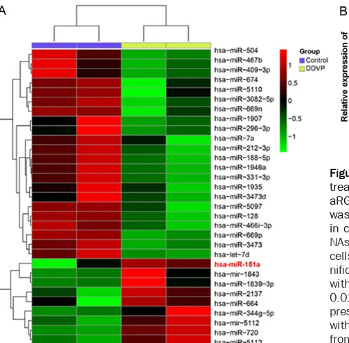
microRNAs profiles in HepaRG cells treated with DDVP
To identify miRNA involved in the mechanism of DDVP treated HepaRG cells, we detected miR-NAs expression profiling of HepaRG cells treat-ed with DDVP. By using microarray analysis, we compared miRNA expression profiles in DDVP treated cells to control. miRNAs were consid-ered as differentially expressed when differ-ences in expression levels were significant both in Student’s t test (P < 0.01) and analysis of microarray test (q value < 5%). Among the indi-vidual miRNAs displayed on the microarray, there were 22 miRNAs significantly up-/down-regulated in HepaRG cells with DDVP treat-ment. The most up-regulated miRNA was miR-181a (Figure 2A). Next, we carried out real-time PCR to detect the expression of miR-181a in HepaRG cells treated with differential concen-tration of DDVP, ranging from 10 to 600 μM. Results showed that miR-181a was significant-ly up-regulated in cells treated with DDVP, with an increase dose-dependent manner (P < 0.01) (Figure 2B). Summarizing, these data demon-strated that miR-181a is up-regulated in DDVP treated HepaRG cells, suggesting miR-181a might involve in DDVP induced HepaRG cell toxicity.
The effect of miR-181a on DDVP induced cell apoptosis and ROS production
HepaRG cells were pretreated with miR-181a
inhibitor/inhibitor NC for 12 h before DDVP treatment for 24 h (300 μM). Cell viability was significantly inhibited in DDVP + inhibitor NC group (**P < 0.01) when compared with the control. However, miR-181a inhibitor significa- ntly reversed DDVP-induced cell viability inhibi-tion (##P < 0.01, DDVP + miR-181a inhibitor vs.
DDVP + inhibitor NC group, Figure 3A). Further- more, miR-181a significantly inhibited DDVP-induced HepaRG cells apoptosis (##P < 0.01,
Figure 3B), caspase-3 activity increase (##P <
0.01, Figure 3C), and ROS production (##P <
0.01, Figure 3D). In contrast, different phenom-enon occurred in HepaRG cells treated with miR-181a mimics. miR-181a mimics promoted
the DDVP induced cell viability inhibition (#P <
0.05, Figure 3E), enhanced DDVP-induced He-
paRG cells apoptosis (##P < 0.01, Figure 3F),
upregulated the caspase-3 activity and ROS
production (#P < 0.05 for both, Figure 3G, 3H).
down-regulationhas a remedy effect on DDVP-indu- ced cell toxicity, while miR-181a overexpres-sion augments the DDVP-induced hepatic cell apoptosis and ROS production.
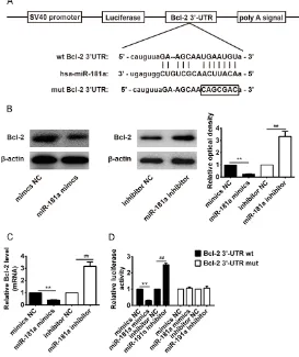
miR-181a directly targets Bcl-2 mRNA
We used bioinformatic tools (Targetscan and miRBase) to identify the target gene of miR-181a. As shown in Figure 4A, Bcl-2 is predicat-ed as the theoretical target gene of miR-181a. Furthermore, to validate whether Bcl-2 is a direct target gene, the 3’-UTR region of Bcl-2 was fused to a luciferase system. Western blot assay showed that the expression of Bcl-2 protein was significantly decreased after miR-181a mimic treatment (**P < 0.01, Figure 4B), while expression of Bcl-2 protein levels were significantly increased after miR-181a inhibit- or treatment, compared to the NC group (##P <
0.01, Figure 4B). Similar phenomenon was also found at the mRNA levels of Bcl-2 (**P < 0.01,
##P < 0.01, Figure 4C). Figure 4D showed that
miR-181a obviously suppressed the lucifera- se activities of the 3’-UTR segment of Bcl-2, whereas miR-181a inhibitor significantly in-
creased the luciferase activities of the 3’-UTR segment of Bcl-2, but not those of the construct containing a mutant binding site (Bcl-2 3’-UTR-mut), compared to the NC group.
Bcl-2 downregulation inhibited the remedy ef-fect of miR-181a inhibitor on DDVP induced cell toxicity
Since Bcl-2 was the target gene of miR-181a, and miR-181a inhibitor has the protection effect of DDVP. Herein, we knock down Bcl-2 to evaluate its protection function of miR-181a on the DDVP-induced cell toxicity, including cell viability, apoptosis, ROS production, and so on. We found that the downregulation of Bcl-2 re- versed the miR-181a inhibitor protective effect on the HepaRG cell viability, when compared with non-transfection treatment group (##P <
0.01, si-Bcl-2 + miR-181a inhibitor + DDVP vs. miR-181a inhibitor + DDVP group, Figure 5A). In the above experiment, we found that miR-181a inhibitor could rescue the increased apoptosis portion, caspase-3 activity and ROS production caused by DDVP treatment. However, when transfected with si-Bcl-2, these effects
weak-Figure 3. The effect of miR-181a on DDVP induced cell apoptosis and ROS production. HepaRG cells were pre-treated with miR-181a inhibitor/inhibitor NC for 12 h before DDVP treatment for 24 h (300 μM). A. The cell viability in different groups was analyzed by using CCK-8 kit. Cell viability was significantly inhibited in DDVP + inhibitor NC group and DDVP + miR-181a inhibitor group when compared with the controls. However, miR-181a inhibitor re-versed DDVP-induced cell viability inhibition. B. The cell apoptosis was detected by flow cytometry. miR-181a inhibi-tor weakened DDVP-induced cell apoptosis. C, D. miR-181a inhibiinhibi-tor decreased DDVP-induced caspase-3 activity and ROS production. E. Cell viability was analyzed by using CCK-8 kit. miR-181a mimics promoted DDVP-induced cell viability inhibition. F. miR-181a mimics upregulated DDVP-induced cell apoptosis. G, H. miR-181a mimics enhanced DDVP-induced caspase-3 activity and ROS production.*P < 0.05,**P < 0.01, vs. control. #P < 0.05,##P < 0.01, vs.
[image:5.612.91.519.73.278.2]Downregulation miR-181a protects Dichlorvos-induced apoptosis
ened, as can be seen from the increased level of apoptosis portion (##P < 0.01, si-Bcl-2 +
miR-181a inhibitor + DDVP vs. miR-miR-181a inhibitor + DDVP group, Figure 5B), caspase-3 activation and expression (##P < 0.01, si-Bcl-2 + miR-181a
inhibitor + DDVP vs. miR-181a inhibitor + DDVP group, Figure 5C, 5E, 5F), as well as increased patterns of ROS production (##P < 0.01, si-Bcl-2
+ miR-181a inhibitor + DDVP vs. miR-181a in- hibitor + DDVP group, Figure 5D). These results suggested bcl-2 downregulation participated in the protection role of miR-181a involved in DDVP induced hepatic cell apoptosis and ROS production. Summarizing, DDVP induced cell
[image:6.612.91.364.76.401.2]that activation of caspase-3 is essential to the apoptosis of hepatic cells [17, 18]. Bcl-2 is known as an anti-apoptosis molecular [19]. In the present study, we observed a significant increase of activation and cleavage of cas-pase-3, as well as decreased Bcl-2 in DDVP treated HepaRG cells. Our finding is consistent with the reports published by Sunkaria et al. (2012), who found that caspase-3 activation was increased in DDVP treated primary rat microglia [6]. Furthermore, cell apoptosis de- tected by flow cytometry showed that DDVP induced significantly apoptosis of HepaRG cells. And also, our study found that the DDVP
Figure 4. Bcl-2 is the direct target of miR-181a. A. A putative miR-181a-bind-ing site exists in the 3’-UTR of Bcl-2 mRNA and point mutations were gener-ated in the binding site. B. The expression of Bcl-2 protein was analyzed by Western blotting. β-actin was used as control. C. mRNA expression of Bcl-2 was detected by real-time PCR after treatment with miR-181a mimics or miR-181a inhibitor (n=3). **P < 0.01 vs. mimics NC group; ##P < 0.01 vs.
inhibitor NC group. D. The luciferase reporter plasmid containing wild-type or mutant Bcl-2 3’-UTR was cotransfected into HepaRG cells with miR-181a mimic/inhibitor or miR-NC. Luciferase activity was determined by the dual lu-ciferase assay and shown as the relative firefly activity normalized to Renilla activity. Each bar represents the mean and s.d. of three independent experi-ments. **P < 0.01 vs. mimics NC group; ##P < 0.01 vs. inhibitor NC group.
cytoxicity might or at least th- rough miR-181a upregulation by binding its target gene Bcl- 2.
Discussion
Since DDVP exposure could pose a serious threaten to human health, and recently studies reported that it could induce the damage and dys-function of liver [10, 11], how-ever, the precise molecular me-chanism of DDVP induced hepatic cell toxicity is not re- ported in the literature. In the present study, we firstly found that the HepaRG cells treated with DDVP significantly inhibit-ed cell viability, basinhibit-ed which, we propose that hepatic cells might undergo apoptotic cell death when exposed to DDVP for a certain time period. To confirm this, studies of pro-teins involved in the apoptotic pathway and apoptosis assays were carried out.
induced significantly ROS production in Hepa- RG cells. Thus, the present study showed that DDVP can inhibit HepaRG cell viability and ul- timately apoptotic cell death, and ROS pro- duction.
MicroRNAs (miRNAs) are a kind of short length RNA molecules regulating the target gene expression, thereby playing role in biology pro-cesses, including proliferation, differentiation, and death [15]. Emerging studies have demon-strated that miR-181a involved in cell apopto-sis [20-22]. However, to date, levels of hepatic cell-associated miRNAs in response to DDVP exposure has received limited attention. In the current study, we found that miR-181a was sig-nificantly upregulated in HepaRG cells after treatment with DDVP. Furthermore, we treated HepaRG cells with miR-181a inhibitors and mimics, respectively, and found that miR-181a downregulation has a remedy effect on
DDVP-induced HepaRG cells apoptosis and ROS
pro-duction, while miR-181a overexpression aug-ments the DDVP-induced hepatic cell apoptosis and ROS production. It is therefore, suggested miR-181a involved in DDVP induced cell ap- optosis.
[image:7.612.93.518.71.286.2]It is widely acknowledged that microRNAs pl- ay biology function by targeting transcription mRNA [23]. MiR-181a has been reported to inhibitcell viability, induce apoptosis and play function in disease, via binding its target gene [20, 24-26]. Bcl-2 is one of the Bcl-2 family membersand specifically considered an impor-tant anti-apoptotic protein which plays essen-tial roles in the regulation of apoptosis via the mitochondrial pathway [27, 28]. Previous study demonstrated that downregulation of miR-181a protects mice from LPS-induced acute lung injury by targeting Bcl-2 [25]. Bcl-2 is reported to participate in the apoptosis pro-cess of hepatic cells [29]. Accordingly, we spec-ulated that miR-181a target the anti-apoptotic
Figure 5. Bcl-2 downregulation inhibited the protective effect of miR-181a inhibitor on DDVP-induced HepaRG cell cytotoxic. Bcl-2 was knocked down by transfected with si-Bcl-2 to evaluate its function in the miR-181a protected DDVP-induced cell toxicity, including cell viability, apoptosis, ROS production. The cells were divided into following groups: control, DDVP group, miR-181a inhibitor + DDVP group, si-Bcl-2 + miR-181a inhibitor + DDVP group. A. The downregulation of Bcl-2 reversed the protective effect of miR-181a inhibitor on the HepaRG cell viability, when compared with non-transfection treatment group. B. miR-181a inhibitor could rescue the increased apoptosis por-tion caused by DDVP, however, when transfected with si-Bcl-2, these effects diminished, as can be seen from the increased level of apoptosis portion. C. When transfected with si-Bcl-2, theactivity of caspase-3 in HepaRG cell was significantly increased when compared with miR-181a inhibitor + DDVP group. D. The ROS production, valued as relative luciferase units, in HepaRG cells transfected with si-Bcl-2 was significantly increased, when compared with miR-181a inhibitor + DDVP group. E. The expression of caspase-3 and Bcl-2 in HepaRG cells with different treatments was detected by western blot analysis, respectively. F. The caspase-3 and Bcl-2 expression value was determined by relative optical density, respectively. *P < 0.05; **P < 0.01 vs. control group. ##P < 0.01, si-Bcl-2 +
Downregulation miR-181a protects Dichlorvos-induced apoptosis
Bcl-2 in HepaRG cells. In this study, we identi-fied that miR-181a targeted 3’-UTR of Bcl-2. MiR-181a mimic decreased Bcl-2 expression at both transcription and translation levels, while miR-181a inhibitor could promote Bcl-2 expr- ession.
In order to evaluate whether miR-181a partici-pate in DDVP-induced cell apoptosis by binding its target gene Bcl-2, we carried out the follow-ing study. Bcl-2 was knockdown and cell apop-tosis and ROS production were determined. Results showed that downregulation of Bcl-2 reversed the protective effects of miR-181a inhibitor on DDVP treated HepaRG cells, as the decreased cell viability, the increased cell ap- optosis portionand the increased ROS produc-tionwere observed in si-Bcl2 transfected cells, when compared with non-transfection controls. It is, therefore, suggested that DDVP induced cell apoptosis by miR-181a via binding its tar-get gene Bcl-2.
In conclusion, our study revealed that miR-181a inhibitor could protect hepatic cells from DDVP induced oxidative stress and apoptosis by targeting bcl-2. However, our study still have some limitations, further investigations remain to be carried out to verify this point.
Disclosure of conflict of interest
None.
Address correspondence to: Caifeng Gu and Feng Zhang, The Centre of Emergency and ICU, Jinshan Hospital Affiliated to Fudan University, No. 1508, Longhang Road, Shanghai 201508, China. Tel: +86-21-57281687; E-mail: caifenggu@yeah.net (CFG); jgyyzf@126.com (FZ)
References
[1] Costa LG. Current issues in organophosphate toxicology. Clin Chim Acta 2006; 366: 1-13. [2] Hai DQ, Varga SI and Matkovics B.
Organo-phosphate effects on antioxidant system of carp (cyprinus carpio) and catfish (Ictalurus nebulosus). Comp Biochem Physiol C Pharma-col ToxiPharma-col Endocrinol 1997; 117: 83-88. [3] He XH, Wu JY, Li CS, Su ZY, Ji XF, Han Y, Wang
SQ and Zhang J. Evaluation of respiratory dys-function in a pig model of severe acute dichlor-vos poisoning. Chin Med J (Engl) 2012; 125: 3612-3618.
[4] Duarte T, Martin C, Baud FJ, Laprevote O and Houze P. Follow up studies on the respiratory pattern and total cholinesterase activities in dichlorvos-poisoned rats. Toxicol Lett 2012; 213: 142-150.
[5] Taylor JT, Davis E, Dabisch P, Horsmon M, Mat-son K, Crouse C and Mioduszewski R. Acute toxic effects of inhaled dichlorvos vapor on re-spiratory mechanics and blood cholinesterase activity in guinea pigs. Inhal Toxicol 2008; 20: 465-472.
[6] Sunkaria A, Wani WY, Sharma DR and Gill KD. Dichlorvos exposure results in activation in-duced apoptotic cell death in primary rat mi-croglia. Chem Res Toxicol 2012; 25: 1762-1770.
[7] Binukumar BK, Bal A, Kandimalla RJ and Gill KD. Nigrostriatal neuronal death following chronic dichlorvos exposure: crosstalk betw- een mitochondrial impairments, alpha synu-clein aggregation, oxidative damage and be-havioral changes. Mol Brain 2010; 3: 35. [8] Gupta SC, Siddique HR, Mathur N, Mishra RK,
Mitra K, Saxena DK and Chowdhuri DK. Ad-verse effect of organophosphate compou- nds, dichlorvos and chlorpyrifos in the repro-ductive tissues of transgenic drosophila me- lanogaster: 70 kDa heat shock protein as a marker of cellular damage. Toxicology 2007; 238: 1-14.
[9] Anderson RH and Wahlstrom RC. Effects of en-ergy intake and dichlorvos during gestation on reproductive performance of gilts and some chemical characteristics of the offspring. J Anim Sci 1970; 31: 907-916.
[10] Binukumar BK, Bal A, Kandimalla R, Sunkaria A and Gill KD. Mitochondrial energy metabo-lism impairment and liver dysfunction follow-ing chronic exposure to dichlorvos. Toxicology 2010; 270: 77-84.
[11] Zhao SX, Zhang QS, Kong L, Zhang YG, Wang RQ, Nan YM and Kong LB. Dichlorvos induc- ed autoimmune hepatitis: a case report and review of literature. Hepat Mon 2015; 15: e25469.
[12] Bui-Nguyen TM, Baer CE, Lewis JA, Yang D, Lein PJ and Jackson DA. Dichlorvos exposure results in large scale disruption of energy me-tabolism in the liver of the zebrafish, danio re-rio. BMC Genomics 2015; 16: 853.
[13] Romero-Navarro G, Lopez-Aceves T, Rojas-Ochoa A and Fernandez Mejia C. Effect of di-chlorvos on hepatic and pancreatic glucoki-nase activity and gene expression, and on insulin mRNA levels. Life Sci 2006; 78: 1015-1020.
[15] Cupp AS, Matthews J, Huff-Lonergan E, Spur-lock DM and McLean D. Cell biology sympo-sium: the role of microRNA in cell function. J Anim Sci 2009; 87: E19-20.
[16] Ben Salem I, Boussabbeh M, Graiet I, Rhouma A, Bacha H and Essefi SA. Quercetin protects HCT116 cells from Dichlorvos-induced oxida-tive stress and apoptosis. Cell Stress Chaper-ones 2016; 21: 179-186.
[17] Jaeschke H, Fisher MA, Lawson JA, Simmons CA, Farhood A and Jones DA. Activation of cas-pase 3 (CPP32)-like proteases is essential for TNF-alpha-induced hepatic parenchymal cell apoptosis and neutrophil-mediated necrosis in a murine endotoxin shock model. J Immunol 1998; 160: 3480-3486.
[18] Zhang XL, Liu L and Jiang HQ. Salvia miltior-rhiza monomer IH764-3 induces hepatic stel-late cell apoptosis via caspase-3 activation. World J Gastroenterol 2002; 8: 515-519. [19] Lacronique V, Mignon A, Fabre M, Viollet B,
Rouquet N, Molina T, Porteu A, Henrion A, Bouscary D, Varlet P, Joulin V and Kahn A. Bcl-2 protects from lethal hepatic apoptosis in-duced by an anti-Fas antibody in mice. Nat Med 1996; 2: 80-86.
[20] Zhai X, Meng R, Li H, Li J, Jing L, Qin L and Gao Y. miR-181a modulates chondrocyte apoptosis by targeting glycerol-3-phosphate dehydroge-nase 1-like protein (GPD1L) in osteoarthritis. Med Sci Monit 2017; 23: 1224-1231.
[21] Ren L, Zhu R and Li X. Silencing miR-181a pro-duces neuroprotection against hippocampus neuron cell apoptosis post-status epilepticus in a rat model and in children with temporal lobe epilepsy. Genet Mol Res 2016; 15. [22] Zhu DX, Zhu W, Fang C, Fan L, Zou ZJ, Wang
YH, Liu P, Hong M, Miao KR, Liu P, Xu W and Li JY. miR-181a/b significantly enhances drug sensitivity in chronic lymphocytic leukemia cells via targeting multiple anti-apoptosis ge- nes. Carcinogenesis 2012; 33: 1294-1301.
[23] Filipowicz W, Bhattacharyya SN and Sonen-berg N. Mechanisms of post-transcriptional regulation by microRNAs: are the answers in sight? Nat Rev Genet 2008; 9: 102-114. [24] Rang Z, Wang ZY, Pang QY, Wang YW, Yang G
and Cui F. MiR-181a targets PHLPP2 to aug-ment AKT signaling and regulate proliferation and apoptosis in human keloid fibroblasts. Cell Physiol Biochem 2016; 40: 796-806.
[25] Li W, Qiu X, Jiang H, Han Y, Wei D and Liu J. Downregulation of miR-181a protects mice from LPS-induced acute lung injury by target-ing Bcl-2. Biomed Pharmacother 2016; 84: 1375-1382.
[26] Luo C and Qiu J. MiR-181a inhibits cervical cancer development via downregulating GRP- 78. Oncol Res 2017; [Epub ahead of print]. [27] Czabotar PE, Lessene G, Strasser A and
Ad-ams JM. Control of apoptosis by the BCL-2 pro-tein family: implications for physiology and therapy. Nat Rev Mol Cell Biol 2014; 15: 49-63.
[28] Wang Y, Brahmakshatriya V, Lupiani B, Reddy SM, Soibam B, Benham AL, Gunaratne P, Liu HC, Trakooljul N and Ing N. Integrated analysis of microRNA expression and mRNA transcrip-tome in lungs of avian influenza virus infected broilers. Am J Clin Pathol 2012; 13: 336-346. [29] Mitupatum T, Aree K, Kittisenachai S,