Original Article
Percutaneous compression cannulated
screw fixation for ankle fractures
Changhua Li, Maoxiu Peng, Guangmao Lin, Wenliang Chen, Weiliang Wang
Department of Orthopedics, Ruian People’s Hospital, Ruian, Wenzhou, Zhejiang Province, P. R. China
Received November 18, 2017; Accepted December 23, 2017; Epub March 15, 2018; Published March 30, 2018
Abstract: Objective: To dissect the efficacy of percutaneous compression cannulated screw fixation in the treat
-ment of ankle fractures. Methods: From January 2014 to June 2016, 100 patients with unilateral ankle fractures admitted to the Department of Orthopedics of our hospital were recruited as participants in this study. They were randomly assigned by a random number table to the experiment group or the control group, with 50 cases in each group. The patients in the experiment group received percutaneous compression cannulated screw fixation, while those in the control group underwent open reduction and internal fixation. Intraoperative blood loss, operation du -ration, the time to initiate restricted weight-bearing ambulation and hospitalization of the patients were compared
between the two study groups. Regular postoperative follow-up visits were performed for assessment of the fracture healing rate, healing duration and the rates of complications; at 1 year after operation, the American Orthopaedic Foot and Ankle Society (AOFAS) Ankle-Hindfoot Scale was employed to evaluate the recovery of ankle functions.
Results: The intraoperative blood loss, operation duration, the time to begin restricted weight-bearing ambulation
and hospitalization were more markedly improved in the experiment group than in the control group (All P<0.05); the fracture healing rate and mean fracture healing duration were remarkably different (P<0.05); the rate of post
-operative complications was significantly lowered in the experiment group (8% vs 26%, P<0.001). At 1 year after operation, the results of the AOFAS Ankle-Hindfoot Scale manifested the proportion of the patients with good or excellent postoperative ankle recovery was greater in the experiment group (88%) than in the control group (72%) (P=0.046). Conclusion: Percutaneous compression cannulated screw fixation featured shorter operation duration, less intraoperative blood, shorter hospital stay, faster fracture healing, a lower rate of complications and better recovery of ankle functions than open reduction and internal fixation in the treatment of ankle fractures. Thus, it is
worth of wide application.
Keywords: Ankle fracture, percutaneous compression cannulated screw fixation, open reduction and internal fixa
-tion, clinical efficacy
Introduction
Clinically, ankle fracture, a common trauma treated in the Department of Orthopedics, is mostly caused by direct or indirect violence, which poses a great threat to the health and quality of life in patients [1-3]. It is crucial to choose a proper treatment method for the rehabilitation of patients after fracture [4, 5]. Surgical treatment is the main clinical method. The conventional open reduction and internal fixation is one major method for the treatment of ankle fractures [6]. It allows full exposure, simple operation, effective reduction of the fracture site and good fixation. However, open reduction and internal fixation results in great surgical trauma and a high rate of
complica-tions [7, 8]. Therefore, to explore the best surgi -cal method is still the hotspot of studies on ankle fracture.
-sion screw fixation and that of conventional open reduction and internal fixation in the treat -ment of ankle fractures, and to lay an experi -mental basis for the treatment of ankle fractures.
Materials and methods
General data
From January 2014 to June 2016, a total of 100 patients with unilateral ankle fractures admitted to the Department of Orthopedics in our hospital were enrolled as subjects. All the enrolled patients signed written informed con-sent and this study was approved by the Hospital Ethics Committee. The patients older than 18 years old were included in this study if they had no history of ankle fracture, and had newly-closed fracture as confirmed by clinical symptoms and imaging review. Patients were excluded if they had contraindications to or were unwilling to receive spinal anesthesia, had severe organ dysfunctions in the liver or the kidney, open fracture, comminuted fracture, pathological fracture, contraindications to sur-gery associated with coagulopathy or were unable to follow in this study. One hundred patients were randomly assigned to the experi -ment group (n=50) or the control group (n=50). The patients in the experiment group were treated with percutaneous cannulated com-pression screw fixation, whereas those in the control group were given open reduction and internal fixation.
ends of the fracture and the debris in the joint cavity. The fracture fragments were reset with the use of towel forceps, with small fragments fixed with Kirschner wires and big fractures fixed with screws. Good reduction of the frac -tures were confirmed by a C-arm device, fol -lowed by incision layered-suturing and com -pressive bandaging, with concomitant external plaster casting.
Percutaneous cannulated compression screw fixation: With the patients under spinal anes-thesia, manual traction reduction or poking reduction was performed on them in the order of medial, lateral and posterior malleolus ankle fractures under the guidance of C-arm fluoros -copy, so that the fracture sites were close to or in full compliance with the conditions of ana-tomical reduction. When the ankle articular sur-face was flattened, a Kirschner wire of 8 mm was inserted for fixation. Subsequently, along the Kirschner wire, some proper compression cannulated screws were implanted inside the fractures for internal fixation. Good reduction of the fractures were confirmed by a C-arm device, followed by incision layered-suturing and com -pressive bandaging, concomitant with external plaster casting.
Outcome measures
[image:2.612.91.358.96.286.2]Primary outcome was the fracture healing rates of the two groups. The patients received follow-up radiography to examine the fracture healing rates within 3 months after surgery. The Table 1. Comparison of general data of the patients between
the two treatment groups
Variable Experiment group Control group t/χ2 P
Case 50 50
Male/Female (n) 30/20 35/15 1.099 0.295
Age (year) 36.4±3.9 34.0±2.6 0.887 0.425 Ankle fracture (Right/left, n) 28/22 32/18 0.667 0.414 Cause of injury (n)
0.914 0.633 Traffic injury 22 23
Sports injury 10 13
Fall injury 18 14
Weber-A0 fracture type
0.980 0.613
Type A 10 9
Type B 32 36
Type C 8 5
Time from injury to surgery (h) 8.7±1.2 11.9±1.7 2.664 0.056
Methods
American Orthopaedic Foot and Ankle Society (AOFAS) Ankle-Hindfoot Scale was applied to assess the recovery of ankle functions at 1 year after operation. Scores range from 1 to 100 points, with more than 90 points indicating excellent outcome, 75 to 89 good outcome, 60 to 74 fair outcome, and less than 60 poor out-come. The proportion of patients with good or excellent postoperative ankle recovery was compared between the two groups.
Secondary outcomes included intraoperative blood loss, operation duration, the time to
initi-Of the 100 eligible patients, 65 were male, and 35 were female, with a mean age of 35.2±3.7 years. Left ankle fracture occurred in 60 pat-ients and right ankle fracture in 40. Traffic inju -ry was present in 45 patients; sports inju-ry in 23 patients; fall injury in 32 patients, with the mean time from injury to surgery of (10.3±1.5) h. Nineteen patients had Weber-A0 fractures of Type A; 68 had Weber-A0 fractures of B type; 13 had Weber-A0 fractures of Type C. The gen -eral data of patients including gender, age, right or left ankle fractures, the causes of inju-ry, the time from injury to surgery and fracture type were generally well balanced between the groups (P>0.05), so they were comparable (Table 1).
Intraoperative blood loss, operation duration, the time to initiate restricted weight-bearing ambulation and hospital stay
[image:3.612.91.374.111.186.2]The patients in the experiment group had intra-operative blood loss of (17.5±3.7) mL, the oper-ation duroper-ation of 43.7±8.5 min, the time to initi-ate restricted weight-bearing ambulation of 6.4±1.6 weeks and hospital stay of 8.9±1.8 days. Greater improvements in intraoperative blood loss, operation duration, the time to initi-ate restricted weight-bearing ambulation and Table 2. Comparison of intraoperative blood loss, operation
dura-tion, the time to initiate restricted weight-bearing ambulation and hospital stay between the two groups
Variable Case (n) IBS (mL) OD (min) TILWBA (week) HS (d) Experiment group 50 17.5±3.7 43.7±8.5 6.4±1.6 8.9±1.8 Control group 50 50.4±4.5 70.1±12.8 9.5±1.9 13.6±2.2 t 9.781 2.976 2.776 3.696
P 0.001 0.041 0.027 0.008
Note: IBS denotes intraoperative blood loss; OD Operation duration; ILWBA time to initiate restricted weight-bearing ambulation; HS hospital stay.
ate restricted weight-bearing ambulation, hospital stay, fr-acture healing duration and the rate of complications of the patients, all of which were compared between the two groups.
Statistical analysis
All data were analyzed using the SPSS software, version 2.0. Measurement data were presented as mean ± stan-dard deviation, with the inde-pendent sample t-test utilized for between-group compari-sons. Enumeration data were expressed as percentages, wi- th the chi-square test applied for between-group compari-sons. P<0.05 was considered significantly different.
Results
[image:3.612.89.375.232.323.2]Patient characteristics Figure 1. Comparison of fracture healing rate and healing duration between
the two groups, compared with the control group, *P<0.05; A: Fracture heal -ing rate; B: Fracture heal-ing duration.
Figure 2. The total postoperative complications rates in the two groups, compared with the control group,
[image:3.612.90.287.389.518.2]hospital stay of the patients were observed in the experiment group patients than in the con-trol group (All P<0.05, Table 2).
Fracture healing rate and duration
The fracture healing occurred in 96% (mean healing duration, 2.7±0.6 months) of the patients in the experiment group and 82% (mean healing duration, 3.6±0.7 months) in of the patients in the control group. The rates (χ2=5.369, P=0.020) and the mean duration of
fracture healing (t=2.319, P=0.037) differed markedly between the two groups (Both P<0.05, Figure 1).
Postoperative complications
Of 50 patients in the experiment group, no inci-sion infection was observed; internal fixation loosening or rupture occurred in 2 patients; ankle joint stiffness occurred in 2, with 8% of the patients having postoperative complica-tions; of 50 patients in the control group, inci-sion infection occurred in 3 patients; internal fixation loosening or rupture occurred in 4; ankle joint stiffness occurred in 6, with 26% of the patients having postoperative tions. The rate of total postoperative complica-tions in the experiment was substantially differ -ent from that of the control group (χ2=14.439,
P=0.000; Figure 2).
The proportion of patients with good or excel-lent postoperative ankle recovery
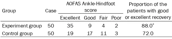
At 1-year follow-up after surgery, among the 50 patients in the experiment group, 35 had excel-lent postoperative ankle recovery and 9 had good recovery, with 88.0% of them achieving good or excellent postoperative ankle recovery; among the 50 patients in the control group, 19 had excellent postoperative ankle recovery and 17 had good recovery, with 72% of them achiev
-The ankle joint is a weight-bearing joint, primar-ily composed of the talus and the lower end of the tibia and fibula. A common ankle fracture is the intra-articular fracture. Poor reduction of ankle fracture tends to result in traumatic ankle arthritis in patients, with numb, stiff and painful ankle, or even difficulty in ambulation. Th-erefore, a good understanding of the anatomic principle with regard to recovery of the frac -tured ankle joint to normal state in the treat-ment of ankle fracture is essential to promote healing and functional recovery of the fractured ankle [11].
[image:4.612.86.375.98.164.2]Currently, open reduction and internal fixation is a common technique for treating ankle frac-ture, which has been accepted by a large num -ber of clinicians. Nevertheless, the results from multiple follow-up studies indicate that open reduction and internal fixation still has such dis -advantages as large incisions and postopera-tive scar hypertrophy affecting the appearance of the fractured limbs; damages to the soft tis-sues around the fracture sites and to the rich vascular network on the periosteum, which is prone to the complications including exposure of the internal fixation and wound infection; pro -longed time to ambulation, which is not condu-cive to callus formation and postoperative recovery [12-16]. As a result, the technique is hard to accept among patients in recent years. Percutaneous compression cannulated screw fixation is a newly-developed minimally invasive surgery. Under the guidance of C-arm fluoros -copy, the fracture of the patient was firstly treated with the use of manipulative reduction and then cannulated screws were percutane-ously implanted for internal fixation. This proto -col integrates the advantages of both closed reduction and external fixation and open reduc -tion and internal fixa-tion, and overcomes the disadvantages of the two techniques. Ad- ditionally, it minimizes secondary damages to Table 3. Comparison of AOFAS Ankle-Hindfoot score between two
groups
Group Case
AOFAS Ankle-Hindfoot
score patients with good Proportion of the or excellent recovery Excellent Good Fair Poor
Experiment group 50 35 9 4 2 88.0*
Control group 50 19 17 11 3 72.0
Note: *P<0.05 for the comparison between the experiment group and with the control group
ing good or excellent postop-erative ankle recovery. The proportion of patients with good or excellent postopera-tive ankle recovery in the experiment group was greater than that of the control gr- oup (χ2=4.000, P=0.046), as
the soft tissues surrounding the fracture site, promoting the healing of ankle fracture. Percutaneous compression cannulated screw fixation is a preferred technique for patients with severe local contusion, poor physical con -dition and intolerance to a long time surgery. However, the technique is ineffective in the treatment of old medial malleolus fractures including vertical compression and comminut-ed mcomminut-edial condyle fractures [17]. It is notcomminut-ed that percutaneous compression cannulated screw fixation has a strict requirement for the clinician to have a good understanding of the indications to the treatment. For the patients with ankle fractures, the proper surgical meth-od should be chosen based on their respective fracture types.
In the current study, we enrolled 100 patients with unilateral ankle fractures to compare the clinical efficacy of open reduction and internal fixation and percutaneous compression cannu -lated screw fixation in the treatment of ankle fractures. The findings of the study indicated that greater improvements in intraoperative bleeding, operation duration, the time to initi-ate restricted weight-bearing ambulation, hos-pital stay and fracture healing duration, a high -er fracture healing rate, a low-er rate of postoperative complications and a larger pro-portion of patients with good or excellent post-operative ankle recovery were noted in the patients with percutaneous compression can-nulated screw fixation as compared with those with open reduction and internal fixation. The above results are basically consistent with those reported by Weinraub et al. and Barnes H et al. [18, 19]. According to another study, in the treatment of patients with medial malleo-lus fractures, closed reduction and internal fix -ation was associated with better and earlier postoperative ankle functional recovery than open reduction and internal fixation [20]. This study suggests that percutaneous compres -sion cannulated screw fixation is more effec -tive than open reduction and internal fixation, which may be due to the small incision which precludes extensive periosteal stripping and damages to the blood supply at the fracture site. All this minimized the surgery-induced trauma. In addition, the use of pressurizing cannulated screws at the broken end increased the torsional and shear strength and ensured stability of internal fixation, which is contribute
to the implementation of earlier functional exer-cise in patients [21, 22].
In conclusion, percutaneous compression can-nulated screw fixation and open reduction and internal fixation were employed for clinical treatment of ankle fractures, but the former was associated with smaller trauma, a lower rate of complications, and a more active role in early function recovery in the ankles of patients. However, there are some limitations in this study, such as a single center study, a small sample size, short follow-ups, as well as inabil-ity to confirm the long-term outcomes of the above surgical techniques in treatment of ankle fractures. Additional studies warrant for further validation.
Disclosure of conflict of interest
None.
Address correspondences to: Maoxiu Peng, De-
partment of Orthopedics, Ruian People’s Hospital,
No.168, Ruifeng Avenue, Ruian, Wenzhou 325200, Zhejiang Province, P. R. China. Tel: +86-0577-65866013; Fax: +86-0577-+86-0577-65866013; E-mail: maoxiupeng93@163.com
References
[1] Goost H, Wimmer MD, Barg A, Kabir K, Valder
-rabano V and Burger C. Fractures of the ankle
joint: investigation and treatment options. Dtsch Arztebl Int 2014; 111: 377-388.
[2] van Zuuren WJ, Schepers T, Beumer A, Sierev
-elt I, van Noort A and van den Bekerom MPJ. Acute syndesmotic instability in ankle frac -tures: a review. Foot Ankle Surg 2017; 23: 135-141.
[3] Toth MJ, Yoon RS, Liporace FA and Koval KJ. What’s new in ankle fractures. Injury 2017; 48:
2035-2041.
[4] Yeung DE, Jia X, Miller CA and Barker SL. Inter -ventions for treating ankle fractures in
chil-dren. Cochrane Database Syst Rev 2016; 4:
Cd010836.
[5] Rammelt S. Management of ankle fractures in
the elderly. EFORT Open Rev 2016; 1:
239-246.
[6] Sun R, Li M, Wang X, Li X, Wu L, Chen Z and
Chen K. Surgical site infection following open reduction and internal fixation of a closed an -kle fractures: a retrospective multicenter
co-hort study. Int J Surg 2017; 48: 86-91. [7] White TO, Bugler KE, Appleton P, Will E, Mc
versus standard open reduction and internal
fixation for fixation of ankle fractures in elderly patients. Bone Joint J 2016; 98-b: 1248-1252. [8] Schairer WW, Nwachukwu BU, Dare DM and
Drakos MC. Arthroscopically assisted open re
-duction-internal fixation of ankle fractures: sig
-nificance of the arthroscopic ankle
drive-through sign. Arthrosc Tech 2016; 5: e407-412.
[9] Kim MB, Lee YH, Kim JH, Lee JE and Baek GH.
Lateral transmalleolar approach and
minis-crews fixation for displaced posterolateral frag -ments of posterior malleolus fractures in
adults: a consecutive study. J Orthop Trauma
2015; 29: 105-109.
[10] Smith M, Medlock G and Johnstone AJ.
Percu-taneous screw fixation of unstable ankle frac -tures in patients with poor soft tissues and
significant co-morbidities. Foot Ankle Surg
2017; 23: 16-20.
[11] Qin C, Dekker RG, Blough JT and Kadakia AR. safety and outcomes of inpatient compared
with outpatient surgical procedures for ankle
fractures. J Bone Joint Surg Am 2016; 98:
1699-1705.
[12] Zwipp H, Rammelt S, Endres T and Heineck J. High union rates and function scores at mid -term followup with ankle arthrodesis using a four screw technique. Clin Orthop Relat Res 2010; 468: 958-968.
[13] Lovy AJ, Dowdell J, Keswani A, Koehler S, Kim
J, Weinfeld S and Joseph D. Nonoperative ver-sus operative treatment of displaced ankle fractures in diabetics. Foot Ankle Int 2017; 38: 255-260.
[14] Gonzalez TA, Macaulay AA, Ehrlichman LK,
Drummond R, Mittal V and DiGiovanni CW.
Ar-throscopically assisted versus standard open reduction and internal fixation techniques for
the acute ankle fracture. Foot Ankle Int 2016; 37: 554-562.
[15] Bariteau JT, Hsu RY, Mor V, Lee Y, DiGiovanni CW and Hayda R. Operative versus nonopera -tive treatment of geriatric ankle fractures: a
medicare part a claims database analysis.
Foot Ankle Int 2015; 36: 648-655.
[16] Pires RE, Mauffrey C, de Andrade MA, Figueire
-do LB, Giordano V, Belloti JC and -dos Reis FB. Minimally invasive percutaneous plate osteo
-synthesis for ankle fractures: a prospective observational cohort study. Eur J Orthop Surg
Traumatol 2014; 24: 1297-1303.
[17] Casstevens C, Le T, Archdeacon MT and Wyrick
JD. Management of extra-articular fractures of
the distal tibia: intramedullary nailing versus plate fixation. J Am Acad Orthop Surg 2012;
20: 675-683.
[18] Weinraub GM, Levine P, Shi E and Flowers A. Comparison of medial malleolar fracture heal-ing at 8 weeks after open reduction internal
fixation versus percutaneous fixation: a retro
-spective cohort study. J Foot Ankle Surg 2017;
56: 277-281.
[19] Barnes H, Cannada LK and Watson JT. A clini
-cal evaluation of alternative fixation tech
-niques for medial malleolus fractures. Injury
2014; 45: 1365-1367.
[20] Sagi HC, Shah AR and Sanders RW. The func
-tional consequence of syndesmotic joint mal
-reduction at a minimum 2-year follow-up. J Or -thop Trauma 2012; 26: 439-443.
[21] Cicekli O, Ozdemir G, Uysal M, Bicici V and Bin
-gol I. Percutaneous cannulated screw fixation for pediatric epiphyseal ankle fractures.
Springerplus 2016; 5: 1925.
[22] Saini P, Aggrawal A, Meena S, Trikha V and
Mit-tal S. Miniarthrotomy assisted percutaneous screw fixation for displaced medial malleolus