Original Article
miR-215 controls proliferation, invasion, and
apoptosis of human retinoblastoma cells
by regulating RB1 expression
Peng Zhang1*, Xiuli Tao1*, Pei Li1, Luxin Ma2
1Department of Ophthalmology, The Fifth People’s Hospital of Jinan, Jinan 250022, China; 2Department of
Ophthalmology, Shandong Provincial Hospital Affiliated to Shandong University, Jinan 250022, China. *Co-first
authors.
Received November 27, 2017; Accepted June 20, 2018;Epub September 15, 2018; Published September 30, 2018
Abstract: Purpose: The aim was to determine the effect of miR-215 on retinoblastoma cells proliferation, invasion, and apoptosis. Methods: ACBRI-181, HXO-Rb44 and SO-Rb50 cells were cultured. HXO-Rb44 cells were transfect- ed. qRT-PCR, Western blot, MTT assay, transwell assay, flow cytometry analysis, and luciferase reporter assay were conducted. In vivo studies with nude mice were performed. Results: miR-215 in retinoblastoma cells was signifi-cantly up-regulated (P < 0.01). Compared with the Blank group and NC group, significantly higher OD495 value and invasive cell number and significantly lower apoptotic cells percentage was found in the miR-215 mimic group (P < 0.05), while the exact opposite result was found in the miR-215 inhibitor group. RB1 relative expression was sig-nificantly decreased (P < 0.05) in miR-215 mimic group and was significantly increased in miR-215 inhibitor group (P < 0.05). RB1 was the target gene of miR-215. siRNA-RB1 + mimic group was with much higher OD495 value and invasive cell number and much lower apoptotic cells percentage when compared with the siRNA-RB1 group (P < 0.05). Nude mice in the siRNA-RB1 + mimic group had a much higher tumor volume compared with that in the siRNA-RB1 group (P < 0.05). Conclusions: miR-215 affected HXO-Rb44 cells proliferation, invasion, and apoptosis through regulation of RB1 protein expression.
Keywords: miR-215, RB1, retinoblastoma, proliferation, invasion, apoptosis
Introduction
Retinoblastoma was an intraocular cancer that occurred frequently in infancy and childhood. Approximately 9,000 new cases of retinoblas-toma were diagnosed every year in the world [1]. Retinoblastoma seriously affected patients’ visual acuity and in most cases surgery for enu-cleation was necessary [2, 3]. More seriously, once a high-risk retinoblastoma was metasta-sized, the disease was very difficult to control and eventually led to an increased risk of death [4]. Therefore, early diagnosis was very impor-tant to improve the prognosis of patients with retinoblastoma.
With the further development of molecular biology techniques, the discovery of potential tumor biomarkers had realized the prevention and early diagnosis of tumors, which was of
found that miR-215 might affect cell prolifera-tion in gastric cancer by targeting RB1. This view was recognized by Wei et al. [10] and they also found that up-regulation of miR-215 pro-moted the migration and invasion of glioma cells by inhibiting the expression of the RB1. Similar research also revealed a significantly reduced proliferation rate of hepatoma cells in nude mice after miR-215 expression was inhib-ited [11]. Many other studies also confirmed the close relationship between miR-215 and tumors, and miR-215 was also considered as a potential biomarker for the early diagnosis and treatment of these tumors.
Unfortunately, as for the relationship between miR-215 and retinoblastoma, the related re- search was very rare. So in this study, we spec-ulated that miR-215 might play a role in retino-blastoma based on previous studies, and relat-ed mechanism was further researchrelat-ed. This study would provide important clinical guidance for prevention and early diagnosis of retinoblas-toma at molecular level.
Materials and methods
Cell culture, transfection and grouping
Human normal retinal vascular endothelial cell line (ACBRI-181) and retinoblastoma cell lines (HXO-Rb44 and SO-Rb50) were all purchased from American Type Culture Collection. These cells were cultured in DMEM containing 10% fetal bovine serum (FBS) in an incubator at 37°C, 5% CO2, respectively. Then they were col-lected at logarithmic growth phase. After sus-pended with DMEM (10% FBS), these cells were seeded in 6-well plates at a density of 1 * 105/ mL with 1 mL cell suspension each well. For HXO-Rb44 cells, they were transfected by miR-215 mimic and miR-215 inhibitor, and were named as the 215 mimic group and miR-215 inhibitor group, respectively. As NC group, the miR-215 negative mimic was also used to transfect HXO-Rb44 cells. 215 mimic,
miR-215 inhibitor and miR-miR-215 negative mimic were bought from Genepharma company (Shang- hai, China). Furthermore, siRNA/RB1 transfec-tion was performed on HXO-Rb44 cells and these cells were named as siRNA-RB1 group. We also co-transfected HXO-Rb44 cells using miR-215 mimic and siRNA/RB1, which was set at the siRNA-RB1 + mimic group. The siRNA/ RB1 sequence was synthesized by Shanghai Bioengineering Company (China). In addition, HXO-Rb44 cells without any treatment were set as the Blank group. Transfection was per-formed using Lipofectamine 2000 (purchased from the American Company Invitrogen) accord-ing to the instructions. Cells that were success-fully transfected were used for subsequent studies. All these cells were kept in the incuba-tor under the same conditions after seeded in 6-well plates at a density of 1 * 105/mL. A total of 1 mL cell suspension was added into each well, respectively.
Real-time fluorescence quantitative RT-PCR
Total RNA of cells was extracted by using miR-Neasy Mini Kit (obtained from Germany Qia- gen company). Reverse transcriptase kit (bo- ught from Germany Qiagen company) was us- ed to perform reverse transcription reaction. PCR amplification reactions were conducted in PCR instrument with a 20 μL reaction system, including 1 μL template cDNA, 1 μL forward primer and 1 μL reverse primer. The reaction conditions were as follows: degeneration at 95°C for 10 seconds, reannealing at 58°C for 20 seconds, extension at 72°C for 34 seconds. The reaction was cycled 40 times. U6 was used as an internal reference. The forward and reverse primer sequences of miR-215 and U6 were shown in Table 1. Data were analyzed using the 2-ΔΔCt method.
MTT assay
[image:2.612.91.288.95.164.2]Cells were incubated for 24, 48 and 72 hours and then 20 μL of MTT solution (5 mg/mL, Si- gma) was added into each well for additional 4 hours of incubation at 37°C. Total 150 μL dimethyl sulfoxide (DMSO) solutions was added into each well after the upper liquid was dis-carded, and the plate was shaken for 10 min-utes to promote formazan crystals dissolution. Finally, the absorbance value of each well was measured at 495 nm by enzyme-linked immu-nosorbent assay.
Table 1. Primer sequences of miR-215 and internal reference
Name of primer Sequences
Transwell experiment
Cells were resuspended in serum-free medi- um and then were seeded in the upper ch- amber of a 6-well MatrigelTM Invasion Chamb- er (BD Biosciences, San Diego, CA, USA) coat- ed with Matrigel. Medium containing 10% FBS was added into the bottom chamber. After incu-bation for 48 hours, cells that passed through the membrane were fixed with 10% formalde-hyde and stained by 0.05% crystal violet. The number of cells that passed through the mem-brane was counted under microscope. The greater the number of cells, the greater the invasive ability.
Flow cytometry analysis
After 48 hours of incubation, cells were har-vested and washed with PBS and fixed by ice-cold 70% ethanol. Then these cells were centri-fuged after incubation for 12 hours at 4°C, and were resuspended with PBS. Ethidium bromide (50 μg/mL) and RNaseA (100 μg/mL) was added before incubated in the dark at 4°C for 30 minutes. Cell cycle analysis was conduct-ed by flow cytometry.
Western blot
Cells to be tested were collected after 48 ho- urs of incubation and total protein was extract-ed and electrophoresextract-ed. After electrophoresis, the protein was transferred to the PVDF mem-brane using wet transfer method. Skimmed milk powder (5%) was used to block the mem-brane for 2 hours at room temperature. Then the membrane was placed in incubations with primary antibodies (rabbit rat primary anti-bodies, 1:1000, Invitrogen) overnight at 4°C. HRP-labeled secondary antibody (1:5000) was added to incubate for 1 hour at room tempera-ture after the membrane washed by TBST buf-fer for three times. After the same TBST clean-ing procedure, chemiluminescence and data analysis were performed. U6 was used as an internal reference.
Luciferase reporter assay
[image:3.612.89.286.69.230.2]Target Scan determined that the RB1 3’UTR was the targeting binding sites of RB1 and miR-215 (Figure 6A). The RB1 3’UTR was insert- ed into psiCHECK-2 vector after amplified by PCR. The mutated- and wild-type sequences of the miR-215 binding sites were designed and inserted into psiCHECK-2 report vectors, re- spectively. Co-transfection of HXO-Rb44 cells was performed by using miR-215 mimic and mutated-type psiCHECK-2 Report vectors or wild-type psiCHECK-2 Report vectors, and was set as MT + mimic group or WT + mimic group, respectively. In addition, miR-215 negative mi- mic and mutated-type psiCHECK-2 Report vec-tors or wild-type psiCHECK-2 report vecvec-tors were also used to co-transfect HXO-Rb44 cells, and were named as MT + NC group or WT + NC group, respectively. Cells were collected after 48 hour incubation and assayed by using Dual Luciferase Assay (Promega).
Figure 1. Up-regulation of miR-215 expression in reti-noblastoma cells. *P < 0.01 when compared to the expression of miR-215 in ACBRI-181 cells.
[image:3.612.91.286.292.463.2]In vivo transplantation experiment
Fifteen nude mice were housed in a sterile isolator at room temperature. Feed, water and other items in contact with the mice were al- so autoclaved. After 2 days of feeding, cells of the Blank group, siRNA-RB1 group, and siR-NA-RB1 + mimic group were collected in loga- rithmic growth phase and were made into cell suspension at a density of 1 * 107/mL. A tot- al volume of 200 μL cell suspension was sub- cutaneously injected into the mice. Tumor vol-ume was measured weekly and was continu-ously observed for 5 weeks. All operations were in line with the ethical standards of animal experiments.
Statistical analysis
All data were processed using SPSS 18.0 sta-tistical software, and expressed as mean ± SD. Data were analyzed using t-test and P < 0.05 was considered statistically significant.
Results
Upregulation of miR-215 in retinoblastoma cells
The results of qRT-PCR showed that the rela- tive expression of miR-215 in HXO-Rb44 ce- lls and SO-Rb50 cells was significantly higher than that in ACBRI-181 cells (P < 0.01), which indicated that expression of miR-215 in retino-blastoma cells was significantly up-regulated (Figure 1).
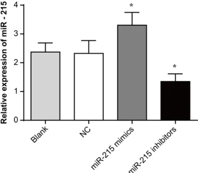
Expression of miR-215 in HXO-Rb44 cells after transfection
Compared with the Blank and NC groups, the relative expression of miR-215 in the miR-215 mimic group was significantly increased (P < 0.05) and significantly decreased miR-215 rela-tive expression was found in miR-215 inhibitor group (P < 0.05). These results demonstrated that miR-215 expression was successfully reg-ulated after transfection (Figure 2).
Effect of miR-215 expression on proliferation and invasion of HXO-Rb44 cells
Of the Blank group, NC group, miR-215 mimic group, and miR-215 inhibitor group, there was no significant difference in OD495 value am- ong all groups at 24 hours. However, at 48 and 72 hours, compared to the Blank gro- up and the NC group, the OD495 value of miR-215 mimic group was significantly increased (P < 0.05) and the OD495 value of the miR-215 inhibitor group was significantly decreased (P < 0.05) (Figure 3A). This result suggested that up-regulation of miR-215 promoted prolifera-tion of HXO-Rb44 cells, and inhibiprolifera-tion of miR-215 expression could attenuate HXO-Rb44 cell proliferation.
[image:4.612.91.523.70.230.2]In addition, at 48 hours, the number of cells passing through the membrane of the miR-215 mimic group was significantly higher than that of the Blank and NC groups (P < 0.05), and at the same time, significantly lower cell number passing through the membrane of miR-215
inhibitor was found (P < 0.05) (Figure 3B), which indicated that up-regulation of miR-215 could enhance the invasion ability of HXO-Rb44 cells, while down-regulation of miR-215 could weaken HXO-Rb44 cells invasion ability.
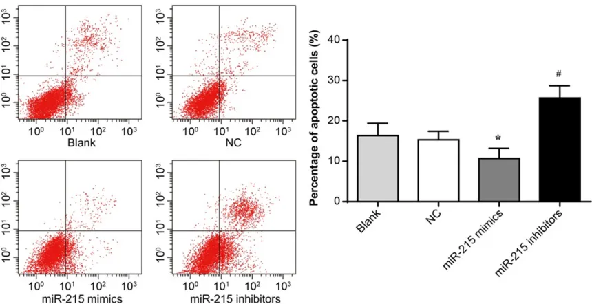
Effect of miR-215 expression on apoptosis of HXO-Rb44 cells
At 48 hours, compared with the Blank group (16.32 ± 2.49%) and the NC group (15.43 ± 1.70%), the percentage of apoptotic cells in the miR-215 mimic group was significantly decreased (10.67 ± 2.05%) (P < 0.05) while that in the miR-215 inhibitor group (25.67 ±
2.49%) was significantly increased (P < 0.05) (Figure 4). miR-215 expression had a negative regulatory effect on the apoptosis of HXO-Rb44 cells.
miR-215 regulated RB1 protein expression
[image:5.612.91.521.70.291.2]Compared with the Blank group and the NC group, the relative expression of RB1 protein in miR-215 mimic group was significantly dec- reased (P < 0.05), while that in miR-215 inhibi-tor group was significantly increased (P < 0.05) (Figure 5). miR-215 negatively regulated RB1 protein expression.
Figure 4. Effect of up-regulation or down-regulation of miR-215 expression on apoptosis of HXO-Rb44 cells. *P < 0.05 when compared with the Blank and NC groups; #P < 0.01 when compared with the Blank and NC groups.
[image:5.612.93.521.347.523.2]RB1 was the target gene of miR-215
Target Scan, an online prediction software, identified the 3’-UTR region as the target site
for the binding of RB1 and miR-215 (Figure 6A). Luciferase reporter assay was performed to further confirm the targeting between RB1 and miR-215 and the results showed no significant
[image:6.612.85.522.73.646.2]Figure 6. miR-215 directly targeted RB1. A. Target Scan pre-dicted the target site for RB1 and miR-215; B. Dual luciferase reporter gene activity assay. *P < 0.05 when compared with the WT + NC group.
difference in luciferase activity between the MT + mimic group and the MT + NC group. How- ever, significantly decreased luciferase activity occurred in the WT + mimic when compared with the WT + NC group (P < 0.05), and lucifer-ase activity was significantly reduced by about 58% (Figure 6B).
miR-215 affected proliferation, invasion, and apoptosis of HXO-Rb44 cells through regula-tion of RB1 protein expression
MTT assay showed that no significant differ-ence was found in OD495 value among the Blank group, the RB1 group, and the siRNA-RB1 + mimic group at 24 hours (P > 0.05). However, at 48 and 72 hours, compared with the Blank group, the OD495 value of the
siRNA-above results showed that miR-215 affected proliferation, invasion, and apoptosis of HXO-Rb44 cells through regulation of RB1 protein expression.
miR-215 affected proliferation, invasion, and apoptosis related proteins expression by regu-lation of RB1
[image:7.612.89.376.71.237.2]In this research, we further studied miR-215 effects on proliferation, invasion, and apopto-sis related proteins expression. We observed that compared with the Blank group, cells of the siRNA-RB1 and siRNA-RB1 + mimic groups were both with much higher c-myc and MMP2 relative expression, as well as obviously lower caspase-3 relative expression (P < 0.05). Fur-
Figure 8. miR-215 affected proliferation, invasion, and apoptosis related proteins expression by regulation of RB1. A. Western blot analysis of c-myc, MMP2, and caspase-3 proteins; B. Statistical histogram of c-myc, MMP2, and caspase-3 proteins relative expression. **P < 0.01 when compared with the Blank group; #P < 0.05 when compared with the Blank or siRNA-RB1 group.
Figure 9. Up-regulation or down-regulation of miR-215 regulated the growth of HXO-Rb44 tumors in nude mice. A. Tumor tissues of each group at 5 weeks; B. Tumor volume of each group at 1, 2, 3, 4 and 5 weeks. *P < 0.05 when compared with the Blank group; #P < 0.05 when compared with the Blank or siRNA-RB1 group.
RB1 group and the siRNA-RB1 + mimic group was significant-ly higher (P < 0.05). At these two time points, the siRNA-RB1 + mimic group had a much higher OD495 value when compared with the siR-NA-RB1 group (P < 0.05) (Figure 7A).
Transwell assay illustrated that the number of cells pa- ssing through the membrane of the RB1 and siRNA-RB1 + mimic groups was sig-nificantly higher than that of the Blank group (P < 0.05) at 48 hours. In contrast, the number of cells across the membrane of the siRNA-RB1 + mimic group was significant-ly higher than that of the siRNA-RB1 group (P < 0.05) (Figure 7B).
[image:7.612.89.375.331.436.2]thermore, we also noted that dramatically increased c-myc and MMP2 relative expression and decreased caspase-3 relative expression was found in the siRNA-RB1 + mimic group when compared with the siRNA-RB1 group (P < 0.05) (Figure 8A, 8B). This indicated that miR-215 affected proliferation, invasion, and apop-tosis related proteins expression by regulation of RB1.
Nude mice in vivo transplantation experiments
There was no significant difference in tumor size among the Blank group, the siRNA-RB1 group, and the siRNA-RB1 + mimic group at 1, 2, and 3 weeks. However, at 4 and 5 weeks, the tumor volume of the siRNA-RB1 group and the siRNA-RB1 + mimic group was significantly higher than that of the Blank group (P < 0.05). Furthermore, nude mice in the siRNA-RB1 + mimic group had a much higher tumor volume compared with nude mice in the siRNA-RB1 group (P < 0.05) (Figure 9A and 9B). These results further confirmed that miR-215 affect-ed proliferation, invasion, and apoptosis of HXO-Rb44 cells through regulation of RB1 pro-tein expression.
Discussion
Much evidence suggested that miRNAs played an important role in tumor development [2, 12, 13]. They could play a biological role in cancer promotion or tumor suppression, and could be used as an effective biomarker for early diagno-sis and prognodiagno-sis of tumors [14-16]. In this paper, the function of miR-215 in retinoblasto-ma was researched through in vitro as well as
in vivo studies, and the results revealed that miR-215 was up-regulated in retinoblastoma cells. Further study demonstrated that miR-215 affected the proliferation, invasion, and apoptosis of retinoblastoma as well as c-myc, MMP2 and caspase-3 proteins relative expres-sion by negatively regulating expresexpres-sion of RB1. It regulated RB1 expression at mRNA and pro-tein levels through binding to the 3’-UTR region. Research findings involving miR-215 expres-sion level were not consistent. Several existing studies showed that miR-215 expression in some types of cancers was down-regulated [8, 17]. For example, researchers have found that miR-215 was acted as a tumor suppressor in colorectal cancer, human non-small cell lung
cancer, and epithelial ovarian cancer [18-20]. However, other studies have revealed that up-regulation of miR-215 expression was occurred in some other tumors, including glioma and gastric cancer [9, 21-25]. Zang et al. [24] observed that miR-215 was frequently overex-pressed in gastric cancer and it promoted gas-tric cancer cells migration and invasion through regulating FOXO1. Another study also illustrat-ed the similar role of miR-215 in gastric cancer [26]. Our results suggested that miR-215 was up-regulated in retinoblastoma cells. Overex- pression of miR-215 was associated with sig-nificantly increased HXO-Rb44 cells prolifera-tion ability as well as invasion ability, while dra-matically decreased proliferation ability and invasion ability were found when the miR-215 was down-regulated by transfection with miR-215 inhibitors. Furthermore, the data also illus-trated an exact opposite relationship between the HXO-Rb44 cells apoptotic capacity and miR-215 expression level. Dramatically decre- ased percentage of apoptotic HXO-Rb44 cells was occurred when expression of miR-215 was up-regulated while down-regulation of miR-215 expression resulted in an increase in the per-centage of apoptotic HXO-Rb44 cells. Our results demonstrated that miR-215 was an oncogene in retinoblastoma, which could be used as a potential biomarker of retinoblasto-ma development.
miR-215 could enhance the proliferation of gastric cancer cells via inhibiting the expres-sion of RB1 [9]. Our work further proved this mechanism in retinoblastoma that miR-215 was through the negative regulation of RB1 to achieve its effect on retinoblastoma cells prolif-eration, invasion, and apoptosis as well as their related protein expression, such as c-myc, MMP2, and caspase-3. C-myc, MMP2, and cas-pase-3 could directly affected proliferation, invasion, and migration of tumor cells, respec- tively.
In conclusion, this study explored the role and mechanisms of miR-215 in affecting the devel-opment of retinoblastoma through in vitro study and in vivo study with nude mice. The results showed that miR-215 was significantly up-regu-lated in retinoblastoma cells and it affected retinoblastoma cell proliferation, invasion, and apoptosis as well as c-myc, MMP2, and cas-pase-3 protein expression by negatively regu-lating the expression of RB1. miR-215 could be used as a potential biomarker for the clinical treatment retinoblastoma. This paper had important guiding significance for the clinical treatment of retinoblastoma. Clinicians could use some drugs that had specific inhibitory effects on miR-215 to treat patients with retinoblastoma.
Disclosure of conflict of interest
None.
Address correspondence to: Luxin Ma, Department of Ophthalmology, Shandong Provincial Hospital Affiliated to Shandong University, 324 Jing Wu Wei Qi Road, Jinan 250021, Shandong, China. Tel: +86-531-68773176; Fax: +86-+86-531-68773176; E-mail: wyyk7073@163.com
References
[1] Dimaras H, Kimani K, Dimba EA, Gronsdahl P, White A, Chan HS and Gallie BL. Retinoblasto-ma. Lancet 2012; 379: 1436-1446.
[2] Fabian ID, Naeem Z, Stacey AW, Chowdhury T, Duncan C, Reddy MA and Sagoo MS. Long-term visual acuity, strabismus, and nystagmus outcomes following multimodality treatment in group d retinoblastoma eyes. Am J Ophthalmol 2017; 179: 137.
[3] Abramson DH, Fabius AW, Issa R, Francis JH, Marr BP, Dunkel IJ and Gobin YP. Advanced unilateral retinoblastoma: the impact of
oph-thalmic artery chemosurgery on enucleation rate and patient survival at MSKCC. PLoS One 2015; 10: e0145436.
[4] Fabian ID, Stacey AW, Chowdhury T, Duncan C, Karaa EK, Scheimberg I, Reddy MA and Sagoo MS. High-risk histopathology features in pri-mary and secondary enucleated international intraocular retinoblastoma classification group D eyes. Ophthalmology 2017; 124: 851. [5] Wijnhoven BP, Hussey DJ, Watson DI, Tsykin A,
Smith CM and Michael MZ. MicroRNA profiling of Barrett’s oesophagus and oesophageal ad-enocarcinoma. Br J Surg 2010; 97: 853. [6] Karaayvaz M, Pal T, Song B, Zhang C,
Georga-kopoulos P, Mehmood S, Burke S, Shroyer K and Ju J. Abstract 2263: prognostic signifi-cance of miR-215 in colon signifi-cancer. Cancer Re-search 2011; 71: 340-347.
[7] Chiang Y, Song Y, Wang Z, Liu Z, Gao P, Liang J, Zhu J, Xing C and Huimian XU. microRNA-192, -194 and -215 are frequently downregulated in colorectal cancer. Exp Ther Med 2012; 3: 560. [8] Li S, Gao J, Gu J, Yuan J, Hua D and Shen L.
MicroRNA-215 inhibits relapse of colorectal cancer patients following radical surgery. Med Oncol 2013; 30: 549.
[9] Deng Y, Huang Z, Xu Y, Jin J, Zhuo W, Zhang C, Zhang X, Shen M, Yan X and Wang L. MiR-215 modulates gastric cancer cell proliferation by targeting RB1. Cancer Lett 2014; 342: 27-35. [10] Wei Y, Sun J and Li X. MicroRNA-215 enhances
invasion and migration by targeting retinoblas-toma tumor suppressor gene 1 in high-grade glioma. Biotechnol Lett 2017; 39: 197-205. [11] Liu F, You X, Chi X, Wang T, Ye L, Niu J and
Zhang X. Hepatitis B virus X protein mutant HBxΔ127 promotes proliferation of hepatoma cells through up-regulating miR-215 targeting PTPRT. Biochem Biophys Res Commun 2014; 444: 128-134.
[12] Zhu L, Zhao J, Wang J, Hu C, Peng J, Luo R, Zhou C, Liu J, Lin J and Jin Y. MicroRNAs are involved in the regulation of ovary develop-ment in the pathogenic blood fluke schistoso-ma japonicum. PLoS Pathog 2016; 12: e1005423.
[13] Chandra S, Vimal D, Sharma D, Rai V, Gupta SC and Chowdhuri DK. Role of miRNAs in de-velopment and disease: lessons learnt from small organisms. Life Sci 2017; 185: 8-14. [14] Zhao Y, Liu X, Zhong L, He M, Chen S, Wang T
and Ma S. The combined use of miRNAs and mRNAs as biomarkers for the diagnosis of pap-illary thyroid carcinoma. Int J Mol Med 2015; 36: 1097-1103.
and prognosis of pancreatic cancer. Clin Chem 2012; 58: 610.
[16] Huang J, Liu J, Chen-Xiao K, Zhang X, Lee WN, Go VL and Xiao GG. Advance in microRNA as a potential biomarker for early detection of pan-creatic cancer. Biomark Res 2016; 4: 20. [17] Wijnhoven BP, Hussey DJ, Watson DI, Tsykin A,
Smith CM, Michael MZ; South Australian Oe-sophageal Research Group. MicroRNA profiling of Barrett’s oesophagus and oesophageal ad-enocarcinoma. Br J Surg 2010; 97: 853-861. [18] Hou Y, Zhen J, Xiaodong XU, Zhen K, Zhu B,
Pan R and Zhao C. miR-215 functions as a tu-mor suppressor and directly targets ZEB2 in human non-small cell lung cancer. Oncol Lett 2015; 10: 1985-1992.
[19] Vychytilova-Faltejskova P, Merhautova J, Mach-ackova T, Gutierrez-Garcia I, Garcia-Solano J, Radova L, Brchnelova D, Slaba K, Svoboda M, Halamkova J, Demlova R, Kiss I, Vyzula R, Conesa-Zamora P and Slaby O. MiR-215-5p is a tumor suppressor in colorectal cancer target-ing EGFR ligand epiregulin and its transcrip-tional inducer HOXB9. Oncogenesis 2017; 6: 399.
[20] Ge G, Zhang W, Niu L, Yan Y, Ren Y and Zou Y. miR-215 functions as a tumor suppressor in epithelial ovarian cancer through regulation of the X-chromosome-linked inhibitor of apopto-sis. Oncol Rep 2016; 35: 1816.
[21] Wang C, Chen Q, Li S, Zhao Z, Gao H, Wang X, Li B, Zhang W and Yuan Y. Dual inhibition of PCDH9 expression by miR-215-5p up-regula-tion in gliomas. Oncotarget 2017; 8: 10287. [22] Meng X and Shi B. miR-215 functions as an
oncogene in high-grade glioma by regulating retinoblastoma 1. Biotechnol Lett 2017; 39: 1351-8.
[23] Xu YJ and Fan Y. MiR-215/192 participates in gastric cancer progression. Clin Transl Oncol 2015; 17: 34.
[24] Zang Y, Wang T, Pan J, Wu R, Ge H, Qu B and Zuo X. miR-215 promotes cell migration and invasion of gastric cancer cell lines by target-ing FOXO1. Neoplasma 2017; 64: 579-587. [25] Na L, Zhang QY, Zou JL, Li ZW, Tian TT, Dong B,
Liu XJ, Ge S, Yan Z and Jing G. miR-215 pro-motes malignant progression of gastric cancer by targeting RUNX1. Oncotarget 2016; 7: 4817. [26] Li N, Zhang QY, Zou JL, Li ZW, Tian TT, Dong B,
Liu XJ, Ge S, Zhu Y and Gao J. miR-215 pro-motes malignant progression of gastric can- cer by targeting RUNX1. Oncotarget 2016; 7: 4817-4828.
[27] Dyson NJ. RB1: a prototype tumor suppressor and an enigma. Genes Dev 2016; 30: 1492. [28] Indovina P, Pentimalli F, Casini N, Vocca I and
Giordano A. RB1 dual role in proliferation and apoptosis: cell fate control and implications for cancer therapy. Oncotarget 2015; 6: 17873. [29] Hu W, Feng Z, Teresky AK and Levine AJ. p53
regulates maternal reproduction through LIF. Nature 2007; 450: 721-724.
[30] Shrestha M and Park PH. p53 signaling is in-volved in leptin-induced growth of hepatic and breast cancer cells. Korean J Physiol Pharma-col 2016; 20: 487-498.
[31] Charni M, Molchadsky A, Goldstein I, Solomon H, Tal P, Goldfinger N, Yang P, Porat Z, Lozano G and Rotter V. Novel p53 target genes secreted by the liver are involved in non-cell-autono-mous regulation. Cell Death Differ 2016; 23: 509-520.