Development of optical bio-sensing methods for detection
of toxins produced by algae.
AL-AMMAR, Rukaiah.
Available from Sheffield Hallam University Research Archive (SHURA) at:
http://shura.shu.ac.uk/19231/
This document is the author deposited version. You are advised to consult the
publisher's version if you wish to cite from it.
Published version
AL-AMMAR, Rukaiah. (2015). Development of optical bio-sensing methods for
detection of toxins produced by algae. Doctoral, Sheffield Hallam University (United
Kingdom)..
Copyright and re-use policy
See
http://shura.shu.ac.uk/information.html
Sheffield Hallam University Research Archive
L e a r n m y c h i u u h u h m c s u u i iw~ .v. «
A d se tts C e n tre , City C a m p u s S heffield 81 1WD
id Hailam University nlormc.f'!on Services
i
Centre, City Cam pus iheffielci S 1 f W P _ProQuest Number: 10694111
All rights reserved INFORMATION TO ALL USERS
The qu ality of this repro d u ctio n is d e p e n d e n t upon the q u ality of the copy subm itted. In the unlikely e v e n t that the a u th o r did not send a c o m p le te m anuscript and there are missing pages, these will be note d . Also, if m aterial had to be rem oved,
a n o te will in d ica te the deletion.
uest
ProQuest 10694111
Published by ProQuest LLC(2017). C op yrig ht of the Dissertation is held by the Author. All rights reserved.
This work is protected against unauthorized copying under Title 17, United States C o d e M icroform Edition © ProQuest LLC.
ProQuest LLC.
789 East Eisenhower Parkway P.O. Box 1346
Development of Optical Bio-sensing Methods for
Detection of Toxins Produced by Algae
Rukaiah Al-Ammar
A thesis subm itted in partial fulfilm ent o f the requirem ents o f
Sheffield Hallam University
For the degree o f Dector o f Philosopy
DECLARATION
I hereby declare that this thesis submitted for the degree o f PhD is the result o f my own
research and that this has not been submitted for a higher degree to any University or
institution.
Signed
ABSTRACT
The main aim o f this work is to develop an optical method for the detection o f microcystin-LR, one o f the most dangerous toxins released by cyanobacteria. Photosynthetic cyanobacteria (also known as blue-green algae) exist in any type o f water (including drinking water) and possess a serious threat to humans and animals, and generally to the environment. Microcystin-LR (MC-LR) is perhaps the most toxic from a large family o f cyanotoxins; it is known to cause liver damage and also acts as a carcinogen. The World Health Organization has set the limit o f lpg/1 for MC-LR in drinking water. However existing detection methods, such as ELISA, cannot provide such high sensitivity. The relatively low molecular weight o f MC-LR (995.2 m/mol) makes it difficult to detect using conventional QCM and SPR based analytical methods. Therefore, the development o f highly sensitive, reliable, and (at the same time) inexpensive and easy-to-use methods o f detection o f microcystin is o f very high importance today. The method o f Total Internal Reflection Ellipsometry (TIRE) is particularly attractive for the above task considering its high sensitivity and particular suitability for detection o f low molecular weight analytes. In this work MC-LR was detected in direct immunoassay with MC10E7 monoclonal antibodies raised against MC-LR in mouse, using TIRE as a detection method. Also the TIRE method was used to study the protein- protein interaction (the protein chaperon with its receptors in the chloroplast) and to detect other types o f toxins such as Aflatoxin B 1 .
At first, the TIRE immunoassay was calibrated using aqueous solutions with known concentration o f MC-LR. The study o f binding kinetics o f MC-LR to MC10E7 yielded the association constant close to 108 1/mol which is typical for highly specific immune reactions. The detection limit for MC-LR between O.lng/ml and lng/m l was achieved using the TIRE detection. Then, the concentration o f the MC-LR toxin produced naturally by algae m icrocystis aeruginosa was evaluated using calibration data obtained for solutions spiked with known concentrations o f MC-LR. The role o f environmental factors (temperature, pH, nutrition contents, and salinity) on the efficiency o f production o f MC-LR by m icrocystis aeruginosa was studied. Purification o f solutions contaminated with MC-LR (both commercial and naturally produced) was achieved using MnCC>3 microparticles coated with polyelecrolytes and functionalised with
ACKNOW LEDGM ENTS
I would like to express my deepest appreciation to my Director o f study, Dr. Alexei
Nabok, for all o f his encouragement, being patient and offering truly valuable support
during this work, and for his useful advice. I greatly enjoyed the learning process and
worthwhile research studies under him. His wide knowledge was a great value to me.
I would like to thank the additional members o f my PhD committee, Dr. Abbass
Hashim, and Dr. Tom Smith, for their fantastic guidance and important discussions
regarding this work.
Thanks are also due to all MERI support staff, especially Veny for his technical help
and support throughout this work. I would also like to thank Zaki for his help and
Hakin. It has been a pleasure working with my colleges in the Laboratory.
Most importantly, I would like to give special thanks to my husband, Maytham, for his
unconditional love and support. His prayers and belief in my ability always encouraged
me to move forward. Thank you.
Finally, I want to say thank you to all my children, Aya, Noralhuda, Sara,and Fatima for
their loving support, patience and understanding throughout my study.
LIST OF ABBREVIATION
MC-LR microcystin-LR
AFT Aflatoxin
Ab Antibody
SEM Scanning electron microscopy
AFM Atomic force microscopy
BSA Bovin Serum Albumin
DNA Deoxyribonucleic acid
ELISA Enzyme-linked immunosorbent assay
IgG Immunoglobulin G
Ka Association constant
Kd Dissociation / affinity constant
LC Liquid chromatography
M n C 03 Magnesium carbonate
MS Mass spectroscopy
PAH Poly(allylamine) hydrochloride
PSS Poly-styrene sulfonate
SPR Surface Plasmon Resonance
TIRE Total internal reflection ellipsometry
WHO World health organization
RACE Rapid Amplification o f cDN A Ends
IP injections Interapretoneal injection
LIST OF PUBLICATIONS
Journals publications:
1. R. A l-Am m ar, A. N abok, A. Hashim, (2014) optical detection and purification o f contam inated substances m icrocystin-LR produced by algae. Sensors & Actuators: B. Chem ical, (in press).
2. R Al-A m m ar, A Nabok, A Hashim and T Smith. (2013) Optical detection o f
m icrocystin produced by cyanobacteria. J. Phys.: Conf. Ser. 450, (1), 012006.
3. A. N abok, V. Erokhin, S. Erokhina, A. Szekacs, M. K. M ustafa & R. A l-A m m ar.
(2013) Extraction o f m ycotoxins from aqueous solutions using functionalized
polyelectrolyte-coated m icroparticles. BioNanoSci, 3 (1), 79-84.
4. V. Kriechbaum er, A. N abok, M.K. M ustafa, R. A l-Am m ar, A Tsargorodskaya,
D.P. Smith, B.M. Abell. (2012) Analysis o f protein interactions at native chloroplast m em branes by ellipsom etry, PLoS One.; 7(3):e34455.
C onference papers:
1. A. N abok, V. K riechbaum er, M.K. M ustafa, A .Tsargorodskaya, R.A l-A m m ar, B.
Abell, D. Sm ith.The study o f interaction o f chaperones with respective receptors assem bled on the surface. 12th European Conference on O rganized Films, ECOF 12 conference, 17-20 July, 2011, Sheffield, UK.
2. R. Al- Am m ar, A. Nabok, A. Hashim & Prof. T. Smith, 21 M ay2011.
Detection o f w ater Pollution using Bio-Sensing Technologies (poster), student day Sheffield Hallam University.
3. R. Al- Am m ar, A. Nabok, A. Hashim & T. Smith, 23Dec 2011.Optical detection o f
M icrocystin-LR produced by cyanobacteria, BM RC ,Sheffield Hallam U niversity, Sheffield UK.
4. R. A l-Am m ar, A. Nabok, A. Hashim, T. Smith, Detection o f M icrocystin in Direct Im m unoassay Format Com bined with Total Internal Reflection Ellipsom etry, l l lh
European Conference on Optical Chem ical Sensors and Biosensors,
EU RO PTRO D E XI, 1-4 April, 2012, Barcelona, Spain.
5. R.A l-Am m ar, A. Nabok, A. Hashim, T. Smith, Optical detection o f toxins released by cyanobacteria, 14th International conference on organized m olecular films, ICOM F 14 (LB 14), 10-13 July, 2012, Paris, France.
6. R.Al-Am m ar, A. N abok, A. Hashim, T. Smith, Detection o f m icrocystin in direct im m unoassay form at combined with Total Internal Reflection Ellipsom etry. Institute o f Physics, Sensors & their A pplications XVII, 16-18 Septem ber, 2013, Dubrovnik, Croatia
7. R. Al-Ammar, A. Nabok, T. Smith, Optical detection o f microcystin produced by
cyanobacteria and purification o f contaminated substances using functionalised
polyelectrolyte micro-particles, 12th European Conference on Optical Chemical
Sensors and Biosensors (EUROPTRODE XII), 13-16 April, 2014, Athens, Greece
8. R.Al-Ammar, A. Nabok, A. Hashim, T. Smith. Detection o f water pollution using
bio-sensing technologies, MERI, student day. (Presentation). 20 June 2012.
Sheffield Hallam University.
9. R. Al- Ammar, A. Nabok, A. Hashim, T. Smith. Optical detection o f
Microcystin-LR produced by cyanobacteria, BMRC, 17 Dec 2012, Sheffield Hallam University,
Sheffield UK.
10. R. Al- Ammar, A. Nabok, A. Hashim & T. Sm ith. Production o f microcystin by
cyanobacteria.Purification o f substances Containing microcystin with
functionalized microparticales.BMRC/ MERI Winter Poster Even 22 Dec 2013.
Prizes:
1- Third prize - poster in student day (2011). Sheffield Hallam University R. Al- Ammar, A. Nabok, A. Hashim & T. Smith’
Detection o f water Pollution using Bio-Sensing Technologies (poster).
2- Third prize (poster), BMRC/ MERI Winter Poster Event (2013). Sheffield Hallam University. R. Al- Ammar, A. Nabok, A. Hashim & T. Smith. Production o f microcystin by cyanobacteria.Purification o f substances containing microcystin
with functionalized microparticales.BMRC/ MERI Winter Poster Even 2013.
I was the reviewer With IEEE Sensors Journal for the paper:
Part-Per-Trillion Level Detection o f Microcystin-LR using a Periodic Nanostructure.(2014).
Contents
I. Declaration
II. A cknow ledgem ent
III. Dedication
IV. A bstract
V. List o f Publications
VI. List o f A bbreviations
Chapter Topics P.
1. IN T R O D U C TIO N
1.1 Problem outline 1
1.2 Detection o f m icrocystin. 2
1.3 M ycotoxins: detection and purification o f contam inated substances. 3
1.4 Protein-protein interaction. 4
1.5 Aims and objectives. 5
2. B A C K G R O U N D OF ALG A E AND TO X IN S PR O D U C E D BY ALG A E
2.1 Algae 7
2.2 Cyanobacteria 10
2.3 M icrocystin-LR. 12
2.4 Presence o f M icrocystins in Aquatic System s 16
2.5 Liver toxicity o f m icrocystin-LR 17
2.6 Health-Based Criteria for Safe Exposure to M icrocystin 17
2 .7 Domestic Anim al Poisonings 18
2.8. Effects o f M icrocystins on Fish and W ildlife 19
2.9 Detection o f M icrocystin-LR 21
2.10 Current Treatm ent Technologies for M icrocystin-LR 22
3. TH E C O NC EPT OF BIO SEN SO R S
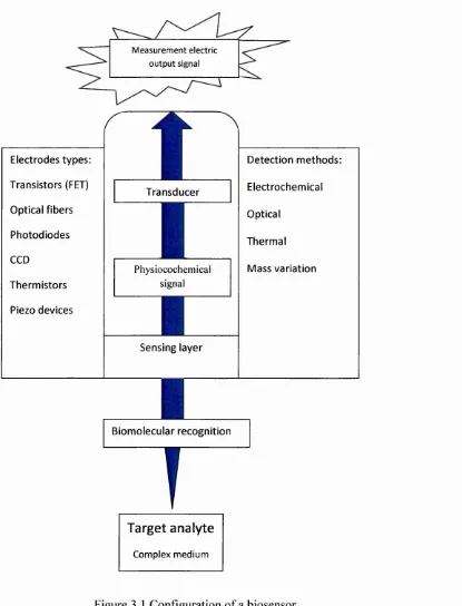
3.1 Biosensors: definitions and classification 24
3.1.1. Importance o f biological sensing system 26
3.1.2 Biosensors world market 27
3.1.3 The need for label-free detection 27
3.2 Im m unosensors 28
3.2.1 A vidity and Affinity 29
3.2.2 A ntibody - Antigen interaction 30
3.2.3 A ntibody imm obilization 34
3.3 A ntibodies production and purification 35
3.4 Optical biosensors and detection o f Immune reaction 36
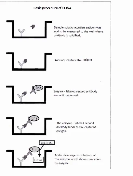
3.4.1 ELISA method 37
3.4.2 SPR (BIA CO RE) 37
3.5 Sum m ary 39
O PTICA L IM M U N O SEN SIN G M ETH O DS: TO TA L IN T ER N A L
R E FLE C T IO N EL LIPSO M ET R Y (TIRE)
4.1 Theoretical background o f evanescent wane technique 40
4.1.1 Ellipsom etry 42
4.1.2 Surface Plasm on Resonance (SPR) 44
4.2 Total Internal Reflection Ellipsom etry (TIRE) 47
4.2.1 TIRE experim ental set up 51
4.2.2 M ethodology o f TIRE experim ents 54
4.2.3 TIRE kinetics analysis 56
COM PLEM ENTARY EXPERIM ENTAL TECHNOLOGUES
5.1 UV-vis. A bsorption Spectrophotom etry 58
5.2 Scanning electron m icroscopy (SEM ) 60
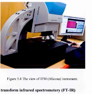
5.3 Infinite Focus M icroscopy (IFM ) 62
5.4 Fourier Transform Infrared Spectrom etry FT-IR 62
5.5 H em ocytom etery 65
5.6 Fluorescence M icroscopy 67
5.7 Langm uir-B lodgett trough 68
5.8 Edwards E360A evaporation unit 70
FU R TH ER D E V E LO PM EN T OF TIRE M ETH O D FO R O T H E R
A P PLIC A TIO N S
6.1 Protein-protein interaction 72
6.1.1 Study o f chaperone interaction with receptors 73
6.1.2 Sample preparation 73
6.1.3 TIRE m easurem ents and data fitting 75
6.1.4 Analysis o f chaperone binding kinetics 79
6.2 The study o f Aflatoxin B1 binding to polyelectrolyte capsules 81
6.2.1 Functionalization o f m icrocapsules 81
6.2.2 UV-vis absorption spectra study 83
6.2.3 Conclusions 87
7.
OPTICAL DETECTION OF M ICROCYSTIN-LR
7.1 Detection o f M icrocystin LR using TIRE 89
7.1.1 Sam ples preparation and TIRE m easurem ents 89
7.1.2 TIRE study o f the imm une reaction between MC LR and M C10E7 90
7.1.3 The study o f M C-LR and M CI 0E7 binding kinetics 93
7.2 Fourier Transform Infrared spectrom etry (FT-IR) m easurem ents 97
7.3 UV-vis spectral study o f m icrocystin-LR 98
7.4 Detection o f M C-LR produced by cyanobacteria; effect 100
o f environm enta factors on algae and toxin production.
7.4.1 Selection o f m icroorganism and growth o f algae 100
7.4.2 The growth in optim al condition 101
7.4.3 Growth m icrocystis aeruginosa under increased 105
and decreased nutrient levels in broth and chanching
the environm ental condition
7.4.4 Extraction o f M C-LR 108
7.4.5 Effect o f salinity on the algae growth 112
7.5 TIRE detection o f naturally produced m icrocystin-LR 114
7.6 M icrocystin-LR cleansing with functionalized polyelectrolyte m icro
particles 117
7.7 Conclusion 117
8.
THE USE OF FUNCTIONALIZED M ICROPARTICALES
AS ABSORBENT FOR M ICROCYSTIN-LR
8.1 M icro encapsulation 118
8.2 Polyelectrolyte m icrocapsules 119
8.3 Preparation o f m icroparticles 121
8.3.1 UV-vis m easurem ent 123
8.3.2 TIRE m easurem ents 124
8.4 Com parison between purification o f natural and com m ercial toxin by
m icrocapsules 126
8.5 Rem oving the core o f the m icrocapsules 127
8.6 M icrocystin-LR removal by electrolysis 133
Conclusions 135
9.
CONCLUSIONS AND FUTURE W O RK
9.1 Conclusions 136
9.2 Future work
137
References
138
LIST OF FIGURES
Figure Page
Figure 2.1 Chemical structure o f microcystin-LR. 14
Figure 3.1 Configuration o f a biosensor. 25
Figure 3.2 Affinity and Avidity. 30
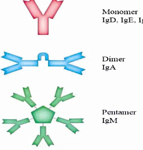
Figure 3.3 Diagram o f the basic unit o f immunoglobulin (antibody). 32
Figure 3.4 the antibody structure. 32
Figure 3.5 Chemical structures o f (a) PAH and (b) PSS; 34
(c) Multilayered film o f alternated layers o f PAH
And PSS deposited on negatively charged surface.
Figure 3.6 (a) various orientations o f antibodies on surface, 35
(b) Improvement o f antibody orientation
using proteins Protein A (or G)
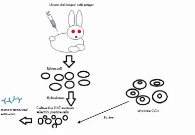
Figure 3.7 Production o f antibodies. 36
Figure 3.8 ELISA methods. 38
Figure. 4.1 (a) Reflection and transmission o f light at the nj-n2 interface; 41
(b) Total internal reflection; (c) The formation o f evanescent wave.
Figure 4.2 Schematic diagrams o f evanescent waves propagating along a
metal-Dielectric interface. 41
Figure 4.3 The formation o f evanescent wave at total internal 42
reflection at the Boundary between two
media with different wave motion properties.
Figure 4.4 Changes in polarization o f light reflected from the surface. 42
Figure 4.5 The schematic o f rotating analyzer spectroscopic ellipsometry. 44
Figure 4.6 Kretschmann (a) and Otto (b) configurations o f an Attenuated Total
Reflection setup for coupling surface plasmons. 45
Figure 4.7 (a) Typical set-up o f an SPR biosensor. 46
Figure 4.8 Experimental setup for surface Plasmon resonance
enhanced ellipsometry.
49
Figure 4.9 Typical TIRE spectra bare gold film on glass. 50
Figure 4.10 (a) J.A. Woollam M 2000 Ellipsometry 51
Figure 4.11 The flow-chart o f data analysis in TIRE. 52
Figure 4.12 Typical time dependencies o f \\f and A extracted
from dynamic TIRE scane at selected wavelength.
55
Figure 5.1 Electronic transitions in organic materials. 59
Figure 5.2 Operation principle o f UV-vis Absorption Spectrophotometry. 59
Figure 5.3 Operation principle o f SEM. 61
Figure 5.4 The view o f IFM (Alicona) instrument. 62
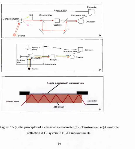
Figure 5.5 (a) the principles o f a classical spectrometer. 64
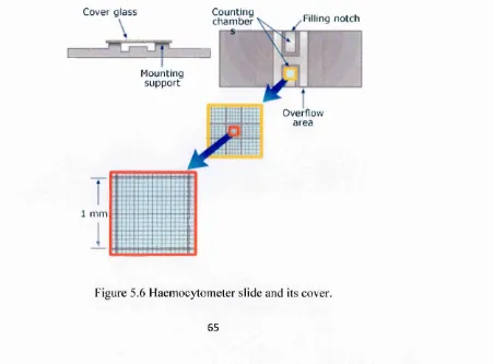
Figure 5.6 Haemocytometer slide and its cover. 65
Figure 5.7 Standerd Haemocytometer chamber. 66
Figure 5.8 Example o f using Haemocytometer for counting cells. 66
Figure 5.9 Schematic diagram o f Fluorescence Microscope 68
Figure 5.10 Schematic diagram o f Langmuir trough for
isotherm measurements
69
Figure 5.11 Schematic diagram o f surface pressure - area
per molecule isotherm.
70
Figure 6.1 (a) typical n-A diagram o f chloroplast
membranes on a water surface;
XIII
(b) Recording n during Langmuir-Schaefer
deposition o f chloroplast.
Figure 6.2 Spectral shift caused by binding
o f Hsp70 chaperones.
Figure 6.3 Hsp70 (a) and Hsp90 (b) chaperones binding
to receptors.
Figure 6.4 Dynamic TIRE measurements and
evaluation o f the association constants.
Figure 6.5 IFM image o f microcapsules functionalized
with antibodies to Aflatoxin B1
Figure 6.6 SEM image microcapsule with antibodies
to Aflatoxin B l.
Figure 6.7 U v-vis absorption spectra o f 1 OOpg/ml
aqueous solution o f AFT B 1.
Figure 6.8 Spectra o f AFT B l solutions o f 20mg/ml (a)
and 50 mg/ml (b) Concentrations mixed with capsules
and incubated for a different time.
Figure 6.9 The dependence o f 362nm band intensity o f
AFT B l on the incubation time.
Figure 7.1 (a) Series o f TIRE spectra recorded after each deposition step,
(b) Calibration curve for MC-LR.
Figure 7.2 Spectral shift without deposited protein A or G.
Figure 7.3 Typical dependencies o f T^t) and A(t)
Evaluation o f the during MC-LR & MC10E7 binding.
Figures 7.4 the evaluation o f the association constant Ka
-Figures 7.5 Binding kinetics yields the association constant Ka.
9 4
96
Figure 7.6 the nagetive test for blocking spaces between 96
the antibody and protien A.
Figure 7.7 Spectral shift for negative control without antibody. 97
Figure 7.8 (a FT-IR spectra o f 1 MC-LR, antibody 98
M C 10E7, and M C 10E7/ MC-LR .
Figure 7.9 UV -vis spectra o f MC-LR solution. 99
Figure 7.10 Optimal condition 25C & sun light. 102
Figure 7.11 Calibration curve for microcystis aeruginosa cells. 103
Figure 7.12 Peak o f commercial toxin in U V -vis at 238 nm. 104
Figure 7.13 Calibration curves for toxin (microcystin-LR) in OD. 104
Figure 7 .1 4 Calibration curves for microcystin-LR by the UV -vis. 105
Figure 7 .1 5 MC-LR release after m icrocystis aeruginosa grown 106
in cultures with increase and decrease nutrient.
Figure 7.16 Cell growths with change in environmental conditions. 107
Figure 7 .1 7 Effect o f environmental conditions on 108
MC-LRreleased by M.aeruginosa.
Figure 7.18 M C -L R extracted from culture 109
which increased 5ml nutrient in it.
Figure 7.19 (a) M icrocystis aeruginosa growth in optimal condition. 111
Figure 7.20 Effect o f salinity on rate growth o f Microcystis aeruginosa . 112
Figure 7.21 a series o f dilutions was made from natural MC-LR. 114
Figure 7.22 kinetic reactions between natural MC-LR and
antibody MC10E7.
Figure 7.23 Calibration curve obtained from TIRE measurements.
Figure 7.24 Correlation between the optical density (absorbance) and
the concentration o f MC-LR.
Figure 8.1 Formation o f polyelectrolyte capsules.
Figure 8.2 Typical SEM image o f hollow microcapsule.
Figure 8.3 SEM images o f 4 pm microcapsules at different
resolutions: scale
(a) 200pm ,(b) 5pm, and (c) 1pm.
Figure 8.4 6 pm microcapsules coted with after (PAH/ PSS)
shell and functionalized with antibodies.
Figure 8.5 U V -vis absorption spectra o f MC-LR (25.9 ug/ml)
during treatment with functionalized microparticles.
Figure 8.6 TIRE measurements taken on commercial
MC-LR samples before and after treatment with
functionalized capsules.
Figure 8.7 TIRE data, thickness increment o f natural MC-LR and
After treatment with microcapsules..
Figure 8.8 Effect o f microparticle treatment on concentration
o f MC-LR: Abs. at238nm .
Figure 8.9 standerd curves for MC-LR
after treatment with capsules.
XVI
115
116
116
119
120
121-122
123
124
125
125
126
Figure 8.10
Figure 8.11
Fig. 8.12
Figure 8.13
Figure 8.14
Figure 8.15
(a Microcapsules with core MnCC>3 6pm
and eight layers (PAH/ PSS).
(a) Microcapsules with core MgCC>3
tested in FT-IR.
(a) microcapsules stained with SYTO-9
Green Fluorescence stain, and (b) Propidium iodide
red Fluorescence stain.
Processes o f fill microcapsules with organic dye.
Absorption spectra o f Microcystin-LR samples
during electrolysis.
LIST OF TABLES
Table Page
Table 4.1. Four-layer TIRE model. 53
Table 6.1 Four-layer TIRE model. 77
Table 6.2 Calculation o f the absorbance reduction caused by 87
adsorption o f AFT on functionalized microparticles.
Table 7.1 TIRE data fitting for the immune reaction between 92
microcystin-LR and MC10E7antibody.
Table 7.2 Concentration o f microcystin-LR released 110
by m icrocystis aeruginosa
under different invironmental conditions.
T able 7.3 the increase in thickness for natural MC-LR. 115
CHAPTER 1
INTRODUCTION
1.1
Problem outline
The presence o f Cyanobacteria (blue-green algae) in surface w ater is o f increasing
concern in Iraq as well as in other parts o f the world. C yanobacteria under the
m icroscope com e into view as small, unicellular organism s, some o f which form
colonies and thus reach sizes visible to the naked eye as tiny green particles. These
organism s are usually finely dispersed throughout the w ater and m ay cause considerable
turbidity if they reach high densities. Human activities (e.g., extensive agriculture,
inadequate sewage treatm ent, ru n o ff from roads) have led to excessive fertilization
(eutrophication) o f m any w ater bodies. This has led to the excessive propagation o f
algae and Cyanobacteria into fresh w ater and thus has had a considerable im pact upon
recreational w ater quality. In hot clim ates, Cyanobacterial dom inance is m ost
pronounced during the sum m er m onths, which coincides with the period w hen the
dem and for recreational w ater is highest. Even though m any species o f freshw ater algae
reproduce quite intensively in eutrophic w aters, they do not accum ulate to form dense
surface bloom s o f extrem ely high cell density, as do some cyanobacteria w hich
naturally produce harm ful com pounds, called cyanotoxins, due to cell lysis during
C yanobacterial bloom s. These toxins m ay cause m ass m ortality o f wild and dom estic
anim als, farm ed fish and shellfish, as well as hum an illnesses such as nervous system
injury or liver damage, and in extrem e cases, death [1].
M icrocystins are the m ost frequently occurring class o f cyanotoxins, o f w hich
m icrocystin-LR is know n to be one o f the m ost toxic cyanotoxins in w ater resources [2].
W hen in contact with skin or consum ed, m icrocystin-LR can lead to skin irritation or
liver injure and m ay initiate liver tum our-prom oting activity [3]. Due to these adverse
health effects, the W orld Health O rganization (W H O ) has established a provisional
guideline o f 1 part per billion ppb (1 pg/L) for m icrocystin-LR in drinking w ater [4].
1.2
Detection o f microcystin
The contam ination o f w ater resources with m icrocystin-LR produced by some algae
species has prom pted the developm ent o f detection m ethods for recognition and
quantification o f toxins. The m ost widespread analytical method for the determ ination
o f m icrocystin-LR in drinking or raw w ater is high-perform ance liquid chrom atography
(HPLC); it is precise but requires expensive equipm ent, com plex procedures, and a long
period o f analysis.
The other m ethods are liquid chrom atography com bined w ith m ass spectrom etry
(LC/M C), thin-layer chrom atography (TLC), and capillary electrophoresis (CE) [5].
A lthough the sensitivity o f chrom atographic techniques is very high (typically in the
range from lp g /L to lp g /L ), these m ethods require tim e-consum ing sam ple preparation
procedures that usually need pre-concentration o f sam ples prior to LC analysis. A m ajor
problem in quantitative analysis o f M Cs is the lack o f standards [6]. Enzym e-linked
im m unosorbant assay (ELISA) m ethod has been widely em ployed to m onitor
m icrocystin-LR at levels below lp g /L [7]. Protein phosphatase inhibition assays (PPIA )
[8] m ay yield false positives if the enzym e is inhibited by other com pounds present in
the sample. Recently, a com m ercial flow dipstick m ethod (M icrocystin Im m unoStrip)
was introduced which is based on colloidal gold particles functionalized with specific
antibodies [9]. Surface plasm on resonance (SPR) has become a w idely used analytical
technique which has led to the developm ent o f an SPR based im m unosensor for the
determ ination o f M C-LR [10]. Furtherm ore, the Quartz crystal m icrobalance (Q CM )
m ethod was developed in com bination with Au nanoparticle am plified sandw iched
im m unoassay lowering the detection lim it for (M C-LR) to 0.1 lpg/L [11]. A nother
detection m ethod using color-changeable polydiacetylene vesicle achieved the lp g /L
detection level [12]. Some studies have focused on developing and im proving
antibodies against (M C-LR), e.g. m onoclonal and polyclonal antibodies [13]. R ecently
developed m ethod o f total internal reflection ellipsom etry (TIRE), w hich records
sim ultaneously tw o param eters A and 'P related to the am plitudes and phases o f p- and
s-com ponents o f polarised light, has been particularly suitable for detection o f low
m olecular w eight m olecules [14], and it could be utilized for detection o f M C-LR.
A nother interesting technology o f polyelectrolyte m icrocapsules [15], functionalized
with specific antibodies, could be used for detection o f m icrocystin-LR as well as for
purification o f substances contam inated with m icrocystin-LR, also these technique w as
used for purification o f substances contam inated w ith m icotoxin.
1.3 M ycotoxins detection and purification o f contaminated substances
M ycotoxins contam inate the diet o f a large proportion o f the w o rld 's population,
especially in low incom e and developing countries. In 1985 the W orld Health
O rganization (W H O ) estim ated that approxim ately 25% o f the w o rld 's grain was
contam inated with m ycotoxins.
M ycotoxins are toxic chem ical com pounds found in certain fungi that can grow on
crops in the field, after harvest, or during storage. Since they are produced by fungi,
m ycotoxins are associated with m ouldy crops. N ow adays there are hundreds o f
m ycotoxins o f different chem ical structures and different m odes o f action, but only five
o f them are regularly found in staple foods and anim al foodstuffs such as grains and
seeds. These m ycotoxins are aflatoxins, zearalenone, ochratoxins, fum onisins and
deozynivalenol/nivalenol.
M ost m ycotoxins are hydrophobic m olecules o f low m olecular w eight and thus are not
soluble in w ater but in organic solvents such as m ethanol, chloroform , acetone, or
acetonitryl. All m ycotoxins are dangerous to hum an and anim al health in connection
with high hepato- and nephro-toxicity, and have carcinogenic, genotoxic, cytotoxic, and
m utagenic actions [16, 17].
A flatoxin, a com m on and naturally w idespread m ycotoxin that is produced by
Aspergillus fungi species, m ost notably A. flavus and A. paraciticus, contam inates a
variety o f staple food. The favorable host plants for aflatoxin are grain cultures and
cereals (maize, rice, wheat, etc.), spices (chili and black pepper, coriander, ginger), high
oil content nuts (alm ond, pistachio, walnut, coconut, Brazil nut) as well as coffee and
cocoa beans, and fruit products [18-20]. It can colonize and contam inate grains before
harvest or during storage. The toxin can also be found in m ilk and m ilk products o f
anim als that are fed w ith contam inated food [21-23]. W ithin the aflatoxin group, the
m ost dangerous toxin is aflatoxin B l (LD50 = 6.5-16.5 m g/kg). The toxicity o f AF-B1
is ten tim es that o f potassium cyanide, 68 tim es that o f arsenic and 416 tim es that o f
m elanin [24].
In addition to a w ide range o f analytical m ethods for detection o f m ycotoxins [25-27],
the method o f TIRE was successfully adapted for detection o f these low m olecular
w eight analytes [28, 29]. In this w ork, however, the m ain focus was not on the detection
m ycotoxins but on purification o f substances contam inated with m ycotoxins. The
technology o f m icroparticles functionalized with antibodies to aflatoxin B l w as further
exploited in this work. The TIRE m ethod along with UV-vis absorption spectroscopy
was used here as an analytical tool for evaluation o f aflatoxin Bl concentration in liquid
sam ples before and after such treatm ent.
1.4 Protein-protein interaction
An additional task in this project was the further developm ent o f the TIRE m ethod
through its application to studying protein-protein interaction. Som e o f the types o f
biological interactions are well studied, nam ely immune reactions (i.e. binding o f
antigen to specific antibodies), enzym e reactions (decom position o f small m olecules
catalyzed by enzym es), hybridization o f single DNA strands due to hydrogen bonding,
etc. However, the interaction o f proteins in general is not w ell-studied and not fully
understood. Very often, we associate m olecular binding which does not have a feasible
explanation with so-called non-specific binding.
In this project, we attem pted to study in m ore detail the interaction betw een chaperones
and their specific receptors. Chaperone proteins play an im portant role in cells
protecting proteins from high tem peratures and other cellular stresses, stabilizing
protein structure and preventing them from aggregation and degradation. It was recently
suggested that m olecular chaperones, such as heat shock proteins Hsp70 and H sp81, can
form com plexes with freshly translated proteins and thus prevent their aggregation [30].
Furtherm ore, the recent finding o f chaperone receptors in plants [31, 32] indicates m ore
specific involvem ent o f m olecular chaperones in protein targeting. The study o f the
m echanism s o f protein targeting m ay have a substantial impact in a num ber o f
applications, including the origin o f neurological diseases. It was shown recently in our
research group that the novel receptor O EP61, extracted from leaves is capable o f
specific binding o f Hsp70 while not binding to Hsp81 [33]. In this work, the interaction
o f chaperones with different receptors (including OEP61) electrostatically im m obilized
on the surface was accessed with the TIRE m easurem ents. A part from confirm ing the
binding properties o f OEP61, this work showed clear separation o f specific and no-
specific binding, in term s o f both the sensor response (i.e. thickness increm ent) and the
affinity o f binding.
1.5 Aims and objectives
The m ain aim o f this research was to develop an effective technology for the detection
o f toxins released by algae, particularly m icrocystin, using optical biosensing
techniques. A nother important problem to be addressed is the purification o f waters
contam inated with algae was achieved.
The research project focuses on the developm ent o f optical bio-sensing m ethods for
detection o f m icrocystin in both purified form and naturally produced by algae. The
optical m ethods o f spectroscopic ellipsom etry in total internal reflection configuration
(TIRE) [34-35], UV -visible absorption, and lum inescent spectroscopy w ere utilized for
the detection o f m icrocystin-LR. The imm une sensing principles were exploited in this
study in conjunction with the TIRE transducing technique. The antibodies specific to
m icrocystin-LR were im m obilized on solid surfaces (Au, Si, glass) using the technique
o f electrostatic deposition via interm ediate layers o f polyelectrolytes (PAH or PSS) and
Protein A (or G) [36]. Polyelectrolyte m icroparticles [37] m ade o f PA H /PSS
consecutive layers and m odified with antibodies specific to M C -LR w ere used for
detection o f M C-LR, as well as for the purification o f w ater contam inated w ith M C-LR.
To achieve the project aims, the follow ing objectives were identified:
• To develop further the m ethods o f polyelectrolyte m icroparticles and TIR E through
a series o f experim ents on detection o f aflatoxin Bl and purification o f substances
contam inated with aflatoxin, as well as in the study o f interaction o f chaperon
proteins with their specific receptors.
• To adapt the TIRE m ethod for detection o f m icrocystin (M C -LR ) involving the
im m obilization o f M C 10E7antibodies specific to M C-LR on the surface o f gold
alongside a series o f TIRE spectral m easurem ents. The TIRE im m une sensing part
o f the project will begin with the use o f com m ercially available M C -L R and w ill be
followed by the detection o f M C-LR produced by algae (see the next task).
• To grow different algae species and to extract m icrocystin from the algae culture.
To study the role o f external factors, i.e. pH, tem perature, light, nutrients,
stim ulating the production o f m icrocystin.
• To prepare polyelectrolyte microcapsules from CaCC>3 or MgCC>3 core particles by
consecutive coating with layers o f PAH and PSS and functionalise them with
antibodies specific to MC-LR.
• To study the structure and morphology o f polyelectrolyte thin films and capsules
modified with active bio-m olecules (antibodies) using complementary physical
methods o f optical microscopy, IFM, SEM, AFM, TEM, FT-IR and Raman
spectroscopy.
• To develop a methodology o f purification o f substances containing M C-LR (in both
purified and natural forms) using functionalized microparticles (see the task above)
in conjunction with optical methods o f TIRE and U V -visible absorption and
fluorescence spectroscopy.
CHAPTER 2
BACKGROUND OF ALGAE AND TOXINS
PRODUCED BY ALGAE
2.1 Algae
Algae are an enorm ous group o f various organism s from different phylogenetic groups,
representing m any taxonom ic divisions generally, and algae can be referred to as plants;
they are usually photosynthetic and aquatic organism s, though w ithout true roots, stem s,
leaves, and vascular tissue and have sim ple reproductive structures. They are distributed
w orldw ide in the sea, freshw ater, and w astew ater. M ost are m icroscopic, but some are
extrem ely large, e.g. som e m arine seaw eeds can exceed 50 m in length. M icro-algae
include a huge group o f photosynthetic, heterotrophic organism s w hich have a potential
for excellent cultivation as energy crops. They can be cultivated under difficult agro-
clim atic conditions and are able to produce a wide range o f com m ercially interesting by
products, such as fats, oils, sugars and functional bioactive com pounds. In addition,
some aquatic species release toxins in water. The algae have chlorophyll and can
produce their own food through the process o f photosynthesis [38]. R ecently they have
been classified in the kingdom o f protista, w hich include a variety o f unicellular and
some sim ple m ultinuclear and m ulticellular eukaryotic organism s that have cells w ith a
m em brane-bound nucleus. A lm ost all the algae are eukaryotes and conduct
photosynthesis within m em brane-bound structures called chloroplasts, w hich contain
DNA. The exact nature o f the chloroplasts is different am ong the different lines o f algae.
There are known m any groups o f algae:
(a) Charophyta, (green algae), or Chlorophyta and the Streptophyta. There are about
4,300 species o f m ostly m arine organism s, both unicellular and m ulticellular. The latter
include the sea lettuce, while the other group within the V iridiplantae are the m ainly
freshw ater or terrestrial Streptophyta (or Charophyta), which contain several groups o f
green algae plus the stonew ort's and land plants. (The names have been used differently,
e.g. Streptophyta to m ean the group w hich excludes the land plants, Charophyta for the
stonew ort alone or the stonew ort's plus the land plants.) Streptophyte algae are either
unicellular or form m ulticellular filam ents, branched or unbranched. The genus
Spirogyra is a filam entous streptophyte alga fam iliar to many, as it is often used in
teaching and is one o f the organism s responsible for the algal "scum" which pond-
ow ners so dislike. G enerally Chlorophyta is a division o f the kingdom o f protista
consisting o f the photosynthetic organism norm ally known as green algae. This diverse
species can be unicellular, m ulti-cellular, coenocytes (having m ore than one nucleus in a
cell), or colonial. Chlorophyta are m ainly aquatic or m arine, a few types are terrestrial,
occurring on m oist soil, on the trunks o f trees, on m oist rocks and in snow banks.
Various species are highly specialized [39].
(b) Diatom s (brow n algae) are unicellular organism s o f the kingdom Protista,
characterized by a silica shell o f often intricate and beautiful sculpturing. M ost diatom s
exist singly, although some jo in to form colonies. They are usually yellow ish or
brownish, and are found in fresh- and saltwater, in m oist soil, and on the m oist surface
o f plants. Fresh-w ater and m arine diatom s appear in greatest abundance early in the year
as part o f the phenom enon known as the spring bloom, which occurs as a result o f the
availability o f both light and (w inter-regenerated) nutrients. They reproduce asexually
by cell division. W hen aquatic diatom s die they drop to the bottom , and the shells, not
being subject to decay, collect in the swam p and eventually form the m aterial know n as
diatom aceous earth. Diatom s can occur in a m ore com pact form as a soft, chalky,
lightw eight rock, called diatom ite. Diatom ite is used as an insulating m aterial against
both heat and sound, in m aking dynam ite and other explosives, and for filters, abrasives,
and sim ilar products. Diatom s have deposited m ost o f the earth’s lim estone, and m uch
petroleum is o f diatom origin. The surface ooze o f a pond, channel, or loch will alm ost
alw ays yield some diatom s.
(c) Euglenophyta is a small phylum o f the kingdom protista, consisting o f m ainly
unicellular aquatic algae. Some euglenoids have chloroplasts w ith photosynthetic
pigm ents; others are heterotrophic and can sw allow or ingest their food. Propagation
occurs by longitudinal cell division. They m ainly live in freshw ater environm ents.
Euglena is the m ost featured genus, ordinary in ponds and pools, particularly w hen the
w ater has been polluted by overflow s from fields or lawns on w hich fertilizers have
been used. There are about 1000 species o f euglenoids.
(d) Rhodophyta (red algae), a phylum o f the kingdom Protista consisting o f the
photosynthetic organism s usually known as red algae. M em bers o f the division have a
characteristic clear red or purplish colour imparted by accessory pigm ents called
phycobilins. The red algae are m ulticellular and are characterized by a great deal o f
branching, but w ithout differentiation into com plex tissues. M ost o f the w orld's
seaw eeds belong to this group. A lthough red algae are found in all oceans, they are m ost
com m on in w arm -tem perate and tropical clim ates, w here they m ay occur at greater
depths than any other photosynthetic organism s. M ost o f the coralline algae, which
secrete calcium carbonate and play a m ajor role in building reefs, belong here. Red
algae are a traditional part o f oriental cuisine. There are 4000 know n m arine species o f
red algae; a few species occur in freshw ater [40].
(e) Cyanobacteria, or blue-green algae, are a phylum o f prokaryotic aquatic bacteria that
obtain their energy through photosynthesis. They are often referred to as blue-green
algae, even though it is now known that they are not related to any o f the other algal
groups, which are all eukaryotes. Cyanobacteria m ay be single-celled or colonial.
D epending upon the species and environm ental conditions, colonies m ay form
filam ents, sheets or even hollow balls. Som e filam entous colonies show the ability to
differentiate into three different cell types. Despite their name, different species can be
red, brown, or yellow; bloom s (dense m asses on the surface o f a body o f w ater) o f a red
species are said to have given the Red Sea its name. There are tw o m ain sorts o f
pigm entation. M ost cyanobacteria contain chlorophyll a, together with various proteins
called phycobilins, w hich give the cells a typical blue-green to grayish-brow n colour. A
few genera, however, lack phycobilins and have chlorophyll b as well as a, giving them
a bright green colour.
(f) D inoflagellata is a large group o f flagellate protistis. Some species are heterotrophic,
but many are photosynthetic organism s containing chlorophyll. V arious other pigm ents
may m ask the green o f these chlorophylls. O ther species are endosym bionts o f m arine
anim als and protozoa, and play an im portant part in the biology o f coral reefs. O ther
dinoflagellates are colourless predators on other protozoa, and a few form s are parasitic.
Reproduction for m ost dinoflagellates is asexual, through sim ple division o f cells
follow ing m itosis. The din flagellates are im portant constituents o f plankton, and as
such are prim ary food sources in w arm er oceans. M any form s are phosphorescent, being
largely responsible for the phosphorescence visible at night in tropical seas. There are
approxim ately 2000 species o f D inoflagellates.
(g) Chrysophyta is a large group o f eukaryote algae, com m only called golden algae,
found m ostly in freshw ater. O riginally they w ere taken to include all such form s, except
the diatom s and m ulticellular brown algae, but they have since been divided into several
different groups based on pigm entation and cell structure. In m any chrysophytes the cell
w alls are com posed o f cellulose w ith large quantities o f silica. Form erly classified as
plants, they contain the photosynthetic pigm ents chlorophyll a and c. U nder some
circum stances they will reproduce sexually, but the usual form o f reproduction is cell
division.
(h) Phaeophyta is a phylum o f the kingdom Protista consisting o f those organism s com m only called brown algae. M any o f the world's fam iliar seaw eeds are m em bers o f
phaeophyta. Like the chrysophytes brown algae derive their colour from the presence, in
the cell chloroplasts, o f several brownish carotenoid pigm ents, such as fucoxathin. W ith
only a few exceptions, brown algae are m arine, grow ing in the colder oceans o f the
world, m any in the tidal zone, where they are subjected to great stress from w ave
action.O thers grow in deep water. There are approxim ately 1500 species o f Phaeophyta.
The scientists believe that the evolution o f plants has resulted in increasing levels o f
com plexity, from the earliest algal m ats, through bryophytes, lycopods and ferns, to the
com plex gym nosperm s and angiosperm s o f today. The groups w hich appeared earlier
continue to thrive, especially in the environm ents in which they evolved. Evidence
suggests that an algal scum form ed on the land 3,500 billion years ago.
2.2 Cyanobacteria
C yanobacteria are organism s traditionally included am ong the algae, but they have a
prokaryotic cell structure. Cyanobacteria, also known as blue-green algae, blue-green
bacteria or Cyanophyta, are very prehistoric organism s that are not really algae. They
photosynthesize like algae, but they are actually bacteria. Scientists refer to them as
“cyanobacteria” to acknow ledge that they are bacteria. “C yan” m eans “blue” , w hich
refers to the fact that these organism s often appear blue-green in colour phylum o f
bacteria that obtain their energy through photosynthesis. The name cyanobacteria com e
from the colour o f the bacteria. Planktonic cyanobacteria are a natural com ponent in
m ost surface waters o f the world. C yanobacteria are m ostly know n for the critical
insights they have provided into the origins o f life. The fossil record shows that
cyanobacteria already existed 3.5 billion years ago. Cyanobacteria played a m ajor role
in the oxygenation o f the air by converting carbon dioxide and w ater with using solar
energy into glucose and releasing oxygen in photosynthesis process. A nother im portant
aspect is the existence o f cyanobacterial strains dangerous to hum an and anim al health.
Som e species o f cyanobacteria produce toxins [41]. The blue-green algae split into two
m ajor groups, the planktonics and the m at-form ers. The planktonic blue-greens are
m icroscopic and cause the typical pea-soup green colour to water. The m ost com m on o f
the planktonic blue-green are Anabaena, A phanizom enon, and M icrocystis (com m only
referred to as Annie, Fannie, and M ike!). In addition to causing w ater to turn green, they
can rise to the surface o f calm or static waters and form surface scum. This yellow ish-
green scum form ation is typical o f m ost planktonic blue-greens. The form ation o f
surface scum tends to block the light to the other types o f algae and aquatic plants that
live deeper in the w ater colum n. By shading out their com petitors, blue-greens can
com pletely dom inate a body o f w ater Blue-green algae (one o f eleven groups o f algae)
are m icroscopic plants that grow m ainly in brackish ponds and lakes throughout the
w orld [42]. O f the m ore than 1500 known species, some are useful as food, w hile others
have been reported to cause gastroenteritis and hepatitis. N orm ally they appear green
and som etim es may turn bluish when dying. Taste and odor problem s com m only occur
with large concentrations o f blue-green algae and some species are capable o f producing
toxins.
Blue-green Algae Blooms. W hen conditions are optim al, including light, tem perature,
levels o f nutrients (i.e., phosphorous and nitrogen, and the ratio o f the tw o), and lack o f
w ater turbulence, blue-green algae can quickly m ultiply into a bloom . B lue-green algae
bloom s are likely to occur m ore often in w arm er m onths, some bloom s occur in w ater
bodies, exposure to the blue-green algae and their toxins can pose risks to hum ans, pets,
livestock and wildlife. Exposure to blue-green algae can cause rashes, skin and eye
irritation, allergic reactions, gastrointestinal upset, and other effects. A t high levels,
exposure can result in serious illness or death. Risks to people m ay occur w hen
recreating in w ater in w hich a blue-green when algae bloom is present, or from the use
o f drinking w ater that uses a surface w ater source in which a blue-green algae bloom is
present. Exposure depending on the particular C yanobacterium , and the am ount to
w hich one is exposed, blue-green algae have the potential to cause a variety o f adverse
health effects, including liver toxicity (e.g., M icrocystis aeruginosa) and neurotoxicity
(e.g., A nabaena circinalis). M icrocystin toxins m ay also prom ote tum our growth.
D estruction o f Cyanobacteria cells m ay release the toxins into surrounding w aters, so
care m ust be taken in dealing w ith blue-green algae bloom s, and these threats are not
ju st theoretical. Several dog deaths have been reported follow ing exposure to blue-green
algae in w ater bodies, while w orldw ide animal poisonings and adverse hum an health
effects have been reported. The concentration o f cyanobacterial cells in bloom s was
reported to be up to 250,000 cells/m L, which is approxim ately 300 m g/L cyanobacterial
biom ass [43].
C yanobacterial cells have been shown to contain an average o f 0.2 pg o f toxin per cell
[44], ranging from 4 to 605 pg toxin/g dry w eight o f biom ass [45, 46] reported that total
concentrations o f cyanotoxins in highly contam inated w aters is 130-300 pg/L.
Cyanotoxins lead to serious health problem s for hum ans such as irritation o f the skin
(derm atotoxins), cell dam age (cytotoxins), liver dam age (hepatotoxins), and dam age to
the nervous system (neurotoxins) [47]. The consequences o f cyanobacterial bloom s
have been reported in the United States as well as other parts o f the world. For exam ple,
exposure to cyanotoxins has been linked to increased liver cancer in China, the deaths
o f 76 dialysis patients in Brazil, and elevated kidney failure and liver injury in A ustralia
[48, 49]. Recently, harm ful cyanobacterial bloom s have resulted in health alerts in N ew
York, Florida, and N ebraska [50, 51]. In the G reat Lakes, cyanobacterial bloom s have
em erged as a serious problem in the last decade [52].
2.3 M icrocystin-LR
M icrocystin-LR (M C -LR) is the m ost studied and the m ost toxic representative o f the
cyanotoxin fam ily. It is a cyclic peptide, stable and resistant to chem ical hydrolysis or
oxidation near neutral pH. It rem ains potent even after boiling. In natural w ater and in
the darkness it survives for m onths or even years, but at 40°C and pH 1 it m ay break
down in ten weeks [53, 54-59]. Its formal nam e is cyclo[2,3-didehydro-N -m ethylalanyl-
D -alanyl-L-leucyl-(3S)-3-m ethyl-D -(3-aspartyl-L-arginyl-(2S,3S,4E,6E,8S,9S)-3-am ino-
9-m ethoxy-2,6,8-trim ethyl-10-phenyl-4,6-decadienoyl-D -y-glutam yl]. The m olecular
form ula o f M C-LR is C 4 9 H 7 4 N 1 0 O 1 2 .
The chemical structure o f M C-LR shown in Figure 2.1 consists o f tw o variable L-am ino
acids, three D-amino acids (alanine m ethyl-aspartic acid and glutam ic acid) and tw o
unusual am ino acids (M dha) and (Adda), the latter being one im portant for the
biological activity o f the toxin [60, 61]. Concern regarding the effects cyanobacteria on
hum an health has grown in recent years since hum an poisoning incidents attributed to
toxic cyanobacteria have been reported w orldw ide. The use o f w ater from reservoirs
containing cyanobacterial bloom s and insufficient w ater treatm ent are the m ain reasons
o f having m icrocystin in drinking water. M icrocystins, released from Microcystis,
Anabaena, Oscillatoria, and Nostoc are the m ost ubiquitous class o f cyanotoxins [62].
A recent study found that 82% o f 181 samples o f Canadian and U.S. utility w aters
tested were positive for the presence o f m icrocystins [63]. M ore than 60 structural
variants o f m icrocystins have been identified [64], o f which m icrocystin-LR has shown
to be the m ost com m only occurring and one o f the m ost toxic congeners [62,16]. The
chem ical structure o f m icrocystin-LR is shown in Figure 2.1. M icrocystin- LR is a
m onocyclic heptapeptide containing five am ino acids invariant in all m icrocystins, and
two specific am ino acids, Leucine and Arginine, designated “L” and “R ”, respectively
[17]. The size o f m icrocystin-LR is approxim ately 3 nm in diam eter, w ith a m olecular
w eight o f 995.2 [18]. M icrocystin-LR is an am phiphatic m olecule [18,19]. H ydrophilic
functional groups include carboxyl groups on glutam ic acid and m ethylaspartic acid and
the am ino group on arginine, while the ADDA residue is hydrophobic (see Figure 2.1).
The net charge o f m icrocystin-LR is negative ( -1 ) at m ost pH values (3 < PH < 1 2), as
the net result o f the dissociation o f two carboxyl groups and the single positive charge
o f the am ino group [17]. M icrocystin-LR is an extrem ely acute toxin. The lethal dose
(LD50) by the intraperitoneal route ranges from 25 to 150 pg/kg while the oral LD 50 is
5000 pg/kg in mice [24].
Meth\l dekxchvAla
HO,.
HN HH
CH;
n h
NH
CH.
CH
[image:35.612.93.459.31.261.2]HN N il,
Figure 2.1 Chem ical structure o f m icrocystin-LR [15]
There are 80 known toxic variants o f m icrocystins. The toxic effect o f M Cs on hum ans
is m ostly related to the liver dam age (M Cs prom ote the form ation o f hepatic tum ors by
chronic ingestion through food or drinking water). M icrocystin-LR can also affect the
kidney and the gastrointestinal tract. Less acute m icrocystin-LR toxicosis sym ptom s
generally include headaches, blurred vision, abdom inal pain, nausea, and vom iting.
Their toxicity is related to the inhibition o f the protein phosphatises, thus disrupting the
cellular processes [65]. The W old Health Organization (W H O ) has adopted a
provisional guideline value o f 1 pg /L for m icrocystin-LR in drinking w ater (as it is the
m ost toxic and frequent m icrocystin), and m any countries have follow ed this guideline
for drinking w ater and food as well. A fter a drought in February, 1996, all 126 patients
in a haem odialysis unit in Caruaru, north-east Brazil, developed signs and sym ptom s o f
acute neurotoxicity and sub-acute hepatotoxicity follow ing the use o f w ater from a lake
with a m assive growth o f cyanobacteria (blue-green algae). 60 patients died [6 6]
requirem ent o f such sensors w ith increased accuracy, sm aller size, high versatility and
predom inantly less financial outlay are therefore alw ays in dem and, from both
com m ercial and scientific bodies. M icrocystin-LR is cyclic peptides w hich has a
com paratively large natural products, its m olecular w eight (M W ) being 800-1,100,
although it is still small com pared to m any other cell oligopeptides and polypeptides
(proteins) (M W > 10,000) and diam eter (3 nm) .It contains seven am ino acids, w ith the
two term inal am ino acids o f the linear peptide being condensed (joined) to form a cyclic
com pound.
The site o f action o f m icrocystins is the hepatocyte, the com m onest cell type in the
liver. They act by disrupting the cytoskeleton, the adaptable protein fram ew ork that
constantly shapes and reshapes the cell as it responds to the environm ent. The cells die,
and this destroys the finer blood vessels o f the liver, leading to m assive hepatic
bleeding. The m olecular targets are a group o f enzym es called protein phosphatases that
play a role in regulating protein interactions and activities. Very w ell-defined types o f
protein phosphatase (Type 1 and Type 2A) are inhibited very specifically by very low
concentrations o f m icrocystins. M icrocystins also activate the enzym e phosphorylase,
w hich plays a very im portant role in the affairs o f the hepatocyte [67, 6 8] The
com bination o f inhibition and activation is rapidly lethal to the cell. The specificity o f
some o f these toxins m akes them valuable research tools. In aquatic environm ents, these
toxins usually rem ain contained within the cyanobacterial cells and are only released in
substantial am ounts on cell lysis. A long with their high chemical stability and their
w ater solubility, this containm ent has im portant im plications for their environm ental
persistence and exposure to hum ans in surface w ater bodies.M icrocystin-LR cause
dam age to the liver which is rapid and irreversible. Dialysis or liver transplants m ay be
the only effective treatm ents. The antibiotic rifampin shows protective effects in anim al
studies and could be used prophylactically if a genuine threat o f an attack exists [69].
N o plastics were used in the preparation o f the aqueous m icrocystin-L R solutions as
adsorption o f the toxin on polypropylene surfaces is known to occur. The m echanism s
o f m icrocystin binding could be based on either sim ple hydrophobic, electrostatic or
hydrogen bonding interactions or a com bination o f those. M icrocystins are am phipathic
m olecules which are characterized by a cyclic hepatapeptide structure containing a
hydrophobic (3-amino acid (Adda) with a term inal phenyl group and conjugated double
bonds in its side chain. M C-LR contains a num ber o f charged functional groups: i.e. the
guanidine group o f the arginine residue is positively charged, and the tw o carboxylic
acid groups are negatively charged at neutral pH giving a negative net charge at the
tested pH 7. This is consistent with the theory that arom atic nature and conjugated
double bonds in the substrate are im portant for rem oval and that electrostatic
interactions also play a role. M icrocystin-LR (M C-LR) are the m ost frequently reported
cyanotoxin class to cause outbreaks o f m ass poisoning and M C -L R are cyclic
heptapeptides produced nonribosom ally by m icrocystin synthetases In M icrocystis
aeruginosa, genes o f m icrocystin synthetase have been identified and sequenced; this is
a 55-kb gene cluster consisting o f 10 open reading fram es bidirectionally transcribed
from a central prom oter o f 732-bp located in an intergenic region betw een mcyA and
mcyD. Two transcriptional start sites (tsp) for the bidirectional prom oter region have
been identified and appear to be dependent on light conditions How ever, other start sites
have been detected for mcyA and mcyD and putative internal prom oters have also been
determ ined for the genes mcyE, mcyF, mcyG, mcyH, mcyl and mcyJ em ploying RA CE
[54].
2.4 Presence o f microcystins in aquatic systems
O nce released into surrounding w aters, m icrocystins go through a variety o f
biochem ical and geochem ical processes in aqueous environm ents. Five pathw ays
contribute to m icrocystin detoxification [32]: (1) dilution by uncontam inated w ater
m asses, (2) therm al decom position aided by tem perature and pH, (3) photolysis, (4)
biological degradation, and (5) adsorption on particulate m aterials. Therm al
decom position does not significantly contribute to the decom position o f m icrocystins in
natural aquatic environm ents [53] since m icrocystins are non-volatile and relatively
stable com pounds due to their cyclic structure [35]. M icrocystins are know n to be
resistant to pH extrem es and tem peratures up to 300 C° [33]. M icrobial degradation has
been a possible w ay to elim inate m icrocystins, but a lag period o f several days to w eeks
w as required before biodegradation was initiated [55]. The photolysis o f m icrocystins
by sunlight alone was very slow, though the presence o f dissolved natural organic
m atter, such as cyanobacterial pigm ents and hum ic substances, enhanced the
degradation, due to the form ation o f highly oxidizing species [56]. A t least 30 days are
needed to achieve 90% degradation o f m icrocystin-LR by indirect photolysis in lake
w ater [57]. Recent studies docum ent that m icrocystins are strongly adsorbed on soils,
sedim ents and clay particles in natural environm ents [58]. Clay m inerals, in particular,
adsorb m icrocystins effectively, and are proposed as a rem oval technology for
m icrocystins from drinking w ater and for m itigation o f toxins in natural w aters [59]. For
exam ple [40] exam ined the effect o f soil properties on the adsorption o f hepatotoxins
such as m icrocystin-LR and nodularin. They observed significant positive correlations
betw een toxin adsorption and clay and silt contents o f the soils [60] found that clays in
m arine sedim ent, such as kaolinite and m ontm orillonite, played an im portant role in
rem ovin
![Figure 2.1 Chemical structure of microcystin-LR [15]](https://thumb-us.123doks.com/thumbv2/123dok_us/723288.576488/35.612.93.459.31.261/figure-chemical-structure-of-microcystin-lr.webp)