K
ith 9 text-figures inted in Great BritainOSMOTIC REGULATION IN THE CRAB-EATING FROG
(RANA CANCRIVORA)
BY MALCOLM S. GORDON, KNUT SCHMIDT-NIELSEN AND HAMILTON M. KELLY
Departments of Zoology, University of California, Los Angeles and Duke University, Durham, North Carolina
(Received 14 April 1961)
INTRODUCTION
Among the lower vertebrates the amphibians are probably the group in which mechanisms of osmotic and ionic regulation have been most carefully studied and hence are best understood. As is often the case, however, attention has been paid to only a few of the many species of the group. One result of the relatively narrow range of amphibians investigated has been the development of a firm belief that amphibians in general cannot survive for more than a few hours in external media more concentrated than about 300-350 milliosmolar (equivalent to a salinity of 9~IX%o) (Adolph, 1933; Bertin, 1920; Brunacci, 1914; Durig, 1901; Duval, 1928; Overton, 1904; Przylecki, 1922; Rey, 1938). This belief ignores repeated observations in many parts of the world of the occurrences of a variety of amphibia, virtually all anurana, in brackish and even marine environments (Neill, 1958; Ruibal, 1959; Schmidt, 1957).
None of these more or less euryhaline amphibia has been investigated with respect to salinity tolerance, osmoregulatory mechanisms, etc., with the sole exception of the variegated or green toad (Bufo viridis) of Europe and the Middle East. Adult B. viridis have been shown to tolerate, for periods up to at least a month, external salinities as high as 29 %0 (coastal sea water usually has a salinity of about 31 %<,). This amazing resistance to hypertonic media apparently is based neither upon skin impermeability nor upon drinking of external environment (Stoicovici & Pora, 1951)^ Excepting a study of the role of the nervous system in salinity adaptation in this form (Pora & Stoicovici, 1955), nothing conclusive is known about the physiological mechanisms used by B. viridis in accomplishing this feat.
One of the most consistent sources of reports of anurans in saline environments has been the tropics of south-east Asia. The most frequent and spectacular reports concern the crab-eating frog (Rana cancrivora) and several of its close congeners, including the form which systematists consider R. cancrivora's closest relative, the tiger frog (Rana
tigerina) (Neill, 1958).
During the summer of i960, we had an opportunity to visit South Viet Nam and Thailand to investigate in detail the validity of the recurring reports of unusual salt tolerance in these two species. We confirmed the reports for R. cancrivora, but not for
R. tigerina. The present paper describes the unexpected physiological mechanisms by
660 MALCOLM S. GORDON AND OTHERS
The major results of this work have been briefly summarized by Gordon, Schmidt^J Nielsen & Kelly (1961).
MATERIALS AND METHODS
R. cancrivora Gravenhorst is a small- to medium-sized frog, most adults ranging
in weight from about 30 to 50 g. It is difficult to separate morphologically from several closely related forms. True R. cancrivora appears to be restricted to coastal lowland areas between southern South Viet Nam and southern Thailand. Several forms from adjacent areas such as the Philippines, Malaya and Indonesia have been considered to be R. cancrivora at one time or another, but, excepting possibly the Philippine frogs, these other forms are probably distinct species (Bourret, 1942; Taylor, personal communication).
Both adults and partly grown tadpoles (only a few at limb bud stage) of R. cancrivora were collected in the vicinity of the village of An Hin, in the mangrove belt along the north shore of the Gulf of Thailand, about 60 miles south-east of Bangkok, Thailand. Adults are nocturnal and were common after dark under and among the houses of the village, and also among the surrounding mangroves. They were easily captured by hand, in the light from a head lamp. Tadpoles were abundant in water-filled ditches, dug by the villagers a short distance above high-tide marks. The salinity of the water in these ditches varied with the time since the last rain. We were unable to obtain ripe eggs and sperm of R. cancrivora as the breeding season for this frog had apparently passed by the time we arrived (R. cancrivora breeds in June in the Philippines (Alcala, 1955)).
R. tigerina Daudin closely resembles R. cancrivora, but is a medium to large frog,
adults often weighing more than 150 g.
We follow Taylor & Elbel (1958) in using the spelling tigerina for the specific name of this form. Tigerina occurs in lowland areas throughout continental south-east Asia from at least southern China to Malaya and west to Burma (Bourret, 1942) and is one of the commonest frogs in flooded rice fields and roadside ditches.
Adult R. tigerina were purchased from local farmers and fishermen near Nhatrang, about 250 miles north-east of Saigon, South Viet Nam. They are a common food item in this area, hence were readily available—though most abundantly after rains. Our visit to Viet Nam took place in June and early July and most adults purchased contained ripe eggs and sperm.
Solutions used as external media for the frogs were, in Viet Nam, dilutions of coastal sea water ('100% sea water', A = 2-0° C , salinity = 36 %<,). In Thailand, the salinity of the near-shore sea water was somewhat variable and adequate quantities of sea water were not easily obtainable for use in Bangkok. We therefore mostly used solutions of sun-dried sea salt. These solutions were standardized so that ' i o o % laboratory salt water' (l.s.w.) had A = 1-9° C , salinity = 35 %Q. Several parallel experiments were carried out with groups of adult R. cancrivora using equal osmotic concentrations of laboratory salt water and diluted Gulf of Thailand sea water (natural sea water, n.s.w.).
All experiments were carried out at room temperature, which was 27-300 C , both in Thailand and Viet Nam. Frogs were not fed. No indications of physiological differences between frogs of the two sexes were noted.
(1) Survival and change in body weight following transfers from fresh water to various dilutions of sea water, or from one dilution of sea water to another. Frogs were kept, for the duration of such experiments, in small, tared, lightweight covered plastic cups so that they could be weighed without being directly handled. These cups were perforated to allow circulation of water and air. Weighings were done on a 500 g. torsion balance with a sensitivity of 0-05 g. Repeated weighings of single frogs, following immersion in water, draining and removal of excess water by wiping drops from the outside of the box, usually agreed within + o-i g.
(2) Plasma samples were obtained by centrifugation of blood collected with hepari-nized glass capillary tubes (R. cancrivora) or heparihepari-nized syringes (R. tigerind) directly from the hearts of frogs. Determinations were:
(a) Freezing-point depression (A). For R. cancrivora A was determined to a precision
of + o-oi° C. on samples of ~ io"4 mm.3 using the apparatus of Ramsay & Brown (1955). In Nhatrang dry ice was unavailable, therefore for R. tigerina A was measured on several ml. of pooled blood. The samples were placed in an ice-salt mixture at about — 6° C. and their supercooling curves were followed by means of a precision thermometer calibrated to + 0-005° C. which was also used to stir the samples. This crude osmometer, calibrated against known standards, gave results accurate to
±O-O2°C.
(b) Chloride was determined by a Volhard AgNO3-SCN titration on duplicate samples of o-o5o-o-ioo ml. Average precision ± 2 m-equiv./l.
(c) Sodium was determined by flame photometry on duplicate samples of 0-025-o-ioo ml. diluted 100 x with glass distilled water. Average precision + 5 m-equiv./l. These analyses were made only on plasma of R. cancrivora using samples which had been sealed into glass capillaries, then immediately frozen and shipped by air, frozen on dry ice, to California.
(d) Potassium. By flame photometry on same samples as Na. Average precision
± 0-5 m-equiv./l.
(e) Urea. By the micro-diffusion technique of Conway. On duplicate o-ioo ml.
aliquots of the same ioox diluted samples used for Na analyses. Average precision ± 0-02 moles/1. Only traces of NHS in samples.
(3) On urine samples obtained, usually just before blood sampling, via polyethylene catheters inserted into the cloaca: A and Cl (both species); Na, K, urea (on R.
can-crivora samples only). Techniques and precision as for plasma analyses.
(4) Electrical potentials and short-circuit currents across isolated pieces of belly skin using the apparatus of Ussing (1954). Ringer solutions made according to the formula given by Adrian (1956) were used on both sides of the isolated skins. The Cl concentration was adjusted to approximate measured plasma levels in our experi-mental frogs by varying NaCl concentration in the solutions. No Ringer solutions containing urea were used. A Triplett Model 631 portable VTVM was used for potential measurements, with two Beckman 39270 calomel electrodes, previously calibrated against one another. A Simpson Model 260 portable VOM was used with a r j V . flashlight battery, a 72,000 Q variable resistor and two Ag-AgCl electrodes
662 MALCOLM S. GORDON AND OTHERS
for short-circuiting the skins and for current measurements. Precision of potential measurements ± 5 mV. Precision of current measurements + o-oi mA. (490 mm.1 skin area across opening of cell). The isolated skins usually would survive for one to several hours, as shown by their electrical activity. However, only the first measure-ments made within 3—5 min. of the removal of the skin are reported because considerable changes have been shown to occur at the level of intracellular fine structure in surviving pieces of animal tissue isolated from living animals for periods even as short as 5 min. (Sjostrand & Baker, 1958; Hanzon, Hermodsson & Toschi, 1959). In addition, important changes in electrical properties were noted in some of the skins we studied (cf. section of skin potentials).
(5) Evaporative water loss as measured by weight loss from frogs placed in a stream of air moving at known, constant velocity and with nearly constant relative humidity. Air stream velocities were measured with a Bacharach Model MRP 'Florite' air-velocity meter and relative humidities with a Taylor sling psychrometer. Frog body temperatures during the experiments were measured with a Yellow Springs Instru-ment Co. 'Telethermometer', Model 43 TD, using a small animal thermistor probe inserted several cm. into the gut via the cloaca.
(6) Preference of the animals themselves for concentration of their external medium. With R. cancrivora, groups of small frogs, with R. tigerina, individual large frogs, were placed in sand-floored boxes where shallow dishes filled with different sea-water concentrations were set with their edges flush with the sand surface. The positions of the frogs were noted at intervals up to about 24 hr. after the experiment began.
(7) Degree of development of artificially fertilized eggs placed in different con-centrations of sea water. Groups of about twenty fertilized eggs were placed in each concentration and observed for about 48 hr.
RESULTS
Rana cancrivora Survival and weight changes
Data on survival and percentage changes in body weights of adult frogs transferred to various concentrations of external medium from their natural habitat and following various acclimatizations are presented in Figs. 1-4.
Independent of initial state of acclimatization, frogs transferred to concentrations of either natural sea water (n.s.w.) or laboratory salt water (l.s.w.) of up to 50% (18 %Q salinity), survived for 7-12 days with almost no mortality.
Acclimatization to elevated external concentrations improved survival at still higher concentrations. Frogs transferred from fresh water to 70 % l.s.w. died within 12 hr. Acclimatization to 50% l.s.w. for 2-5 days enabled one frog out of five trans-ferred to 60 % l.s.w. to survive until observations ceased after S\ days, while the four other frogs in this group survived for 2-4 days. Direct transfers from 50 to 80 or 100% n.s.w. or l.s.w. were uniformly fatal, but, at least for the 80% transfers, only after longer periods of time than for the fresh water to 70% transfers.
finally transferred to 80% l.s.w. (28 %<, salinity). Six of the eight frogs survived a sequence of 80% for 3 days, 64% for 1 day, then 80% for another 4 days. It is possible that such slowly acclimatized adult R. cancrivora or animals in their natural surroundings can indefinitely tolerate still higher salinities as there was a slight trend toward recovery of original body weight in the six surviving frogs toward the end of the experimental period.
+15
-Fresh water
15
-8 0 %
One frog only
[image:5.451.116.361.139.420.2]48
Fig. 1. Mean percentage weight changes in groups of R. cancrivora taken directly from their natural habitat and transferred either to fresh water or 80 % Ls.w. (salinity a8%j). Vertical lines indicate ± 2 s.E. of the mean in this and all other figures.
Major changes in body weight which occurred after transfers were completed within about 24 hr. Changes in weight of up to 20 % were tolerated and there was usually little sign of return to original weights (allowing for starvation effects in the interim) over periods of about a week. The direction taken by changes in body weight following transfers to media significantly different from initial body-fluid concen-trations (see below) was apparently determined by the osmotic gradients established. This is most clearly shown in Figs. 1 and 4. The skin of R. cancrivora therefore appears to be readily permeable to water. The persistence of weight changes over long periods indicates the possibility that this frog does not drink the external medium even after severe dehydration.
The fairly synchronous cyclic (approximately 24 hr. duration) fluctuations in body weights shown in several graphs, especially Fig. 2, is possibly due to similarly cyclic accumulation of urine and emptying of the bladder. Another possibility is periodic
664 MALCOLM S. G O R D O N AND OTHERS
drinking of external medium followed by periods of slower water loss. The peaks of these fluctuations occurred about mid-day, the low points during the night. All series of weight measurements shown in Fig. 2 were carried out concurrently.
+ 15 r
+10
-25%
- 2 0
[image:6.451.50.400.104.384.2]3 4 Time (days)
Fig. 2. Mean percentage weight changes in groups of R. cancrwora acclimatized to fresh water for at least 3-3 days, then transferred to fresh water (controls), 10, 35, 40 and 70% l.s.w. s.E.'s of means similar to those shown in Fig. 1, but omitted from this figure for clarity.
+ 10
[image:6.451.68.386.435.602.2]The tadpoles of R. cancrivora were studied with respect to survival only. They seem, however, to be even more unusual than their parents.
The salinity of the water in the ditches in which the tadpoles live apparently fluctuates widely. Values as high as 35 %,, and as low as 23 %<, were measured during our brief observation period. Literally thousands of tadpoles were present in these ditches.
+15
+10
[image:7.451.52.398.136.422.2]-20
Fig. 4. Mean percentage weight changes in a group of R. cancrivora acclimatized for 3 days to 40 % l.s.w. and for another 3 days to 60 % l.s.w. They were moved to 80 % l.s.w., and after 3 more days in 80%, changed to 64% for 1 day, then returned to 80% for remainder of experiment.
In transfer experiments a clear difference in responses to l.s.w. and n.s.w. was found. Tadpoles transferred from 23%,, salinity ditch water to 33 %o n.s.w. all survived for at least 5 days. This survival occurred despite the fact that evaporation increased the concentration in the tadpole's container to 39 %, (~i2O% n.s.w.) after only 3 days. However, tadpoles transferred to l.s.w. concentrations higher than 60%, whether from ditch water or tap water, all died.
Plasma and urine concentrations
Results of analyses for A, Cl, Na, K and urea of plasma and urine in variously acclimatized adult R. cancrivora are summarized in Table 1 and Figs. 5 and 6.
666 MALCOLM S. GORDON AND OTHERS
Table i. Plasma and urine concentrations in adult Rana cancrivora
Concentrations [X± s.E. (N)]
State of acclimatization
FW, 2-7 days
25 % l.s.w., 2 days 50 % l.s.w. 50% l.s.w. 50 % n.s.w
75 % l.s.w. 80% l.s.w.
2 days
7 days
, 2 days 1 day
7 days
FW, 2-7 days
25 % l.s.w., 2 days 50% l.s.w.
50 % Ls.w.
50 % n.s.w
75 % l.s.w. 80 % l.s.w.
2 days
7 days
, 2 days 1 day
7 days
A (m-osm./l.)
290 ± io (6)
34°± 15 (4) —
59O± 10 (6) 56o± IO(I)
—
83O±5O(5)
8o±5(5) 185 ± 3 0 (3)
455 ± a o (5) — —
600 ± 7 0 (4)
Cl (m-equiv./L)
Plasma
98 ± 1 0 (7) 122 ± 1 (4)
179 ± 5 (5) 155 ± 5 (6)
146 ± 2 (5)
286 ± 8 (2) 227 ± 9 (5)
Urine
5±i(5) 9±4(4) 33 ± 14 (4)
30 ± 14(6) IO±I(5) 273 ± 5 ( i ) 12 ± 6 (4)
N a (m-equiv./L)
I25±I7(5) 161 ±13 (3)
—
i74±5(i) 191 ±5 (4)
252±I2(4) — 10 (3)* 10 (5)* 5(4)' — 2O±5(l) K (m-equiv./L)
9 ± i ( 5 ) 8 ±0-3(3)
— 6±o-5(i) 8 ±0-8(4)
—
14 ±0-5 (2)
— 2 (3)* — 9(5)# 42(4)* —
21 ± 1 ( 1 )
Urea
(mM./L)
4O± 1 (5)
no± 1 (3) —
3I0±20(l)
28o±i(4) —
35O ± 1 (4)
—
7O(3)#
190(5)* 260 (4)*
—
230 ± 2 0 ( 1 )
• Pooled sample from indicated number of frogs.
the increase in body-fluid concentrations over fresh-water levels is due to urea (approximately 60% of the increased A in frogs in the highest salinities). Urea con-centrations as high as 0-48 M (2-9 %) have been measured in individual frogs. Frogs in fresh water for 3-5 days retain about 0-04 M. urea in their blood, a concentration about ten times higher than normal blood urea levels in fresh-water frogs (Forster,
1954)-Plasma Na and Cl also increase as the concentration of the external medium rises. Salt concentrations in the plasma of frogs in 80% l.s.w. are approximately twice those in the plasma of frogs in fresh water.
The urine of R. cancrivora was hypotonic to the blood independent of state of acclimatization. It was also uniformly low in monovalent inorganic ions. Considerable quantities of metabolites other than urea must also have been present in the urine as the total contribution of Cl, Na, K and urea to urinary A was only about 50% (Fig. 5). Urinary urea concentrations were high (0-23 M in one frog in 80% l.s.w.) and, while never more than about 60—70 % of plasma levels, increased more or less proportionately to plasma levels (Fig. 6).
No measurements were made of rates of urine production, but the relative ease of obtaining urine samples from frogs even in 80% l.s.w., plus the magnitude of the daily cyclic fluctuations in weight of the frogs in most environments, make it seem probable that significant rates of production occur in frogs in all media.
Electrical potentials and short-circuit currents across the skin
were used, respectively, on both sides of the skins of frogs from fresh water, 25 % l.s.w. and 50% l.s.w.
The variability of the skin potential measurements is such that no particular relation-ship to acclimatization concentration is discernible. Excepting two frogs in 25 % l.s.w. and one frog in 50 % l.s.w. all skins generated lasting potentials such that their inner surfaces were positive. This would be consistent with a sodium pump operating in the usual direction for fresh-water frogs, that is, inwards. The three exceptional skin samples mentioned showed reversals in potential. The reasons for this remain obscure.
1000
200 400 600
External concentration (m-osm./l.)
800 1000
Fig. 5. Plasma and urine osmotic concentration, Cl, Na, K and urea in variously acclimatized R. cancrivora. All samples taken after at least 48 hr. acclimatization to each environment. Upper solid line ( ) plasma osmotic concentration, middle line, line of equality between internal and external concentrations, lower dashed line ( ) urine osmotic concen-tration. Right-hand bar in each pair plasma concentrations, left-hand bar urine concentrations. Four groups are frogs in fresh water and 25, 50 and 80 % l.s.w. Arrows along abscissa mark actual acclimatization concentrations. 0 = C1~, § = [n]
668
MALCOLM S. GORDON AND OTHERSin frogs acclimatized to 50 % l.s.w. as it does in frogs in fresh water. Since R.
can-crivora in concentrated media appear to be in the same situation as other amphibia
in fresh water, i.e. hypertonic to the medium, these results are not surprising. It would be very interesting to see if the addition of urea to Ringer solutions used for future studies of ion transport across isolated skins of R. cancrivora and other anurans would have any effects.
800
•^600
I
£400
200
200 400 600 800 Plasma concentration (m-osm./l.)
[image:10.451.74.387.149.416.2]1000
Fig. 6. Relationships between urine osmotic concentration and plasma osmotic concentration ( ), urinary urea and plasma osmotic concentration ( ), and urinary ureaand plasma urea ( + + ) in variously acclimatized Rana cancrivora.
Table 2. Potential and short-circuit current measurements in isolated skins
of Rana cancrivora
State of Skin potentials (mV) acclimatization (inside of skin always positive)
FW 15-2°, 20-25, 4O-4S, 7O-75 25 % l.s.w. o, 15-20, 25-30, 60-65.
50 % 1-s.w. 15-20, 20-35, 25-30, 30-35, 35-40
S h o r t - c i r c u i t c u r r e n t s ( ^ /
O-53, 0-57, 1-39, 3-06 O"2O, 0-41
o-i2, o-iz, o-i6, o-i6,
currents could have been caused by an active outward transport of Cl, rather than the usual inward transport of Na. The resolution of this problem awaits measurement of the ion fluxes across the skin.
Evaporative water loss
Evaporative water losses in a stream of air at 310 C , relative humidity of 55%, at a velocity of 5-7 m.p.h. are summarized in Fig. 7 for two groups of R. cancrivora of four each, acclimatized respectively to 10 and 40% l.s.w. Acclimatization periods were 7 days. Frogs were run in pairs, one from each acclimatization group, the four pairs consecutively in a single afternoon. Initial weights of the frogs were 12-20 g.
Frejh water
[image:11.451.86.376.194.395.2]30 -10 Time (min)
Fig. 7. Mean percentage weight losses due to water evaporation from groups of R. cancrivora ( ) and R. tigerina ( ) placed in moving streams of air at constant velocity, temperature and relative humidity. Cancrivora experiments done in Bangkok on groups acclimatized for several days to 10 and 40 % l.s.w. Tigerina experiments done in Nhatrang on groups similarly acclimatized to fresh water and 25 % n.s.w.
There are no statistically significant differences between the two groups. Apparently no important changes in permeability of the skin of R. cancrivora to water vapour occur in association with acclimatization to higher concentrations of external medium.
Body temperatures of these frogs were measured at intervals during these experi-ments. Within a few minutes after starting the fan which blew air over the frogs, their body temperatures dropped from only a degree or so below environmental temperature to the wet-bulb temperature. They remained at this level until the flow of air was stopped at the end of the experiment.
Environmental salinity preference
670 M A L C O L M S. GORDON AND OTHERS
at intervals of 15-30 min. over the first 4 hr, then at intervals of about an hour (early' morning hours excepted) until 24 hr. had passed.
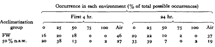
[image:12.451.44.409.193.273.2]There were eleven observation periods during the first 4 hr., thus fifty-five obser-vations of the positions of individual frogs were made during this time for each acclimatization group. There was a total of twenty-two observation periods during the entire 24 hr., for a total of n o position observations for individual frogs for each group. The percentages of these total numbers of possible occurrences accounted for by frogs in each of the possible environmental situations are summarized, for the two groups, in Table 3.
Table 3. Environmental salinity preference by Rana cancrivora
Occurrence in each environment (% of total possible occurrences)
First 4 hr. 34 hr.
Acclimatization , ' , , * , group o 25 50 75 100 Air o as 50 75 100 Air FW 16 20 18 o o 46 29 aa 10 2 o 37 SO%n.s.w. 30 38 13 o 2 37 33 39 7 o 3 19
The number of observations made is inadequate for a detailed analysis of environ-mental salinity preferences, but the data show that frogs in both acclimatization groups preferred external media no more concentrated than 50% n.s.w. The fresh-water frogs appeared to prefer sitting on the sand to everything else available, but other-wise showed no marked preference for a particular environmental concentration—as long as it was below 50%. The frogs acclimatized to 50% n.s.w. appeared to prefer sitting on the sand or in 25 % n.s.w. initially, but shifted as time went on to fresh water and 25 % n.s.w.
This species when pushed can, as an adult, tolerate at least up to 80 % sea water. It apparently prefers a concentration of no more than about half that.
Rana tigerina
The results of our study of water balance and osmoregulatory mechanisms in
R. tigerina are summarized in Figs. 7-9. The principal differences between our
studies of R. cancrivora and R. tigerina are that, in the work on R. tigerina, we made no analyses for Na, K and urea, and that we were able to study the effect of environ-mental salinity on development of fertilized eggs.
Adult R. tigerina tolerate direct transfer and indefinite exposure (i.e. for at least 8 days) to all concentrations of n.s.w. up to 25 % (salinity 9 %<,). 30 % n.s.w. (salinity n %o) is uniformly fatal within 24-48 hr., even after several days acclimatization to
25%-Transfer of this species to any medium other than fresh water results in marked and rapid gains in weight. Return to about original weight takes place after 5 days (Fig. 8).
of external medium will occur even in frogs with their mouths sewn shut. Perhaps the nostrils are used in this situation.
The contrast between the behaviour of R. tigerina and R. cancrivora, transferred to 25 % n.s.w. and l.s.w., respectively, following several days in freshwater, is illustrated in Fig. 8.
+15
+10
t chang
e +
op ±0
- 5
-10
- 1 5
A / V
/
•
A
\ \
V \
\
• i • i
0 1 2 3 4 5 6 7
[image:13.451.52.395.133.406.2]Time (days)
Fig. 8. Comparison between mean percentage weight changes in groups of R. cancrivora ( ) and R. tigerina ( ) each acclimatized to fresh water, then transferred to 25 % sea water. s.E.'s of means omitted for clarity.
Data on A and Cl of plasma and urine in variously acclimatized R. tigerina are presented in Fig. 9. R. tigerina regulates its plasma concentrations fairly well up to 25 % n.s.w., the kidney apparently being a very important organ in this effort. Urinary A and Cl both rise rapidly from levels far below those in the blood, in frogs in fresh water, to near equality with blood levels in frogs in 25 % n.s.w. No measurements of rates of urine production were made, but undisturbed frogs, even in 25% n.s.w., usually accumulated considerable volumes of urine in their bladders.
As in R. cancrivora, no specific relationship was discernible between the magnitudes of the electrical potentials developed by isolated pieces of skin from variously accli-matized frogs and the concentration of the external medium. All such potentials were positive inward, ranging from 10 to 100 mV. in frogs in fresh water, 60 to 110 mV. in frogs in 10% n.s.w. and 25 to 40 mV. in frogs in 25% n.s.w. Isolated skins of
R. tigerina usually survived for several hours. No reversals of potential occurred such
672 MALCOLM S. GORDON AND OTHERS
Measurements of short-circuit currents across isolated skins indicate that the skin* also plays a part in the effort this frog makes in adjusting to saline media. Short-circuit current across the skin was only about 10% of fresh water values in frogs in 25 % n.s.w. Mean values and ranges were 072 (0-45-0-94) /iA./mm.J for four frogs in fresh water, 0-46 (0-29-0-73)/iA./mm.2 for four frogs in 10% n.s.w., and 0-08 (0-08-0-08) fiA./mm.! for two frogs in 25 % n.s.w.
300
S200
100 200 External concentration (m-osm./l.)
300
Fig. 9. Relationships between mean plasma and urine osmotic concentration and Cl and environmental osmotic concentration in variously acclimatized groups of Rana tigerina. Open circles (O) mean values for osmotic concentration, solid circles (•) mean values for Cl. Solid lines ( ) joining points for plasma concentrations, dashed lines ( ) for urine concentrations. Diagonal solid line, line of equality.
As also in R. cancrivora, acclimatization to different media produced no change in evaporation from the skin of R. tigerina (Fig. 7). The difference in relative rates of water loss of the two species (Fig. 7) is partially accounted for by differences in surface to volume ratio in frogs of very different size. R. cancrivora used weighed 12-20 g. initially, R. tigerina weighed 83-143 g. Other contributory differences between the two species are not accounted for.
Experiments testing preference for environmental salinity in groups of R. tigerina acclimatized to fresh water and to 25 % n.s.w. presented the frogs with a range of choices from fresh water to 20 % n.s.w. There were no apparent differences between the two groups, neither showing a marked preference for any specific salinity between fresh water and 15% n.s.w. Fewer than 5% of the observations for each group, however, were frogs in 20% n.s.w.
An estimate of the effect of environmental salinity on embryonic development in
female frogs. These eggs were artificially fertilized with sperm contained in a brei made from excised testes of a ripe male. Groups of about twenty-five eggs were placed in Petri dishes containing 50 ml. of sea water dilutions at 5 % intervals from o to 40%. The dishes were covered and allowed to stand at room temperature. The eggs were examined after 4^, 18 and 38 hr.
Development of embryos to normal-appearing neurula stages occurred in 18 hr. in o, 5 and 10% n.s.w., but percentage of successful development declined from about 50% in fresh water to about 23% in 5% n.s.w. and 10% in 10% n.s.w. Virtually all eggs in all three of these concentrations showed some sign of development.
Sea water concentrations of 15 % and higher completely prevented normal develop-ment beyond what appeared to be early gastrula stages. Some further changes beyond these stages occurred in a small proportion of the eggs in 15 and 20% n.s.w., but the resulting embryos were abnormal. Only 50-75% of all eggs in 15 and 20% n.s.w. showed any signs of development.
In 25 % sea water and above no development occurred in any eggs and most eggs were shrunken in appearance.
DISCUSSION
Approximately 300,000,000 years have passed since the appearance of the earliest forms which palaeontologists recognize as amphibians. Present opinion seems to be that two different subgroups of rhipidistid crossopterygians gave rise independently to the amphibian groups today called urodeles and anurans (Jarvik, 1955). Palaeo-ecological evidence indicates that this transition, for both groups, took place in fresh water environments and almost all amphibian fossils found to date are probably from fresh water areas (Romer, 1945).
The fossil record is far from complete, but it provides evidence that few amphibians have lived in even brackish environments at any time between the Devonian and now (Romer, 1957). Whether or not this situation is due to misinterpretation of palaeo-ecological evidence, it seems reasonable to view the present possession of considerable salinity tolerance by at least two anurans, Bufo viridis and Rana cancrivora, as an indication that the present may be either an early stage in an evolutionary development or an only partial success at invading an environment to which amphibia seem unable to adjust. R. cancrivora would appear to be an excellent possibility as an evolutionary stem form in either case as it has already entered a rich, widely distributed and unexploited (by amphibians) environment—the mangrove swamps of the tropical world. An inspection of the map given by West (1956) indicates that mangrove swamps along the coasts of continents and major island groups may extend over as much as 100,000 miles.
674 MALCOLM S. GORDON AND OTHERS
when subjected to elevated salinities (Brunacci, 1914, 1915, 19170, b) or desiccatio^ (Przylecki, 1922).
The evolution of an ability to tolerate a uraemia sufficient to be of osmotic significance in brackish and marine environments does, however, appear to be a very difficult feat. Urea concentrations comparable to those measured in the plasma of adult R.
cancrivora acclimatized to 80% sea water will denature certain enzymes (Elodi &
Jecsai, i960; Riordan, Bier & Nord, i960) and may affect the oxygen-binding properties of haemoglobin (Rossi-Fanelli, Antonini & Caputo, 1959). The difficulties associated with the osmotically significant use of urea by animals are such that Smith (1936) was prompted to write that a sufficient number of animals had been investigated ' to preclude reasonably the possibility of a physiological uremia [such as that occurring in the elasmobranchs and in the African lungfish]... occurring elsewhere in the animal kingdom.'
The continuing physiological uraemia of adult R. cancrivora possesses at least one precedent in the elasmobranchs. There may, however, be no precedent, at least among the juvenile stages of oviparous vertebrates, for the osmoregulatory mechanism used by the tadpoles of this frog.
The tadpoles of R. cancrivora in our experiments exceeded their parents in salinity tolerance, surviving over the range from fresh water to 120 % sea water. If they accomplish this by means of a physiological uraemia, their nitrogen metabolism is very different from the complete ammoniotelism which is a universal property of all premetamorphosis amphibian larvae studied to date (Brown, Brown & Cohen, 1959; Munro, 1953; Underhay & Baldwin, 1955). In view of the physiology of their parents it seems improbable that they exclude salt by mechanisms similar to those of teleost fishes. Their survival may be due to an exceptional tolerance to increased salt concentration in their body fluids. Whatever these tadpoles do, they will amply repay further investigation.
The uraemia of adult R. cancrivora probably implies that the skin of this frog is relatively impermeable to urea. This appears not to be the case with ordinary fresh-water frogs (Przylecki, Opienska & Giedroyc, 1922). Impermeability of the integu-ment to urea is not, however, a necessary condition for the maintenance of high levels of uraemia. Fresh-water elasmobranchs, for example, maintain plasma urea concen-trations of near o-i M in the face of continuing losses to the environment, more than half of which is due to passive diffusion across the gills (Smith, 1931).
The kidneys of R. cancrivora apparently conserve some of the urea brought to them by the blood, but appear to be rather inefficient in this regard. Urinary urea levels are always below plasma levels, but are still quite high. This is another resem-blance between R. cancrivora and the sharks (Smith 1931, 1936).
Jglomeruli in the kidneys of frogs are normally inactive may be relevant. Note should also be made of the fact that histological examination of the kidneys of R. cancrivora demonstrates numbers of well-developed glomeruli.
Whatever the details of urea-conserving mechanisms in this frog may be, the basic fact remains that R. cancrivora appears to sustain a severe and continuing loss of urea via its urine. An interesting question is the source of all this urea.
The remainder of our data indicate that R. cancrivora, having solved the basic problem of water supply in a marine environment, is otherwise very similar to ordinary frogs in fresh water. It has a water-permeable skin, perhaps does not drink external medium, very probably takes up inorganic sodium and chloride from its environment by active transport across its skin, and readily suffers desiccation in air. It is interest-ing to note that plasma salt concentrations in R. cancrivora in 80 % n.s.w. are much higher than the plasma salt concentrations which are fatal to R. tigerina.
Probably the most striking thing about R. tigerina is the fact that, despite its supposed very close phylogenetic relationship to R. cancrivora, it appears to be a normal fresh-water frog in every way. Its overall salinity tolerance and osmoregulatory responses to high environmental concentration are virtually identical with those of various of its congeners which have been studied in far distant parts of the world
(R. pipiens: Adolph, 1927, 1933; Ruibal, 1959; R. temporaria: Bertin, 1920; Duval,
1928; Jargensen, 1954; Overton, 1904; Rey, 1938; R. esculenta: Brunacci, 1914, 1915, 1917a, b, c; Przylecki, 1922). The same is true for its response to desiccation (Adolph, 1932, 1933; Durig, 1901; Reichling, 1957; Rey, 1937; 1938, Thorson, 1955, 1956). The decrease in active uptake of sodium by the skin of fresh-water frogs maintained in media more concentrated than fresh water has recently been described by Maetz (1959) for R. esculenta.
In closing, note should be made of the possibility that salinity tolerances of amphibia in general may be greater when they are living under natural conditions and not starved in the laboratory. Since urea is an important factor in the tolerance of R. cancrivora to high salinities, and since this frog loses appreciable quantities of urea via its urine, well-fed R. cancrivora especially may tolerate salinities significantly higher than those just fatal to fasting laboratory animals.
SUMMARY
1. The osmotic and ionic regulatory abilities of adults of the euryhaline crab-eating frog (Rana cancrivora) have been studied. Adult frogs tolerated environmental salinities as high as 28 %<, at 300 C. Tadpoles of this form tolerated salinities as high as 39 %o at the same temperature.
2. Changes in body weight of frogs following transfers to different environmental salinities indicate both that the skin of this frog is permeable to water and that these animals do not swallow large volumes of external medium, even in high salinities.
3. Above salinities of about 9%,,, plasma A rises with increasing environmental A. Plasma A is always higher than environmental A. Increases in plasma concentration above fresh-water levels are due partly to increased NaCl concentration (about 40%), partly to increased urea concentration (about 60%). Urea concentrations as high as
676 MALCOLM S. GORDON AND OTHERS
4. Urinary A parallels plasma A, but is always lower than plasma A. Considerable^ quantities of urea are lost via the urine, even though urinary urea levels are below plasma levels.
5. Measurements of short-circuit current indicate that active uptake by the skin of inorganic ions continues in R. cancrivora acclimatized to high salinities.
6. R. cancrivora is no less susceptible to water loss by evaporation from the skin than are other amphibians.
7. In preference experiments R. cancrivora chooses salinities below 18 %„, but shows no strong preference for a particular salinity.
8. Similar observations on osmoregulatory mechanisms in a close relative of R.
cancrivora, the tiger frog (R. tigerina), show that the latter species is similar to ordinary
fresh-water frogs.
9. The striking physiological convergence between R. cancrivora and the elasmo-branch fishes is discussed, as are various possible implications of our data regarding nitrogen metabolism in tadpoles and kidney function in adult frogs.
These studies have been supported by research grants from the U.S. Public Health Service (RG-7114 and H-2228), National Science Foundation (G8802) and the Associates in Tropical Biogeography (Grant No. 54).
Hospitality, aid and advice were generously given to us by many people during the extended expedition involved in this work and we would like to express our appre-ciation to them all. In particular, the following gave us valuable aid: Captain James Faughn and members of the staff of the Scripps Institution of Oceanography-International Cooperation Administration NAGA Expedition; Dr William Shelton and members of the staff of the Education Division, USOM, Saigon; Rector Nguyen Quang Trinh and Mr Vu at the University of Saigon; Professor Nguyen Dinh Hung and the staff of the Institut Oceanographique, Nhatrang; Capt. Amphorn Penyapol and Lt. Thawatchai Thaiyong, Hydrographic Department, Royal Thai Navy, Bangkok; Professor Klum, Messrs Chanonwat and Twesuk Piyukarncharna and other staff of the Department of Biology, Chulalongkorn University, Bangkok; Dr H. A. Fehlmann of the George Vanderbilt Foundation, Bangkok; and Professor Edward Taylor, Department of Zoology, University of Kansas. Cynthia Rosenblum and Gordon Engel supplied valuable technical assistance.
REFERENCES
ADOLPH, E. F. (1927). The excretion of water by the kidneys of frogs: Amer. J. Phytiol. 81,
3I5-24-ADOLPH, E. F. (1932). The vapor tension relations of frogs. Biol. Bull., Woods Hole, 6a, 112-25. ADOLPH, E. F. (1933). Exchanges of water in the frog. Biol. Rev. 8, 224-40.
ADRIAN, R. H. (1956). The effect of internal and external potassium concentration on the membrane potential of frog muscle. J. Physiol. 133, 631-58.
ALCALA, A. C. (1955). Notes on the eggs and egg-laying of some amphibians on Negros Island, Philippines. Silliman J. 2, 103—6.
BERTIN, L. (1920). Les grenouilles peuvent-elles s'adapter a l'eau saumatre? C.R. Soc. Biol., Paris, 83, 1308-9.
BOURRET, R. (1942). Les batraciens de l'lndochine. Mem. Inst. Odanogr. Indochine, 6, 1—547. BROWN, G. W., JR., BROWN, W. R. & COHHN, P. P. (1959). Comparative biochemistry of urea synthesis.
WBBBRUNACCI, B. (1914). SuU" adattamento degli anfibi all' ambiente liquido esterno mediaate la regolazione della pressione osmotica dei loro liquidi intemi. III. Proprieta chimiche e fisico-chimiche dei liquidi intemi di animali tenuti in acqua distillata ed in soluzioni Ringer ipertoniche. R.C. Accad. Lincei, Ser. s, 33 (pt. z), 645-51.
BRUNACCI, B. (1915). Sull' adattamento. . . IV. II tempo entro il quale awiene la regolazione osmotica. R.C. Accad. Lincei, Ser. 5, 34 (pt. 1), 272-6.
BRUNACCI, B. (1917a). Sull' adattamento. . .V. Proprieta chimiche e fisico-chimiche dei liquidi intemi di animali tenuti in soluzioni Ringer isotoniche ed ipotoniche. R.C. Accad. Lincei, Ser. 5, 36 (pt. 1), 180-5.
BRUNACCI, B. (19176). Sull' addattarhento.. .VII. I fenomeni di adattamento nelle rane esculente ibernanti. R.C. Accad. Lincei, Ser. 5, 36 (pt. 1), 252—7.
BRUNACCI, B. (1917c). Influenza dell temperature sulla regolazione osmotica della rana esculenta estiva. R.C. Accad. Lincei, Ser. 5, 36 (pt. 2), 243-7.
CRANE, M. M. (1927). Observations on the function of the frog's kidney. Amer. J. Phytiol. 81, 232-43. DURIG, A. (1901). Wassergehalt und Organfunktion. Pfliiger's Arch. get. Phytiol. 85, 401-504. DUVAL, M. (1928). L'adaptation des grenouilles a l'eau saumatre. Arm. Phytiol. 4, 181—9.
ELODI, P. & JECSAI, G. (i960). Studies on d-glyceraldehyde-3-phosphate dehydrogenase. XV. The effect of urea. Acta phytiol. Acad. Sci. Hungaricae, 17, 175-82.
FORSTER, R. P. (1942). The nature of the glucose reabsorptive process in the frog renal tubule. Evidence for intermittency of glomerular function in the intact animal. J. Cell. Comp. Phytiol. 3O, 55—69. FORSTKR, R. P. (1954). Active cellular transport of urea by frog renal tubules. Amer. J. Phytiol. 179,
372-7-GORDON, M. S., SCHMIDT-NIELSEN, K. & KELLY, H. M. (1961). Osmotic regulation in the euryhaline crab-eating frog (Rana cancrivora) of southeast Asia. Fed. Proc. 30, 208.
HANZON, V., HERMODSSON, L. H. & TOSCHI, G. (1959). Ultrastructural organization of cytoplasmic nucleoprotein in the exocrine pancreas cells. J. Ultrattructure Ret. 3, 216-27.
JARVIK, E. (1955). The oldest tetrapods and their forerunners. Sci. Mon. 80, 141-54.
• J0RGEN8EN, C. B. (1954). On excretion of chloride in sodium chloride loaded frogs and toads. Acta
phytiol. tcand. 30, 171—7.
LOVE, J. K. & LIFSON, N . (1958). Transtubular movements of urea in the doubly perfused bullfrog kidney. Amer. J. Phytiol. 193, 662-8.
MAETZ, J. (1959). Le controle endocrinien du transport actif de sodium a travers la peau de grenouille.
\er Coll. Biol. de Saclay, 185-96.
MUNRO, A. F. (1953). T h e ammonia and urea excretion of different species of amphibia during their development and metamorphosis. Biochem. J. 54, 29—36.
NEILL, W. T . (1958). The occurrence of amphibians and reptiles in saltwater areas, and a bibliography.
Bull. Mar. Sci. Gulf Caribbean, 8, 1-97.
OVERTON, E. (1904). Neununddreissig Thesen iiber die Wasserokonomie der Amphibien und die osmotischen Eigenschaften der Amphibienhaut. Verh. phys.-med. Get. Wtirzb. 36, 277-95. PORA, A. E. & STOICOVICI, F. (1955). Cercetari asupre rolului sistemului nervos de la Bufo viridit in
fenomenele de adaptare la salinitate. Bull, stiint. Acad. Rom&ne, 7, 59—89.
PRZYLECKI, S. J. (1922). L'echange de l'eau et des sels chez les amphibiens. Arch. int. Phytiol. 19,
I48-59-PRZYLECKI, S. J., OPIENSKA, J. & GIEDROYC, H. (1922). L'excretion des substances azotees chez les
grenouilles a difRrentes temperatures. Arch. int. Phytiol. 30, 207-12.
RAMSAY, J. A. & BROWN, R. H. J. (1955). Simplified apparatus and procedure for freezing-point deter-minations upon small volumes of fluid. J. Sci. Inttrum. 33, 372-5.
REICHLING, H. (1957). Transpiration und Vorzugstemperatur mitteleuropaischer Reptilien und Amphi bien. Zool. Jb., Abt. allg. Zool. Phytiol. Tiere, 67, 1-64.
REY, P. (1937). Recherches expdrimentales sur l'economie de l'eau chez les batraciens. I. Ann.
Phytiol. 13, 1081-1144.
REY, P. (1938). Recherches experimentales sur l'economie de l'eau chez les batraciens. I I . Ann. Phytiol. 14, 1-66.
RICHARDS, A. N . & SCHMIDT, C. F. (1924). A description of the glomerular circulation in the frog's kidney and observations concerning the action of adrenalin and various other substances upon it.
Amer. J. Phytiol. 71, 178-208.
RIORDAN, J. F., BIER, M . & NORD, F. F. (i960). On the mechanism of enzyme action. LXX. Urea
denaturation of trypsin and acyltrypsins. Arch. Biochem. Biophyt. 90, 125-31. ROMER, A. S. (1945). Vertebrate paleontology, 2nd. ed. University of Chicago Press. ROMER, A. S. (1957). Amphibians. Mem. Geol. Soc. Amer. (fj, 2, i o n .
ROSSI-FANELLI, A., ANTONINI, E. & CAPUTO, A. (1959). T h e effect of urea on the oxygen equilibrium
of mammalian hemoglobins. Arch. Biochem. Biophyt. 85, 540—9.
^RUIBAL, R. (1959). The ecology of a brackish water population of Rana pipient. Copeia, no. 4, 315-22. • J C H M I D T , K. P. (1957). Amphibians. Mem. Geol. Soc. Amer. 67, 1, 1211-12.
678 MALCOLM S. GORDON AND OTHERS -SCHMIDT-NIELSEN, B. & FORSTER, R. P. (1954). The effect of dehydration and low temperature orW
renal function in the bullfrog. J. Cell. Comp. Phytiol. 44, 233-46.
SJOSTRAND, F. S. & BAKER, R. F. (1958). Fixation by freeze-drying for electron microscopy of tissue cells. J. Infrastructure Res. I, 239-46.
SMITH, H. W. (1930). Metabolism of the lung-fish Protopterut aetkiopicus. J. Biol. Chem. 88, 97-130. SMITH, H. W. (193 I). The absorption and excretion of water and salts by the elasmobranch fishes. I.
Fresh water elasmobranchs. Amer. J. Physiol. 98, 270-95.
SMITH, H. W. (1936). The retention and physiological role of urea in the elasmobranchii. BioL Rev. 11, 40-82.
STOICOVICI. F. & PORA, E. A. (1951). Comportarea la variatiuni de salinitate. Nota XXX. Influenta variatiunilor de salinitate si a factorului ecologic asupra supravietiurii si mediului interior la Bufo viridis in diferitele perioade ale anului. Stud. Cercet. Stiintif., Acad. Rep. Pop. Romane, Fil. Cluj 2,
150-219.
TAYLOR, E. H. & ELBEL, R. E. (1958). Contribution to the herpetology of Thailand. Bull. Sci. Univ. Kans. 38, 103 3-1189.
THORSON, T. B. (1955). The relationship of water economy to terrestrialism in amphibians. Ecology, 36, 100-16.
THORSON, T. B. (1956). Adjustment of water loss in response to desiccation in amphibians. Copeia, no. 4, 230-7.
UNDERHAY, E. E. & BALDWIN, E. (1955). Nitrogen excretion in the tadpoles of Xenopus laevis Daudin.
Biochem. J. 6 1 , 544—7.
UssiNGr H. H. (1954). Active transport of inorganic ions. Symp. Soc. Exp. Biol. 8, 407-22.