Original Article
Curcumin promotes apoptosis of
breast cancer cells by down-regulating
DJ-1-PTEN/PI3K/AKT signaling pathways
Mingzhu Li, Congren Wang, Conglin Lin
Department of Surgical Oncology, Quanzhou First Hospital Affiliated Fujian Medical University, Quanzhou 362000, Fujian, China
Received April 2, 2019; Accepted June 11, 2019; Epub July 15, 2019; Published July 30, 2019
Abstract: Enhanced activity of the PI3K/AKT signal pathway is correlated with tumorigenesis. As a negative regula-tor of PTEN, DJ-1 over-expression is associated with the occurrence of multiple tumors. Curcumin (Cur) is a phenol group compound with anti-tumor effects. Previous studies have shown its role in mediating the PTEN-PI3K/AKT pathway. However, it is still unclear whether Cur regulates DJ-1-PTEN/PI3K/AKT activity and proliferation or apopto-sis of breast cancer cells. Cultured normal human breast epithelial cells MCF-10A, breast cancer cells MDA-MB-231 and MCF-7 were compared for DJ-1 and PTEN mRNA expression using qRT-PCR, and protein expression by Western blot. MCF-7 cells were treated with 0, 5, 10 and 20 μM Cur, and CCK-8 assay measured cell proliferation, followed by Western blot for DJ-1 and PTEN protein expression. MCF-7 cells were divided into control, 20 μM Cur treatment and Cur + PTEN inhibitor SF1670 groups, for comparing DJ-1, PTEN and p-AKT expression. Cell apoptosis and prolif-eration were tested by flow cytometry and EdU staining. Compared to MCF-10A cells, breast cancer cells showed an up-regulation of DJ-1, plus suppressed PTEN expression. MDA-MB-231 cells had higher DJ-1 expression and lower PTEN levels than MCF-7 cells. Cur treatment remarkably inhibited MCF-7 cell proliferation in a dosage-dependent manner. Gradient Cur suppressed DJ-1 expression and increased PTEN. Combined treatment with SF1670 en-hanced phosphorylation activity of AKT, weakened Cur induced inhibition on AKT phosphorylation, and alleviated pro-apoptosis or anti-inflammatory functions of Cur. Cur can inhibit breast cancer cell proliferation and facilitate cell apoptosis via down-regulating DJ-1 expression and modulating PTEN/PI3K/AKT pathway activity.
Keywords: Curcumin, DJ-1, PTEN/PI3K/AKT, cell proliferation, apoptosis, breast cancer
Introduction
Breast cancer (BC) is a malignant tumor origi-nating from breast gland epithelium. BC is the most commonly occurring cancer in females and has the highest incidence among all female cancers [1, 2]. BC is responsible for about 20%~30% of all female systemic tumors, hav-ing a 2%-3% annually increased rate of inci-dence, making it the most threatening malig-nant tumor for women [1, 2]. It is estimated that about 1.5 million people are newly diagnosed with BC, and one third (about 0.5 million) of these patients die from BC [3]. BC
characteris-tics include difficulty in early diagnosis, rapid
progression at terminal stages and high mortal-ity, thus severely affecting patients life health and quality [4, 5].
and facilitating cell apoptosis [11, 12]. Existing evidence has demonstrated that Cur plays a role in the regulation of the PTEN-PI3K/AKT sig-naling pathway [13, 14]. Considering the role of PTEN in the occurrence or development of tumors, whether or not Cur plays a role in treat-ment of breast cancer via the PTEN/PI3K/AKT pathway remains poorly understood. This study investigated if Cur modulates DJ-1-PTEN/PI3K/ AKT pathway activity, and affects BC cell prolif-eration or apoptosis.
Materials and methods
Major reagent and materials
Normal breast epithelial cells MCF-10A, BC cell lines MDA-MB-231 and MCF-7 were purchased from Beina Bio (China). RPMI 1640 and fetal bovine serum (FBS) were purchased from
Hyclone (US). Transfection reagent Lip2000 and Trizol were purchased from Invitrogen (US).
PrimeScriptTM RT reagent Kit was purchased
from Takara (China). Cur was purchased from
Sigma (US). Edu flow cytometry test reagent
was purchased from RioBio (China). Rabbit
anti-human DJ-1, AKT, p-AKT and β-actin poly -clonal antibodies were purchased from Abcam
(US). Rabbit anti-human PTEN polyclonal anti
-body was purchased from CST (US). HRP conju -gated secondary antibody was purchased from
Sangon Bio (China). BCA protein quantification
kit, CCK-8 assay kit and Annexin V/PI cell apop-tosis kit were all purchased from Beyotime Bio (China). PTEN inhibitor SF1670 was purchased
from MedChemExpress (US). FORMA model
3131 incubator was purchased from Thermo
(US).
Cell incubation
MCF-10A, MDA-MB-231 and MCF-7 cells were kept in RPMI 1640 medium containing 10%
FBS, in a 37°C chamber with 5% CO2. After cell growth on the plate, cells were passed at a 1:3~1:4 ratio. Cells in log-growth phase were used for experiments.
Cell treatment and grouping
In vitro cultured MCF-10A, MCF-7 and MDA-MB-231 cells were inoculated into 96-well plates (10,000 cells per well, in 6 parallel wells). After 24 h attached growth, cells were treated
with 0, 5, 10 and 20 μM Cur. Seventy-two hours later, 10 μL CCK-8 solution was added into
each well. After a 4 h reaction, absorbance val-ues (A) were measured at 450 nm wavelength. Relative proliferation activity (%) = (A450of drug treatment group - A450 of empty well)/ (A450 of control group - A450 of empty well) × 100%.
Cur and SF1670 treatment of MCF-7 cells
MCF-7 cells were treated with 10 μM EdU for
120 min, and were assigned into three groups:
control group, Cur (20 μM) treatment group,
and Cur + SF1670 (200 nM) treatment group. After 48 h of incubation, cells were digested by
trypsin. After collection, cells were fixed in 4%
paraformaldehyde for 15~30 min and were neutralized in 2 mg/mL glycine for 5 min. After
PBS rinsing, 500 μL Apollo staining buffer was
added for resuspension of cells, which were then incubated in the dark at room tempera-ture for 10 min. Cells were then centrifuged at 300 g for 5 min, and the staining buffer was discarded. Followed by rinsing in 0.5% Triton X-100 permeabilizing buffer at room tempera-ture for 1~3 rinses, cells were then resuspend-ed in PBST and were measurresuspend-ed with FC500
MCL flow cytometry (Beckman, US).
Cell apoptosis assay
Cells from all treatment groups were digested and collected, followed by PBS rinsing and
cen-trifugation. Cells were resuspended in 100 μL
Annexin V Binding Buffer and were sequentially
mixed with 5 μL Annexin V-FITC and 5 μ PI dyes.
After 15 min room temperature incubation,
400 μL Annexin V Binding Buffer was added for measuring cell apoptosis by flow cytometry.
qRT-PCR for measuring gene expression
RNA was extracted by the Trizol method. cDNA was synthesized from RNA using reverse tran-scription by PrimeScriptTM RT reagent Kit. Using
cDNA as the template, PCR amplification was
performed under the direction of TaqDNA
poly-merase. In a 10 μL system, we added 5.0 μL 2X SYBR, 0.5 μL forward/reverse primer (5 μm/L), 1 μL cDNA and ddH2O up to 10.0 μL. Reverse
transcription conditions were: 50°C for 15 min, and 85°C for 5 min. PCR conditions were: 95°C to denature for 5 min, followed by 40 cycles each consisting of 95°C for 15 sec and 60°C for 1 min. PCR was performed on a Bio-Rad
CFX96 real time fluorescent quantitative PCR
Western blot
Cell precipitations were mixed with 100 μL RIPA
lysis buffer for 15 min to lysis on ice. After 10,000 g centrifugation for 5 min, proteins in the supernatant were transferred to new
Eppendorf tubes. After BCA quantification for quality and quantity for proteins, 40 μg of sam -ple was loaded and separated in 10% SDS-PAGE, and were transferred to a PVDF mem-brane (300 mA, 100 min). The protein membrane was blocked with 5% nonfat milk powder. Primary antibodies (DJ-1 at 1:2,000, PTEN at 1:1,000, AKT at 1:1,000, p-AKT at
1:500, and β-actin at 1:10,000) were added for 4°C overnight incubation. On the next day, the
membrane was rinsed in PBST three times, and HRP conjugated secondary antibody (1:10,000 dilution) was incubated for 60 min at room temperature. The membrane was rinsed in PBST three times and was washed with chro-mogenic reagent. After 2-3 min of dark incuba-tion, the membrane was exposed, developed and scanned for data storage.
Statistical analysis
SPSS 18.0 was used for data statistics. Measurement data were presented as mean ±
standard deviation (SD). Measur- ement data between the two groups were compared by t-test. The comparison of means across
multiple groups was first per -formed by analysis of variance
(ANOVA), followed by Bonferroni
comparison between the two gr-
oups. A statistical significance was defined when P<0.05.
Results
DJ-1 upregulation and PTEN down-regulation in BC cells
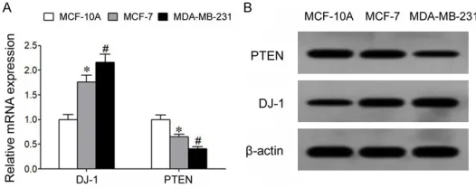
qRT-PCR results showed that com-pared to normal breast gland epi-thelial cells MCF-10A, the BC cell lines MCF-7 and MDA-MB-231
showed significantly elevated DJ-1
[image:3.612.91.354.73.177.2]mRNA expression, whilst PTEN mRNA expression was remarkably decreased. Moreover, MDA-MB2- 31 cells with higher malignancy presented higher DJ-1 expression compared to MCF-7 cells which
Figure 1. Up-regulation of DJ-1 and down-regulation of PTEN in BC cells. A: qRT-PCR assay for mRNA expression. B: Western blot for cellular pro-tein expression. *P<0.05 compared to MCF-10A cells; #P<0.05 com-pared to MCF-7 cells.
had relatively lower malignancy, and PTEN expression was lower than in the MCF-7 cells (Figure 1A). Western blot results showed that BC cell lines MCF-7 and MDA-MB-231 had
sig-nificantly higher DJ-1 protein expression than
normal breast gland epithelial cells MCF-10A, and that PTEN protein expression was remark-ably lower in BC cells (Figure 1B).
Cur treatment remarkably inhibited prolifera-tion activity of BC cells
CCK-8 assay showed that different concentra-tions of Cur had no major effect on proliferation activity of normal breast gland epithelium cells MCF-10A (Table 1). The proliferation activity of
BC cells, however, was significantly inhibited by 5, 10 or 20 μM Cur. Higher Cur dosage further
suppressed proliferation activity. Compared to MDA-MB-231 cells with higher malignancy, MCF-7 cells with relatively lower malignancy presented higher drug sensitivity towards Cur under the same dosage, as presented by lower proliferation activity (Table 1).
Cur suppressed DJ-1 expression and increased PTEN expression in the BC cell line MCF-7
qRT-PCR results showed that Cur treatment remarkably suppressed DJ-1 mRNA expression
Table 1. Effects of Cur on the proliferation activity of MCF-10A, MCF-7 and MDA-MB-231 cells. Cells were treated with differ-ent concdiffer-entrations of Cur followed by analysis of cell
prolifera-tion by flow cytometry
0 μM 5 μM 10 μM 20 μM MCF-10A 100% 96.3±5.1% 105.2±5.4% 93.5±4.9% MCF-7 100% 72.6±3.6%* 51.3±3.1%* 38.9±2.6%*
MDA-MB-231 100% 87.3±4.1%*,# 68.4±3.9%*,# 57.2±3.3%*,#
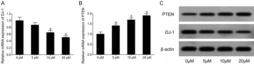
[image:3.612.90.355.296.350.2]in MCF-7 cells, with lower RNA expression at higher dosage (Figure 2A). Compared to 0 μM treatment group the 5, 10 and 20 μM Cur treat
-ed MCF-7 cells had significantly elevat-ed PTEN
mRNA expression, with more potent increase of PTEN mRNA expression in the high dosage groups (Figure 2B). Western blot results sho- wed that compared to the untreated group, Cur treated MCF-7 cells had remarkably decreased DJ-1 protein expression, whilst PTEN protein expression was remarkably increased (Figure 2C).
Cur significantly inhibited BC cell proliferation and induced cell apoptosis whilst SF1670 antagonized Cur effects
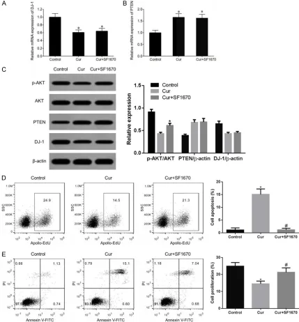
qRT-PCR results showed that Cur treatment remarkably decreased DJ-1 mRNA expression in MCF-7 cells and up-regulated PTEN mRNA expression. Combined treatment using SF1670 did not affect DJ-1 or PTEN mRNA expression (Figure 3A and 3B). Western blot results showed that SF1670 combined Cur treatment did not affect the modulatory effect of Cur on DJ-1 or PTEN protein expression, but remark-ably up-regulated the p-AKT protein expression (Figure 3C). Flow cytometry results showed that Cur remarkably inhibited the proliferation of MCF-7 cells (Figure 3D) and induced cell apop-tosis (Figure 3E). Combined treatment of Cur and SF1670 antagonized the effect of Cur on the inhibition of proliferation of MCF-7 cells and induction of cell apoptosis.
Discussion
PI3K/AKT is a signal pathway that is widely dis-tributed in multiple tissues and cells. When PI3K/AKT is activated, PI3K can catalyze the production of phosphatidylinositol
3,4,5-tri-phosphate (PIP3) from phosphatidylinositol 4,5-trisphophate (PIP2), the former of which can directly phosphorylate and activate AKT to further activate downstream signaling mole-cules to modulate various important biological processes including cell growth, survival and apoptosis via regulating target gene transcrip-tion and expression [15]. Previous studies showed that over-activation of the PI3K/AKT signaling pathway was correlated with the occurrence, progression, metastasis and drug resistance of multiple tumors such as endome-trial carcinoma [16], gastric cancer [17] and pulmonary carcinoma [18]. PTEN is the only tumor suppressor gene that has been discov-ered with dual activities including protein lipase and phosphatase. By dephosphorylation of its substrate PIP3, PTEN can inhibit AKT phosphor-ylated activation by PI3K via PIP3, thus block-ing the PI3K/AKT pathway transduction, and negatively regulating its activity [19]. DJ-1/ Parkinson gene 7 (PARK7) located on human chromosome 1p36.2-36.3, being around 24 kb length, and encoding a 21 kDa protein consist-ing of 189 amino acids [10]. As a negative regu-lator of PTEN, DJ-1 can weaken the inhibitory role on the PI3K/AKT signaling pathway by PTEN, via suppressing its expression and func-tion, thus indirectly activating the PI3K/AKT sig-naling pathway to play a role in alleviating cell apoptosis, facilitating cell proliferation, survival and tumorigenesis, making it one oncogenic factor [10].
Cur is a polyphenol compound with the
charac-teristics of high efficiency, wide spectrum of
anti-tumor functions, gastrointestinal actions and minimal toxic effects, plus
pharmacologi-cal effects including inflammation,
anti-oxidation, and clearance of free radicals. Increasing evidence show that Cur has
[image:4.612.90.522.72.181.2]tumor effects including suppression of tumor cell proliferation, facilitation of cell apoptosis, anti-angiogenesis, anti-invasion and anti-mi- gration [11, 12]. The anti-tumor effects of Cur are drawing increasing attention from people, and has become a hot topic in tumor preven-tion and treatment. Previous studies showed that Cur played a role in modulating the PT- EN-PI3K/AKT pathway [13, 14]. Whether Cur can modulate the PTEN/PI3K/AKT pathway via
DJ-1 as a negative regulator upstream of PTEN, is still unclear. This study thus investigated whe- ther Cur modulates the DJ-1-PTEN/PI3K/AKT pathway activity, and proliferation and/or apop-tosis of BC cells.
Our results showed that compared to normal
breast gland epithelial cell line MCF-10A, BC
cells showed significantly elevated DJ-1
exp-ression plus lower PTEN expexp-ression.
[image:5.612.90.519.73.535.2]tosis, and enhanced the Adriamycin sensitivity of cells via modulating PI3K/AKT signal path-way [22]. However, these studies were per-formed via genetic approaches that interfere with DJ-1 expression, in sharp contrast to our studies. In a study on the effect of DJ-1 expres-sion, Ismail et al, found that Paclitaxel can up-regulate the expression of a negative regulator of cell invasion KLF17 via suppressing DJ-1 expression, which weakens BC cell invasion potency [23]. Yang et al, also showed that bis-demethoxycurcumin can remarkably suppress DJ-1 expression in pancreatic carcinoma cells and facilitate PANC-1 cell apoptosis [24]. These studies, however, were not simultaneously
tar-geting Cur and BCs. Our study revealed that Cur
can affect PTEN/PI3K/AKT pathway activity via suppressing DJ-1 expression, thus exerting anti-proliferation and pro-apoptotic roles of BC cells for anti-tumor effects: all of which have not been previously reported and are thus a novelty of this study. However, the treatment effects of Cur on BC and its in vivo modulatory effects are largely unclear and require further
animal studies to fulfil the weakness of the cur -rent studies.
Conclusion
Cur can exert anti-proliferation and pro-apop-totic roles on BC cells in an anti-tumor manner via suppression of DJ-1 expression to modulate the PTEN/PI3K/AKT pathway activity.
Disclosure of conflict of interest
None.
Address correspondence to: Dr. Mingzhu Li, De- partment of Surgical Oncology, Quanzhou First Hospital Affiliated Fujian Medical University, Anji Road, Chengdong Branch, Quanzhou 362000, Fujian, China. Tel: 0595-28073120; Fax: +86-0595-28073120; E-mail: [email protected]
References
[1] Ferzoco RM and Ruddy KJ. The epidemiology of male breast cancer. Curr Oncol Rep 2016; 18: 1.
[2] Tao Z, Shi A, Lu C, Song T, Zhang Z and Zhao J. Breast cancer: epidemiology and etiology. Cell Biochem Biophys 2015; 72: 333-338.
[3] Liu FC, Lin HT, Kuo CF, See LC, Chiou MJ and Yu HP. Epidemiology and survival outcome of breast cancer in a nationwide study. Oncotar -get 2017; 8: 16939-16950.
MB-231 cells had higher DJ-1 expression than MCF-7 cells, and lower PTEN expression. Results showed that DJ-1 up-regulation played an important role in suppressing PTEN expres-sion and BC pathogenesis, and was related to malignancy of BCs. Klawitter et al, found that lovastatin can enhance PTEN functional activi-ty via suppressing DJ-1 expression, and inhibit AKT phosphorylation to suppress BC cell pro- liferation [19]. Ismail et al, found that compared to non-invasive BC cell lines MCF-7 and T47D cells, the invasive BC cells MDA-MB-231 and
MDA-MB-435 had significantly elevated DJ-1
apop-[4] Leone JP, Haraldsson B, Mott SL, McDowell BD and Chrischilles EA. Treatment patterns and survival of elderly patients with breast cancer brain metastases. Am J Clin Oncol 2019; 42: 60-66.
[5] Tanguy ML, Cabel L, Berger F, Pierga JY, Savi-gnoni A and Bidard FC. Cdk4/6 inhibitors and overall survival: power of first-line trials in met -astatic breast cancer. NPJ Breast Cancer 2018; 4: 14.
[6] Shi B, Deng W, Long X, Zhao R, Wang Y, Chen W, Xu G, Sheng J, Wang D and Cao S. MiR-21 increases c-kit+ cardiac stem cell proliferation
in vitro through PTEN/PI3K/Akt signaling. PeerJ 2017; 5: e2859.
[7] Jiang Y, Chang H and Chen G. Effects of mi-croRNA-20a on the proliferation, migration and apoptosis of multiple myeloma via the PTEN/ PI3K/AKT signaling pathway. Oncol Lett 2018; 15: 10001-10007.
[8] Li NN, Meng XS, Men WX, Bao YR and Wang S. Total flavonoids from oroxylum indicum induce apoptosis via PI3K/Akt/PTEN signaling path-way in liver cancer. Evid Based Complement Alternat Med 2018; 2018: 3021476.
[9] Wang L, Tang B, Han H, Mao D, Chen J, Zeng Y and Xiong M. MiR-155 affects osteosarcoma MG-63 cell autophagy induced by adriamycin through regulating PTEN-PI3K/AKT/mTOR sig -naling pathway. Cancer Biother Radiopharm 2018; 33: 32-38.
[10] Kim RH, Peters M, Jang Y, Shi W, Pintilie M, Fletcher GC, DeLuca C, Liepa J, Zhou L, Snow B, Binari RC, Manoukian AS, Bray MR, Liu FF, Tsao MS, Mak TW. DJ-1, a novel regulator of the tumor suppressor PTEN. Cancer Cell 2005; 7: 263-273.
[11] Su P, Yang Y, Wang G, Chen X and Ju Y. Cur-cumin attenuates resistance to irinotecan via induction of apoptosis of cancer stem cells in chemoresistant colon cancer cells. Int J Oncol 2018; 53: 1343-1353.
[12] Zhang L, Yang G, Zhang R, Dong L, Chen H, Bo J, Xue W and Huang Y. Curcumin inhibits cell proliferation and motility via suppression of TROP2 in bladder cancer cells. Int J Oncol 2018; 53: 515-526.
[13] Chang M, Wu M and Li H. Curcumin combined with glycyrrhetinic acid inhibits the develop-ment of hepatocellular carcinoma cells by down-regulating the PTEN/PI3K/AKT signal-ling pathway. Am J Transl Res 2017; 9: 5567-5575.
[14] Rana C, Piplani H, Vaish V, Nehru B and Sanyal SN. Downregulation of PI3-K/Akt/PTEN path-way and activation of mitochondrial intrinsic apoptosis by diclofenac and curcumin in colon cancer. Mol Cell Biochem 2015; 402: 225-241.
[15] Oudit GY, Sun H, Kerfant BG, Crackower MA, Penninger JM and Backx PH. The role of phos-phoinositide-3 kinase and PTEN in cardiovas-cular physiology and disease. J Mol Cell Cardiol 2004; 37: 449-471.
[16] Guan Q, Zhang Q, Zhang C, Liu Q and Ren QL. HOTTIP regulates progression of endometrial cancer via activating PI3K/AKT pathway. Eur Rev Med Pharmacol Sci 2018; 22: 3727-3733. [17] Ke J, Ma P, Chen J, Qin J and Qian H. LGR6
promotes the progression of gastric cancer through PI3K/AKT/mTOR pathway. Onco Tar -gets Ther 2018; 11: 3025-3033.
[18] Sun DM, Tang BF, Li ZX, Guo HB, Cheng JL, Song PP and Zhao X. MiR-29c reduces the cis-platin resistance of non-small cell lung cancer cells by negatively regulating the PI3K/Akt pathway. Sci Rep 2018; 8: 8007.
[19] Shi B, Deng W, Long X, Zhao R, Wang Y, Chen W, Xu G, Sheng J, Wang D and Cao S. MiR-21 increases c-kit+ cardiac stem cell proliferation
in vitro through PTEN/PI3K/Akt signaling. PeerJ 2017; 5: e2859.
[20] Ismail IA, Kang HS, Lee HJ, Kim JK and Hong SH. DJ-1 upregulates breast cancer cell inva-sion by repressing KLF17 expresinva-sion. Br J Can-cer 2014; 110: 1298-1306.
[21] Kawate T, Iwaya K, Koshikawa K, Moriya T, Ya-masaki T, Hasegawa S, Kaise H, Fujita T, Mat-suo H, Nakamura T, Ishikawa T, Hiroi S, Iguchi-Ariga SM, Iguchi-Ariga H, Murota K, Fujimori M, Yamamoto J, Matsubara O and Kohno N. High levels of DJ-1 protein and isoelectric point 6.3 isoform in sera of breast cancer patients. Can-cer Sci 2015; 106: 938-943.
[22] Zhang GQ, He C, Tao L and Liu F. Role of DJ-1 siRNA in reverse sensitivity of breast cancer cells to chemotherapy and its possible mecha-nism. Int J Clin Exp Pathol 2015; 8: 6944-6951.
[23] Ismail IA, El-Sokkary GH and Saber SH. Low doses of paclitaxel repress breast cancer inva-sion through DJ-1/KLF17 signalling pathway. Clin Exp Pharmacol Physiol 2018; [Epub ahead of print].