Original Article
Reproducibility of quantitative real-time PCR analysis
in microRNA expression profiling and comparisons
with microarray assays in diffuse large
B-cell lymphoma patients
Yan Shi, Tian-Yi Liu, Mei-Yue Song, Lu Chen, Jin Liu, Shan Gao The 2nd Hospital of Harbin Medical University, Harbin, China
Received June 19, 2017; Accepted January 9, 2019; Epub May 15, 2019; Published May 30, 2019
Abstract: Diffuse large B-cell lymphoma (DLBCL), a malignant tumor, accounts for 30% to 40% of Non-Hodgkin lym-phoma (NHL) patients. Dysregulation of microRNA (miRNA) expression has been documented in many tumors. This study aimed to assess miRNA expression in DLBCL to identify novel biomarkers that may improve diagnostic levels. The current study used miRNA microarray analysis to explore miRNA profiles in DLBCL patients, using formalin-fixed paraffin-embedded (FFPE) tissues of 3 samples from DLBCL patients and 3 from normal controls. This study also validated results using real-time quantitative PCR (RT-qPCR) in DLBCL (n=50), Burkitt’s lymphoma (BL, n=40), and normal controls (n=25). Comprehensive analysis provided 12 miRNA expression profiles (miR-181, miR-638, miR-328, miR-1268a, miR-1915-3p, miR-1268b, miR-1825, miR-4788, hsa-miR-4290, hsa-miR-3162-3p, hsa-miR-1202, hsa-miR-4649-3p) of DLBCL showing overexpression from normal tissues (P < 0.05). After candidate miRNAs were selected, qRT-PCR further verified that 3 miRNAs (hsa-miR-181, hsa-miR-683, hsa-miR1268) were significantly overexpressed in DLBCL and BL tissues, compared to normal sam -ples. Results indicate that aberrant miRNA expression was involved in the development and progression of DLBCL. Many of the miRNAs, identified in the current research, are new and have not been reported in previous studies, perhaps due to population differences. In conclusion, present results suggest that these miRNAs have potential use as diagnostic biomarkers, distinguishing DLBCL and BL patients from normal individuals and offering new targets for future therapies.
Keywords: MicroRNA, expression, diffuse large B-cell lymphoma, Burkitt’s lymphoma
Introduction
Non-Hodgkin lymphoma (NHL) is the fifth most common cancer, worldwide, and comprises many different subtypes, including diffuse large B-cell lymphoma (DLBCL), Burkitt’s Lymphoma (BL), Mantle Cell Lymphoma (MCL), Mucosa-Associated Lymphoid Tissue (MALT) Lymphoma, Follicular Lymphoma (FL) and Nodal Marginal Zone Lymphoma (NMZL), and so forth [1]. DLBCL is one of the most malignant lympho-mas in the world, accounting for nearly 40% of non-Hodgkin’s lymphoma cases. It is highly het-erogeneous from both morphological and clini-cal perspectives [2, 3]. Although great progress in diagnosis and treatment technologies has been made, many DLBCL patients still have poor prognosis [4]. Moreover, although several
biomarkers have been identified to better clas -sify and predict outcomes at diagnosis, the detailed mechanisms leading to occurrence and development of diffuse large B cell lym- phoma have not been clarified [5]. MicroRNAs have been shown to have the potential for diag-nosis and prediction of cancer [6-9]. Moreover, microRNA expression profiling could distinguish cancers based on both the cellular nature and developmental stage [10]. Therefore, the cur-rent study aimed to explore altered expression of miRNAs involved in DLBCL, providing more effective therapeutic opportunities.
regions of target miRNAs [11-13]. Currently, about 10-30% of all human genes are regulat-ed by microRNAs [14]. The latent importance of microRNAs in tumors has been discovered because of the aberrant expression of microR-NAs that occurs in cancer-associated genomic regions, suggesting that microRNA dysfunction is a common feature in malignant tumors [15]. An increasing number of studies have discov-ered that aberrant expressed miRNAs are in- volved in a variety of human cancers and medi-ate in initiation, promotion, progression, and resistance to cancer chemotherapy [16-19]. Se- veral studies have suggested that many miR-NAs are highly expressed in DLBCL, such as 21, 106a-5p, 181a-5p, and miR-200c [20-22]. Furthermore, miR-21 expression profiles in tumor cells [8] and serum [9] have been proven to be associated with DLBCL pati- ent prognosis. Microarray-based gene expres-sion profiling is a potential method of investi -gating expression of miRNAs in DLBCL patients. Several groups have performed survival analy-ses on larger DLBCL patient cohorts by micro-arrays, confirming that miRNAs are associated with survival, including miR-21, miR-23a, and miR-222 [8, 23, 24]. However, studies concern-ing miRNA profiles of DLBCL have been scarce. Therefore, the present study was performed to detect miRNA expression profiles of DLBCL patients and normal lymphoid tissues using miRNA microarrays.
The current study reported miRNA expression profiles of 3 DLBCL tumors and 3 normal lym -phoid samples, aiming to identify a DLBCLs specific miRNA signature. A total of 12 miRNAs were found to be differentially expressed in DLBCL. In addition, significantly aberrant miR -NAs (miR-181a-5p, miR-638, miR-1268b) were validated using RT-qPCR in DLBCL, BL, and nor-mal lymphoid samples. Present results reveal- ed that the three miRNAs were more frequently expressed in DLBCL and BL, compared to
nor-For the current study, 50 DLBCL, 40 BL, and 25 normal paraffin-embedded tissues were obtained, between 2014 and 2016, from the Second Affiliated Hospital of Harbin Medical University, Heilongjiang Province, China. Of these, 3 pairs of DLBCL and corresponding nontumorous lymphoid tissues were collected, in parallel, for miRNA microarray analysis. Histopathologic slices were examined via H&E staining and FISH staining, then diagnosed sep -arately by three pathologists. No patients received chemotherapy, radiotherapy, or immu-notherapy before undergoing surgery. Sample collection was approved by the Ethics Committee of Harbin Medical University and written informed consent was obtained from each patient before tissue collection. Clinical information of the patients, including age and gender, are listed in Table 1. Patients with DLBCL had a mean age of 57.94±14.04 (22-84) years, while 27 patients (54%) were male. Patients with BL had a mean age of 56.70± 13.42 (25-81) years, while 19 patients (47.5%) were male. Normal controls had a mean age of 57.84±15.42 (23-85) years, while 14 patients (56%) were male.
RNA extraction and real-time quantitative PCR analysis (qRT-PCR)
[image:2.612.89.338.84.178.2]Total RNA was isolated from DLBCL, BL, and normal lymphoid samples using the miRNA- prep Pure FFPE Kit (Tiangen, Beijing, German, China), according to manufacturer protocol. Next, cDNAs of miRNAs were synthesized with miRcute Plus miRNA First-Strand cDNA Syn-thesis Kit (Tiangen, Beijing, German, China). RT reactions with miRcute Plus miRNA First-Strand cDNA Synthesis Kit contained 2.0 ug total RNA, 10 ul 2× miRNA RT Reaction Buffer, 2 ul miRNA RT Enzyme Mix, and RNase-Free ddH2O2 filled up to 20 ul in each reaction. The RT reaction was conducted under the follow- ing conditions: 42°C for 60 minutes, then 95°C
Table 1. Clinicopathological information of the patients DLBCL (n=50) BL (n=40) Normal (n=25) Age
Range 22-84 25-81 23-85
Mean ± SD 57.94±14.04 56.70±13.42 57.84±15.42 Sex
Male 27 19 14
Female 23 21 11
mal lymphoid samples. Present data suggests that aberrant expression of miRNAs involved in DLBCL and BL may provide useful information in the search of future therapeutic targets.
Materials and methods
for 3 minutes. Afterward, the cDNA products from RT reaction were diluted 20 times. The purities and concentrations of cDNA were checked, spectrophotometrically, using Nano- Drop ND-2000c (Thermo Fisher Scientific, Inc., Wilmington, DE). PCR was performed with miR-cute Plus miRNA qPCR Detection Kit (Tiangen, Beijing, German, China) by an ABI PRISM 7500 fast Sequence Detection System (Applied Bio- systems, Foster City, CA, USA). PCR was carried out with 1.5 ul of the diluted products in 20 ul PCR reaction containing 10 ul 2× miRcute Plus miRNA Premix, 2 ul 50× ROX Reference Dye, 0.4 ul 10× miScript universal primer, and 0.4 ul 10× miScript primer assay. Amplification was performed as follows: 95°C for 15 minutes, fol-lowed by 45 cycles at 94°C for 20 seconds and 60°C for 34 seconds. All reactions were per-formed in triplicate. Expression of U6 was used as endogenous control for detection of miRNA expression profiles. The 2-ΔCt method was used
to quantify expression of miRNAs. Primers were used as follows: miR-181a-5p: 5’-AACAUUCA- ACGCUGUCGGUGAGU-3’; miR-638: 5’-AGGG- AUCGCGGGCGGGUGGCGGCCU-3’; miR-1268b: 5’-CGGGCGUGGUGGUGGGGGUG-3’; U6: 5’-TG- GTGAAGACGCCAGTGGA-3’.
miRNA microarrays
MicroRNAs from all RNA samples were labeled with Cy3 fluorescent dye and were turned into cDNA using RNA Agilent miRNA labeling Kit (Agilent, UK) and Spike Kit (Agilent, UK). Agilent microRNA Hybridization Kit (Agilent, UK) was then applied for hybridizing into Human miRNA Microarray, Release-21.0, 8×60K microarray slides (Agilent, UK) and scanning with Nimble-gen MS200 array scanner. TIFF image files were obtained using Agilent Feature Extraction software to extract original data and quality control (QC) reports. Sample data, passing QC parameters, was subjected to quantile normal-ization and analyzed using GeneSpring GX (Agilent, UK) 13.0 software. MicroRNAs with
P-values less than 0.05 and fold-change values of > 2 and < -2 are determined to be statisti-cally significant.
Statistical analysis
Statistical analyses were conducted using SP- SS software version 20 (IBM, New York, NY, USA) and GraphPad Prism 5.0. Hierarchical clustering analysis was used to describe mi-
RNA expression profiles of 3 paired DLBCL and normal samples. Visualization of differentially-expressed miRNAs was conducted using R sta-tistical language v.3.1.2. Moreover, miRNA ex- pression is expressed as mean ± marked dif-ference (X ± S) using the comparative ΔCt method. Comparisons between miRNA expres-sion levels of the two different groups were con-ducted using Student’s t-test. P-values were two-sided, with less than 0.05 indicating statis-tical significance.
Results
Differentially-expressed miRNAs profiling in DLBCL
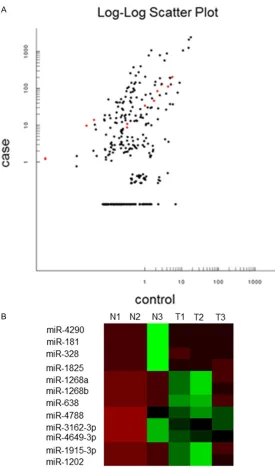
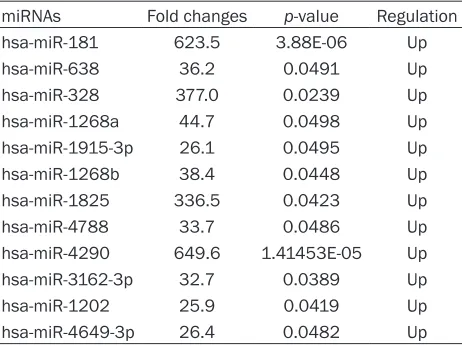
To analyze worldwide miRNA profiles of diffuse large B-cell lymphomas, microarray analysis was conducted using a discovery set comprised of 3 normal tissues and 3 DLBCL samples. To check miRNA variations between DLBCL and normal tissues, this study evaluated data using scatter-plots. A total of 12 miRNAs were upreg-ulated in DLBCL, while other miRNAs seemed to be same in tumorous and normal tissues in this study (Figure 1A). Microarray profiling also indicated that the 12 miRNAs were abnormally expressed, with p values < 0.05 (Student t-test) and at least a 2-fold change. A heat map repre-sentation of the 12 dysregulated miRNAs in DLBCL is shown in Figure 1B. Each of the 12 significantly differentially-expressed miRNAs were upregulated (hsa-miR-181, hsa-miR-638, hsa-miR-328, hsa-miR-1268a, hsa-miR-1915-3p, hsa-miR-1268b, hsa-miR-1825, hsa-miR- 4788, miR-4290, miR-3162-3p, hsa-miR-1202, hsa-miR-4649-3p). Upregulated mi- RNAs are listed in Table 2. hsa-miR-4646-3p was upregulated the greatest, by almost 624-fold (P=3.88E-06).
Expression validation of selected miRNAs us-ing qRT-PCR analysis in DLBCL and BL
func-Figure 1. Analysis of microRNAs differentially expressed between DLBCL and normal controls. A: Scatter plot to show differences in miRNAs expression between DLBCL and normal control samples. The X axis and the Y axis rep-resent the fluorescence intensity of the normal and the case group respec -tively. One dot shows one miRNA. Red represents high relative expression and green represents low relative expression. Marked black is no expression differences between the two groups. B: Hierarchical clustering for differen-tially expressed miRNAs in DLBCL (n=3) versus normal controls (n=3). Red indicates high relative expression and green indicates low relative expres-sion. Missing values are indicated in gray.
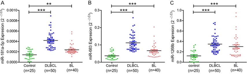
tions were selected for further validation. Quantitative real time PCR (qRT-PCR) was used to validate microarray results among 50 DLBCL, 40 BL, and 20 healthy controls. Comparing DLBCL to control groups,
miR-181a-5p (P < 0.0001), miR-638
(P=0.0001), and miR-1268b
(P < 0.0001) were found to have significantly increased expression levels (Figure 2). This study also confirmed th-at miR-181a-5p (P=0.0058),
miR-638 (P < 0.0001), and
miR-1268b (P < 0.0001) were upregulated in BL, more than in controls. Results of qRT-PCR confirmed that three miRNAs, miR-181, miR-638,
and miR-1268, in DLBDL and BL showed similar change patterns, as shown in micro-array analysis (Figure 2).
Discussion
Diffuse large B-cell lympho- ma (DLBCL) is the most com-mon type of non-Hodgkin’s lymphoma. MicroRNAs may provide a new management model of gene expression. Moreover, miRNA expression levels are closely associa- ted with specific clinical fea -tures of tumors. They can be used to classify normal and tumor tissues and prognostic evaluation. MicroRNAs have a vital function in lymphoma-genesis. However, the effects of miRNAs and their clinical roles in DLBCL have not been completely elaborated. Here, present results indicate that miRNA expression levels were clearly distinguished betw- een DLBCLs and normal tis-sues. This study identified overexpression of hsa-miR- 181, hsa-miR-638, hsa-miR- 328, miR-1268a, hsa-miR-1915-3p, hsa-miR-126-8b, hsa-miR-1825, hsa-miR- 4788, miR-4290, hsa-miR-3162-3p, hsa-miR-1202, and hsa-miR-4649-3p in DLBCLs. S Caramuta et al. used microarray analysis, finding that
[image:4.612.94.369.68.536.2]research discovered that 40 and 20 microRNAs were up- and downregulated in DLBCLs, com-pared to controls (data not fully displayed) [23]. Lim et al. found that 63 miRNA-expression lev-els were increased and 39 miRNA-expression levels were decreased in DLBCLs (data not fully shown), especially expression of miR-28-5p, miR-214-5p, miR-339-3p, and miR-5586-5p,
all associated with good prognosis [26]. Interestingly, most of the miRNA deregulation, in present results, has not been previously reported in DLBCL and/or other malignant tumors. Therefore, present results may provide some novel biomarkers for DLBCL. To further validate current results, real-time quantitative PCR detection of miR-181, miR-638, and miR-1268 expression in diffuse large B-cell lympho-ma and Burkitt’s lympholympho-ma was used. The lat-ter is a common type of non-Hodgkin’s lympho-ma. Compared with the normal group, it was found that expression of 181a-5p, miR-638, and miR-1268b was elevated in diffuse large B-cell lymphoma and Burkitt’s lymphoma. However, whether these molecules are abnor-mally expressed in all non-Hodgkin’s lympho-mas, or all lymphomas in general, remains to be elucidated.
In accord with current results, overexpression of miR-181 has been found in DLBCLs, com-pared to normal B-cells, in Emilia L Lim’s study [26]. Some studies have reported that miR-181 dysregulated expression in many human can-cers, such as acute myeloid-leukemia (AML), luminal breast cancer, non-small cell lung can-cer, and oropharyngeal squamous cell
carcino-expression, resulting in Nipah virus infection by increasing henipavirus-induced membrane fusion. There is also a research certifying that miR-181 activates Akt signaling by inhibiting INPP4B and PHLPP2 phosphatases to promote cancer cell entry S-phase in luminal breast can-cer. Jian’s study has proven that miR-181a-5p significantly decreased MMP-14 luciferase activity, thus miR-181a inhibits cancer cell metastasis by inhibiting MMP-14 expression [31]. Although Emilia L Lim results, along with present results, show that miR-181 was highly expressed and may function as an oncogene in DLBCL. However, the effects of miR-181 on DLBCL cell biological behavior and the poten-tial mechanisms remain unclear. Future studies and discussion are necessary.
[image:5.612.92.323.96.269.2]The current study explored expression of miR-638 in DLBCL, BL patients, and controls using qRT-PCR for the first time. Results suggest that expression levels of miR-638 were upregulated in DLBCL and BL patients, compared to healthy controls. Many studies have suggested that upregulation of miR-638 plays an important role in the development of melanoma, esopha-geal squamous cell carcinoma, and breast can-cer [32, 33]. Ren Y et al. proved that miRNA-638 stimulates autophagy and malignant phe-notypes of esophageal squamous cell carcino-ma and breast cancer cells by suppressing DACT3 [32]. Animesh Bhattacharya et al. indi-cated that miR-638 promoted melanoma pro-gression by inhibiting expression of p53 and p53 downstream target genes, resulting in de- creased of apoptosis and autophagy. However,
Table 2. Fold change and P values of differentially-expressed miRNAs with P < 0.05
miRNAs Fold changes p-value Regulation
hsa-miR-181 623.5 3.88E-06 Up
hsa-miR-638 36.2 0.0491 Up
hsa-miR-328 377.0 0.0239 Up
hsa-miR-1268a 44.7 0.0498 Up
hsa-miR-1915-3p 26.1 0.0495 Up
hsa-miR-1268b 38.4 0.0448 Up
hsa-miR-1825 336.5 0.0423 Up
hsa-miR-4788 33.7 0.0486 Up
hsa-miR-4290 649.6 1.41453E-05 Up
hsa-miR-3162-3p 32.7 0.0389 Up
hsa-miR-1202 25.9 0.0419 Up
hsa-miR-4649-3p 26.4 0.0482 Up
other studies have suggested that miR-638 plays the role of tumor suppressor in cancer. One study confirmed that miR-638 restrains cell proliferation by inhibiting PIM1 genes in osteosarcoma [34]. Zhang’s study demonstrat-ed that miR-638 expression was rdemonstrat-educdemonstrat-ed in hepatocellular carcinoma, suggesting that miR-638 refrained invasion and epithelial-mesen-chymal transition by targeting SOX2 [35]. The different roles of miR-638 in different tumors may be due to actions on different target ge- nes. Therefore, bioinformatics enrichment ana- lysis and corresponding experiments should be conducted to explore that which target genes are regulated by miR-638 and which biological functions are affected in DLBCL and BL cells. Present results demonstrated, for the first time, that miR-1268b is highly expressed in DLBCL and BL. However, the effects and me- chanisms of miR-1268b on tumors remain unknown, as there have been very few stu- dies concerning miR-1268 in tumors. Veronica Colangelo et al. suggested that miR-1268 was upregulated in facioscapulohumeral muscular dystrophy cells, compared to normal cells, indi-cating that miR-1268 may be involved in the regulation of cell proliferation [36]. A previous study showed that miR-1268 was repressed in peripheral blood mononuclear cell obtained from gestational diabetes (GDM) patients, sug-gesting that it may be involved in glucose metabolism in the human body [37]. However, further examination is necessary concerning the effects and mechanisms of miR-1268 on malignant cellular behavior in DLBCL and BL.
Many researchers have shown that many miR-NAs regulate the expression and function of target genes, which participate in occurrence and development of DLBCL. Studies of clinical DLBCL cases have proven that miR-21 overex-pression decreases exoverex-pression of fork-head box protein O1 (FOXO1) and PTEN, resulting in the activation of PI3K/AKT/mTOR pathways and boosting more malignant biological be- havior to DLBCL cells [38]. miR-155 promotes DLBCL cell growth by depressing expression of tumor suppressor gene phosphatidylinositol-3,4,5-trisphosphate 5-phosphatase 1 (SHIP1). Present results, however, should be verified in a larger population of DLBCL, with even more non-Hodgkin’s lymphomas patients [39]. Over- expression of miR-125a/miR-125b directly de- creases the activity of tumor necrosis factor alpha-induced protein 3 (TNFAIP3), thus acti -vating NF-κB signaling pathways and participat -ing in the progression of DLBCL [40]. Accor- dingly, overexpression of these oncomiRNAs is associated with decreased tumor suppressor gene activity and promotes proliferation, inva-sion, and metastasis in DLBCL, BL, and all kinds of non-Hodgkin’s lymphomas. Therefore, GO and KEGG pathway enrichment analysis, clarifying mechanisms in the development of DLBCL and BL, is necessary.
[image:6.612.93.520.73.202.2]In general, the current study determined miRNA expression profiles of FFPE tissues from DLBCL patients, and normal controls. Results con-firmed the association between abnormal expression of 12 specific miRNAs and DLBCL patients. In particular, this study validated that miR-181a-5p, miR-638, and miR-1268b were
upregulated, possibly exerting potential onco-genic roles in DLBCL development. The current study may contribute to identification of poten -tial diagnosis and prognostic biomarkers for DLBCL and BL patients. Further studies should be undertaken to investigate the impact of indi-vidual miRNAs in DLBCL patients, determining the effects on B-cell biology and lymphomagen-esis. Many studies have shown that miRNAs have profound effects on expression of their target genes, suggesting that GO and KEGG Pathway enrichment analysis are necessary to elucidate their mechanisms in DLBCL and BL.
Acknowledgements
This work was supported by the Heilongjiang Postdoctoral Financial Assistance (No. LBH-Z14214). Funders were not involved in study design, data collection and analysis, decision to publish, or preparation of the manuscript.
Disclosure of conflict of interest
None.
Address correspondence to: Yan Shi, The 2nd Hos- pital of Harbin Medical University, Harbin 150000, China. Tel: +8613159867529; 0451-86297425; E-mail: [email protected]
References
[1] Skrabek P, Turner D and Seftel M. Epidemiolo-gy of non-Hodgkin lymphoma. Transfus Apher Sci 2013; 49: 133-138.
[2] Rovira J, Valera A, Colomo L, Setoain X, Rodri-guez S, Martinez-Trillos A, Gine E, Dlouhy I, Magnano L, Gaya A, Martinez D, Martinez A, Campo E and Lopez-Guillermo A. Prognosis of patients with diffuse large B cell lymphoma not reaching complete response or relapsing after frontline chemotherapy or immunochemother-apy. Ann Hematol 2015; 94: 803-812.
[3] Harris NL, Jaffe ES, Stein H, Banks PM, Chan JK, Cleary ML, Delsol G, De Wolf-Peeters C, Falini B, Gatter KC, et al. A revised European-American classification of lymphoid neoplas-ms: a proposal from the International Lympho-ma Study Group. Blood 1994; 84: 1361-1392. [4] Rosenwald A, Wright G, Chan WC, Connors JM,
Campo E, Fisher RI, Gascoyne RD, Muller-Her -melink HK, Smeland EB, Giltnane JM, Hurt EM, Zhao H, Averett L, Yang L, Wilson WH, Jaffe ES, Simon R, Klausner RD, Powell J, Duffey PL, Longo DL, Greiner TC, Weisenburger DD, Sanger WG, Dave BJ, Lynch JC, Vose J,
Armit-age JO, Montserrat E, Lopez-Guillermo A, Gro-gan TM, Miller TP, LeBlanc M, Ott G, Kvaloy S, Delabie J, Holte H, Krajci P, Stokke T and Staudt LM. The use of molecular profiling to predict survival after chemotherapy for diffuse large-B-cell lymphoma. N Engl J Med 2002; 346: 1937-1947.
[5] Dwivedi A, Mehta A and Solanki P. Evaluation of immunohistochemical subtypes in diffuse large B-cell lymphoma and its impact on sur-vival.
[6] Calin GA, Ferracin M, Cimmino A, Di Leva G, Shimizu M, Wojcik SE, Iorio MV, Visone R, Sev-er NI, Fabbri M, Iuliano R, Palumbo T, Pichiorri F, Roldo C, Garzon R, Sevignani C, Rassenti L, Alder H, Volinia S, Liu CG, Kipps TJ, Negrini M and Croce CM. A MicroRNA signature associ-ated with prognosis and progression in chronic lymphocytic leukemia. N Engl J Med 2005; 353: 1793-1801.
[7] Iorio MV, Ferracin M, Liu CG, Veronese A, Spiz -zo R, Sabbioni S, Magri E, Pedriali M, Fabbri M, Campiglio M, Menard S, Palazzo JP, Rosenberg A, Musiani P, Volinia S, Nenci I, Calin GA, Quer-zoli P, Negrini M and Croce CM. MicroRNA gene expression deregulation in human breast can-cer. Cancer Res 2005; 65: 7065-7070. [8] Lawrie CH, Soneji S, Marafioti T, Cooper CD,
Palazzo S, Paterson JC, Cattan H, Enver T, Mag-er R, Boultwood J, Wainscoat JS and Hatton CS. MicroRNA expression distinguishes be-tween germinal center B cell-like and activated B cell-like subtypes of diffuse large B cell lym-phoma. Int J Cancer 2007; 121: 1156-1161. [9] Lawrie CH, Gal S, Dunlop HM, Pushkaran B,
Liggins AP, Pulford K, Banham AH, Pezzella F, Boultwood J, Wainscoat JS, Hatton CS and Har-ris AL. Detection of elevated levels of tumour-associated microRNAs in serum of patients with diffuse large B-cell lymphoma. Br J Hae-matol 2008; 141: 672-675.
[10] Lu J, Getz G, Miska EA, Alvarez-Saavedra E, Lamb J, Peck D, Sweet-Cordero A, Ebert BL, Mak RH, Ferrando AA, Downing JR, Jacks T, Horvitz HR and Golub TR. MicroRNA expres-sion profiles classify human cancers. Nature 2005; 435: 834-838.
[11] Yu X, Li Z and Liu J. MiRNAs in primary cutane-ous lymphomas. Cell Prolif 2015; 48: 271-277. [12] Li Z, Yu X, Shen J, Chan MT and Wu WK. Mi -croRNA in intervertebral disc degeneration. Cell Prolif 2015; 48: 278-283.
[13] Li Z, Yu X, Shen J, Law PT, Chan MT and Wu WK. MicroRNA expression and its implications for diagnosis and therapy of gallbladder can-cer. Oncotarget 2015; 6: 13914-13921. [14] Lewis BP, Burge CB and Bartel DP. Conserved
-dicates that thousands of human genes are microRNA targets. Cell 2005; 120: 15-20. [15] Calin GA, Sevignani C, Dumitru CD, Hyslop T,
Noch E, Yendamuri S, Shimizu M, Rattan S, Bullrich F, Negrini M and Croce CM. Human mi -croRNA genes are frequently located at fragile sites and genomic regions involved in cancers. Proc Natl Acad Sci U S A 2004; 101: 2999-3004.
[16] Huang J, Zhang SY, Gao YM, Liu YF, Liu YB, Zhao ZG and Yang K. MicroRNAs as oncogenes or tumour suppressors in oesophageal cancer: potential biomarkers and therapeutic targets. Cell Prolif 2014; 47: 277-286.
[17] Cui XB, Li S, Li TT, Peng H, Jin TT, Zhang SM, Liu CX, Yang L, Shen YY, Li SG, Li N, Li Y, Hu JM, Ji-ang JF, Suo J, Qi Y, LiJi-ang WH, WJi-ang LH, DJi-ang HW, Li L, Cao WW, Wei Y, Laibo Y, Wu CY, Yuan XL, Zhou H, Zheng Y, Chen YZ and Li F. Target -ing oncogenic PLCE1 by miR-145 impairs tu-mor proliferation and metastasis of esopha-geal squamous cell carcinoma. Oncotarget 2016; 7: 1777-1795.
[18] Cui X, Zhao Z, Liu D, Guo T, Li S, Hu J, Liu C, Yang L, Cao Y, Jiang J, Liang W, Liu W, Wang L, Gu W, Wu C, Chen Y and Li F. Inactivation of miR-34a by aberrant CpG methylation in Ka -zakh patients with esophageal carcinoma. J Exp Clin Cancer Res 2014; 33: 20-26.
[19] Jorgensen LK, Poulsen MO, Laursen MB, Marques SC, Johnsen HE, Bogsted M and Dyb-kaer K. MicroRNAs as novel biomarkers in dif -fuse large B-cell lymphoma--a systematic re-view. Dan Med J 2015; 62: 133-145.
[20] Lim EL, Trinh DL, Scott DW, Chu A, Krzywinski M, Zhao Y, Robertson A, Mungall AJ, Schein J, Boyle M, Mottok A, Ennishi D, Johnson NA, Steidl C, Connors JM, Morin RD, Gascoyne RD and Marra MA. Comprehensive miRNA se-quence analysis reveals survival differences in diffuse large B-cell lymphoma patients. Ge-nome Biology 2015; 16: 1-18.
[21] Wang K, Xu Z, Wang N, Xu T and Zhu M. Mi -croRNA and gene networks in human diffuse large B-cell lymphoma. Oncol Lett 2014; 8: 2225-2232.
[22] Handal B, Enlow R, Lara D, Bailey M, Vega F, Hu P and Lennon A. Investigating the expres-sion of oncogenic and tumor suppressive mi-croRNA in DLBCL. J Assoc Genet Technol 2013; 39: 14-20.
[23] Lawrie CH, Chi J, Taylor S, Tramonti D, Ballabio E, Palazzo S, Saunders NJ, Pezzella F, Boult -wood J, Wainscoat JS and Hatton CS. Expres-sion of microRNAs in diffuse large B cell lym-phoma is associated with immunophenotype, survival and transformation from follicular lym-phoma. J Cell Mol Med 2009; 13: 1248-1260.
[24] Alencar AJ, Malumbres R, Kozloski GA, Advani R, Talreja N, Chinichian S, Briones J, Natkunam Y, Sehn LH, Gascoyne RD, Tibshirani R and Lossos IS. MicroRNAs are independent predic-tors of outcome in diffuse large B-cell lympho-ma patients treated with R-CHOP. Clin Cancer Res 2011; 17: 4125-4135.
[25] Caramuta S, Lee L, Ozata DM, Akcakaya P, Georgii-Hemming P, Xie H, Amini RM, Lawrie CH, Enblad G, Larsson C, Berglund M and Lui WO. Role of microRNAs and microRNA machin-ery in the pathogenesis of diffuse large B-cell lymphoma. Blood Cancer J 2013; 3: 152-164. [26] Lim EL, Trinh DL, Scott DW, Chu A, Krzywinski
M, Zhao Y, Robertson AG, Mungall AJ, Schein J, Boyle M, Mottok A, Ennishi D, Johnson NA, Steidl C, Connors JM, Morin RD, Gascoyne RD and Marra MA. Comprehensive miRNA se-quence analysis reveals survival differences in diffuse large B-cell lymphoma patients. Ge-nome Biol 2015; 16: 18-30.
[27] Butrym A, Rybka J, Baczynska D, Poreba R, Mazur G and Kuliczkowski K. Expression of mi -croRNA-181 determines response to treat-ment with azacitidine and predicts survival in elderly patients with acute myeloid leukaemia. Oncol Lett 2016; 12: 2296-2300.
[28] Strotbek M, Schmid S, Sanchez-Gonzalez I, Boerries M, Busch H and Olayioye MA. miR-181 elevates Akt signaling by co-targeting PHLPP2 and INPP4B phosphatases in luminal breast cancer. Int J Cancer 2017; 140: 2310-2320.
[29] Lee SH, Lee CR, Rigas NK, Kim RH, Kang MK, Park NH and Shin KH. Human papillomavirus 16 (HPV16) enhances tumor growth and can-cer stemness of HPV-negative oral/oropharyn-geal squamous cell carcinoma cells via miR-181 regulation. Papillomavirus Res 2015; 1: 116-125.
[30] Tian F, Shen Y, Chen Z, Li R, Lu J and Ge Q. Aberrant miR-181b-5p and miR-486-5p ex-pression in serum and tissue of non-small cell lung cancer. Gene 2016; 591: 338-343. [31] Roth E and Cao J. miR-181 suppresses
metas-tasis via MMP-14. Aging (Albany NY) 2015; 7: 740-741.
[32] Ren Y, Chen Y, Liang X, Lu Y, Pan W and Yang M. MiRNA-638 promotes autophagy and malig-nant phenotypes of cancer cells via directly suppressing DACT3. Cancer Lett 2017; 390: 126-136.
[34] Wang XX, Liu J, Tang YM, Hong L, Zeng Z and Tan GH. MicroRNA-638 inhibits cell prolifera-tion by targeting suppress PIM1 expression in human osteosarcoma. Tumour Biol 2017; [Epub ahead of print].
[35] Zhang Y, Zhang D, Jiang J and Dong L. Loss of miR-638 promotes invasion and epithelial-mesenchymal transition by targeting SOX2 in hepatocellular carcinoma. Oncol Rep 2017; 37: 323-332.
[36] Colangelo V, Francois S, Solda G, Picco R, Roma F, Ginelli E and Meneveri R. Next-gener -ation sequencing analysis of miRNA expres-sion in control and FSHD myogenesis. PLoS One 2014; 9: e108411.
[37] Collares CV, Evangelista AF, Xavier DJ, Rassi DM, Arns T, Foss-Freitas MC, Foss MC, Puthier D, Sakamoto-Hojo ET, Passos GA and Donadi EA. Identifying common and specific microR -NAs expressed in peripheral blood mononucle-ar cell of type 1, type 2, and gestational diabe-tes mellitus patients. BMC Res Nodiabe-tes 2013; 6: 491.
[38] Go H, Jang JY, Kim PJ, Kim YG, Nam SJ, Paik JH, Kim TM, Heo DS, Kim CW and Jeon YK. MicroR -NA-21 plays an oncogenic role by targeting FOXO1 and activating the PI3K/AKT pathway in diffuse large B-cell lymphoma. Oncotarget 2015; 6: 15035-15049.
[39] Pedersen IM, Otero D, Kao E, Miletic AV, Hother C, Ralfkiaer E, Rickert RC, Gronbaek K and Da -vid M. Onco-miR-155 targets SHIP1 to promote TNFalpha-dependent growth of B cell lympho -mas. EMBO Mol Med 2009; 1: 288-295. [40] Kim SW, Ramasamy K, Bouamar H, Lin AP, Ji