Recognition of Diseases of Leaf using SVM with
Radial Basis Kernel Function
Anuradha Sharma1, Abhilash Sonker2
1, 2
Computer Science & Information Technology, Madhav Institute of Technology & Science, Gwalior (M.P.), India
Abstract: Organic farming obtains admiration in agricultural industry. Several complications get up during farming wherein the disease affected leaf contemplated to be very powerful aspect for the shortage of agricultural products and also upshot the quality of product. The target is to diminish this problem through machine learning technique. This research showcased a methodology for Recognition and Grouping of diseased leaf and healthy leaf by Support vector machine(SVM) classifier with radial basis function. The texture features are take out from the diseased leaf and from the Healthy Leaf images separately for the grouping/classification. Here, we cast-off SVM classifier with RBF will group/classify the ailments such as Cercospora leaf spot,Bacterial blight, Anthracnose, Alternaria alternata, and healthy leaves of several species of plants. Emphasis is given on the initial recognition of diseases.
Keywords: (Support vector machines (svm), diseased and healthy leaf, Digital Image processing(DIP) techniques, ,k-mean clustering )
I. INTRODUCTION
Computers had greatly influenced most of the aspects of life by their incredible technological improvements in expression of strong, reliable and flexible computing devices. In the similar fashion the potential of computer is explored in agricultural field. Agriculture is contemplated to be the utmost important part that directly influence the life as food is the basic need of all living beings. [1] The delivery of food to enormous population needs appropriate quantity of production and for producing sufficient amount of good quality agricultural product it is necessary to protect the growing plants from diseases/ailments produced by several types of pathogens. Observation of plants and their supervision is very essential from initial stage of growth. If the proper observation of leaves are practiced at starting stage itself, further spread of ailments in plants can be dodged. Frequent types of diseases occur on plants which target diverse parts of plant body such as stem, fruit, seed and leaf etc. [2] Leaves are contemplated to be the core/central fragment of the plant. The lifespan of the plant gets highly effected if the leaf is prone to diseases and to check these diseases in each and every plant needs lot of manpower and time, sometimes it is not accurate as well. [3] Consequently, there is requirement of an automated system that will does the task of the agricultural experts. So (DIP) techniques are deployed to achieve the target of recognizing diseased leaf and healthy leaf. In this research work, the four most common diseases of leaves have been recognized named as Cercospora leaf spot ,Bacterial blight, Anthracnose, Alternaria alternata, along with the healthy leaves of several species of plant. Where anthracnose, alternaria alternata and cercospora leaf spot are caused by fungus and Bacterial blight is caused by bacteria. For detection/recognition of plant leaf ailments from images, different kinds of machine training methods are anticipated in recent times but the supreme challenge being encountered is the proficiency and the robustness of the achieved results. In this work there is casting-off IP (Image processing) techniques for the identification/recognition of diseased leaf and healthy leaf. The foremost target of this work is to train/learn the farmers with sufficient and affordable information to increase the productivity along with quality. This paper organized in five sections. The remaining Section is composed as pursues. The related works are talked about in Segment II. In Segment III, proposed methodology is given. The Experimental outcomes is clarified in Segment IV. End is completed in Segment V. The future extent of this work is talked about in the last Section.
II. RELATEDWORKS
Pooja V et al. [4]. Proposed an approach for identifying five common leaf diseases on diverse types of leaf species. The digital prints of the plant are taken and then it is pre-processed. After pre-processing segmentation technique is deployed on the outcome given by pre-processing module. Features are taken out from the Segmented image and on the basis of that classification of diseases are done with an aid of SVM classifier.
This system is cast-off for detection of cotton leaf sickness.
Al Bashish et al [6] showcased a methodology for recognition of leaf and stem diseases. Here k-means method is castoff to split the processed image. After this the Segmented image is given to the trained neural network. Consequences of this work determine the successful grouping/classification of diseases.
P. Krithika and S. Veni [7] showcased k-means clustering method for division of the diseased part from the leaves of cucumber. Gray level co-occurrence matrix method are used for extraction of suitable features of the segmented diseased area of cucumber leaves which were castoff for training & testing of the classifier. Here Multiclass Support Vector Machine classifier was conveyed for characterization of the leaf spot, leaf minor and mosaic infection on the leaves of cucumber.
Shriroop C. Madiwalar and Medha V. Wyawahare [8] showcased a philosophy for discovery of mango leaf ailments. It cast-off the color dependent segmentation on YcbCr color space & the testing also cast-off minimum distance classifier (MDC) & support vector machine (SVM) to group the ailment in their corresponding class such as anthracnose and leaf spot. This research work illustrates, 3 diverse kind of features such as color, GLCM and Gabor features were take-out for the efficiency evaluation of Minimum distance classifier (MDC) & support vector machine (SVM) disease classification of Mango leaves.
Jobin Francis et al. [9] proposed a procedure for pepper leaves disease detection. Threshold based segmentation and masking of green pixels are done for obtaining the author’s purpose. By this method quick wilt and berry spot diseases are recognized. Features are extracted by GLCM technique and for classifying neural network (NN) is used.
Pranjali B. Padol and Prof. Anjali A. Yadav [10] proposed a technique where before segmentation of image Gaussian filter were deployed. Here, Linear SVM classifier were castoff. For classifying leaf diseases 54 features taken into consideration. This work is conducted on Grape leaf image for classification or grouping of powdery mildew & downy mildew diseases.
III. PROPOSEDMETHODOLOGY
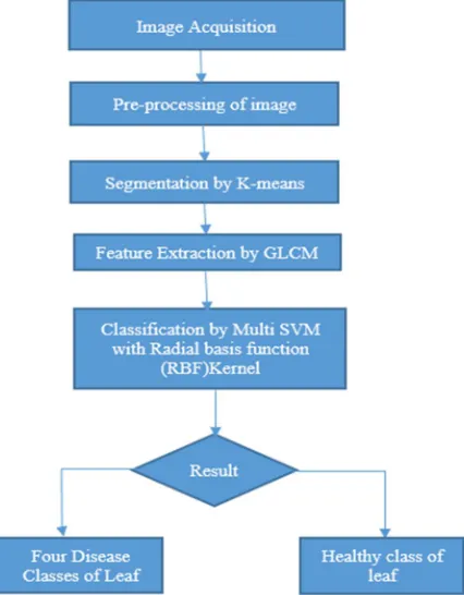
The Proposed method of this paper is symbolized in Fig. 1. The first and foremost tread is to acquire the infected/diseased leaf. Pre-processing is deployed on acquired image to make that image appropriate for disease detection system. The output of pre-Pre-processing module is given to the segmentation module to section the infected part. Further, feature extraction done on segmented image and the values the features are used for training Support vector machine (SVM) along with RBF kernel for final classification.
[image:2.612.205.418.439.712.2]The comprehensive description of various modules involved in this approach are described below-
A. Image Acquisition
The images are taken from a validated dataset available on kaggle.com so as to check the accuracy of our approach. The dataset contains leaves infected by Cercospora leaf spot,Bacterial blight, Anthracnose, Alternaria alternata diseases of several plant species. It also contains healthy leaves of several plant species. Following are sample images from the dataset, they are-
(a) Alternaria alternata (b) Anthracnose
(c) Bacterial Blight (d) Cercospora leaf spot
[image:3.612.161.469.150.505.2](e) Healthy leaf
Fig. 2. Illustrates the dataset images where (a) , (b), (c) ,(d) shows infected leaves from Alternaria alternata, Anthracnose, Bacterial
blight, Cercospora leaf spot, and (e) represents healthy leaves of several plant species respectively .
B. Image pre-processing:
Since the images might not be of similar dimension hence the the images are pre-processed to make it in similar dimension for the system. Also the images needs pre-processing to get appropriate results because results may vary according to the color space ,size and noise in the image. Therefore image pre-processing is essential before going to the further steps.
1) Image Rescaling: In real applications the obtained Image may not be in the same dimension. It is suggested to rescale the attained Images. Here in our work we take the dimension of the images as 256 × 256 × 3. The interpolation method cast-off for resizing. It is bilinear interpolation because in this technique value of four adjacent pixel are averaged to discover the novel pixel value.[11]
= 1 + 2 + 3 + 4
4
Fig. 3. Bilinear interpolation [11]
2) Contrast Enhancement: The enhancement of contrast is essential to differentiate between object and background. [12] An enhancement module is vital to pick the appropriate contrast measure, For this cause, “imadjust()” function of MATLAB is applied for increase the several levels of contrast.
3) Conversion of color space: Conversion of color space is needed for giving a mode to recognize additional spontaneous color information because Red, green, blue (RGB) color space is very device dependent and furthermore it hang on light intensity [13]. The images that includes color variances are easy to execute in CIELAB color model/space as equated to RGB color model because the L*a*b color space is device independent space. The influence of L*a*b shading model is that it is made to genuinely exact the view of human to the light. The Lab colour space permits the users to calculate these visual differences. The format of conversion is in makecform and applycform before converting in Lab color space[14]
The outcome of Image Acquisition & Pre-processing step is passed to the next module.
C. Segmentation
Segmentation is needed to get the ROI(region of interest) i.e. the disease influenced region. k-means algorithm is deployed for this task, here the partition of the whole RGB image is done into three clusters.In our situation three clusters are chosen i.e. k=3 and those three clusters will repesent : the background, diseased part of leaf and healthy portion of leaf. Further, clusters given by k-means technique ae used for selecting region of interest. The appropriate cluster in which diseased leaf and healthy leaf are segmented can be individually choosen for feature extraction. Here, ROI (region of interest) is explicitly given by the user. This selected cluster turn as the input to the next module.
D. Feature Extraction
Features are take out from GLCM (Gray level co-occurrence method) and color feature from the segmented part according to user’s choice. Let i and j are thecoefficients of co-occurrence matrix, P(i,j), is the component inthe matrix at the coordinates i and j and N isthe dimension of the matrix.
1) Mean: Mathematically mean is calculated by formula given below
2) Standard Deviation: Mathematically it is computed by formula given below
3) Energy: Mathematically it is computed by formula given below
5) Homogeneity: Mathematically it is computed by formula given below
6) Correlation: Mathematically it is computed by formula given below
7) Entropy: Mathematically it is computed by formula given below
Extracted features are built on the basis of region of interest (ROI) selected. A number of feature extraction methods such as Mean, Standard deviation, Entropy, Contrast, Energy, Homogeneity etc. [15] are used to take out the desired features. Here the energy and homogeneity decreases with the increasing image quality. Whereas entropy & contrast shows consistent increase with increasing image quality for all the images.
E. Classification
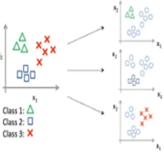
[image:5.612.221.388.514.672.2]The practice of staggering a class on a novel illustration based on learning accomplished by the classifier model in the course of training is called classification. This work describes the grouping of plant ailment into four classes along with one healthy leaf class are done by aid of Multiclass SVM classifier along with Radial basis function kernel. Support vector machine (SVM) is contemplated as a method that require pre-defined learning. In this method, training data is generated with tag built on the trial Image composed. Here, each ailment/disease is contemplated as an isolated class and tags are specified for that reason. This learning data denotes to a group of instances that are cast-off to produce a database for the training procedure. In this type of grouping there is analysis of each training set and an equivalent output is conveyed. The class tags or labels should be allocated to the trial data when the trial data is passed to it. Furthermore, the learning samples are minor/small SVM with RBF kernel gives are effective and accurate classification. Former efforts have also specified that support vector machine gave good results as equated to Neural Network for classification. Hence, we deploy Multiclass support vector machine (Multi-SVM) to execute Supervised Classification. It is a binary classifiers but it can be prepared to knob Multiple classifications. [16] The methodology for this various grouping is (1AA) one-against-all where there is division of n classes in n two-class machines. On the off chance that the three classes are marked as 1,2 and 3, at that point arrangement would be done by looking at class 1 against non-class 1 (class 2,3) or class 2 against non-class 2 (class 1,3) or class 3 against non-class 3 (class 1,2) as portrayed in Fig. 4.
Fig. 4. IAA Method/Approach
IV. EXPERIMENTALRESULTS
For the evaluation of proposed methodology, the sample images taken from various authentic and verified dataset providing websites. Here the instances are divided into training/learning dataset and test dataset. The training dataset images are cast-off to train/learn the Multiclass SVM with RBF kernel classifier whereas the test dataset images are cast-off to compute or predict the efficiency/accuracy of the classifier.
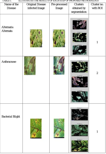
[image:6.612.121.493.186.725.2]The following are the table1 representing the operations performed in our methodology. Column 1 ,2,3,4,5 illustrates operations right from acquiring image to the segmentation of desired cluster to classify it in further step of classification.
TABLE I. ILLUSTRATES THE RESULTS OF EACH STEP OF PROPOSED METHODOLOGY
Name of the Disease
Original Disease infected Image
Pre-processed Image
Clusters obtained by segmentation
Cluster no. with ROI
Alternaria Alternata
1
Anthracnose
2
Bacterial Blight
Name of the Disease
Original Disease infected Image
Pre-processed Image
Clusters obtained by segmentation
Cluster no. with ROI
Cercospora Leaf Spot
2
Healthy Leaf 1
1) Here 269 images are taken to train the network , among them
a) 62 are of Alternaria Alternata category.
b) 62 samples are of type Anthracnose .
c) 52 samples belongs to Bacterial Blight.
d) 62 are of Cercospora Leaf spot.
e) 31 samples are of healthy leaves.
2) Also, we test our proposed module for the same no. of samples used in previous existing work that contains
a) 23 images of Alternaria Alternata category.
b) 50 images are of type Anthracnose.
c) 52 images belongs to Bacterial Blight.
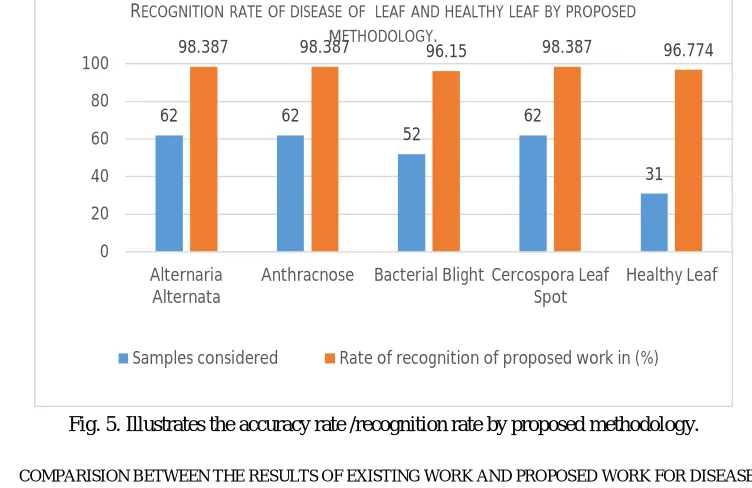
TABLE II. RECOGNITION RATE OF DISEASE OF LEAF AND HEALTHY LEAF BY PROPOSED METHODOLOGY.
Name of
classes Samples considered Recognized Samples Misclassifie d Samples Rate of recognition (%) Alternaria
Alternata 62 61 1 98.387
Anthracnose
62 61 1 98.387
Bacterial
Blight 52 50 2 96.15
Cercospora Leaf Spot
62
61 1 98.387
Healthy Leaf 31 30 1 96.774
[image:8.612.114.490.329.573.2]The above table II gives us the recognition rate of disease of leaf and healthy leaf. By referring the above table we calculate the overall recognition rate found to be 97.617 %.
Fig. 5. Illustrates the accuracy rate /recognition rate by proposed methodology.
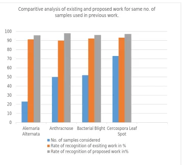
TABLE III. COMPARISION BETWEEN THE RESULTS OF EXISTING WORK AND PROPOSED WORK FOR DISEASE DETECTION.
Name of classes Samples considered
Rate of recognition of existing work in (%)
Rate of recognition of proposed work in (%)
Alternaria Alternata
23 91.3 95.7
Anthracnose
50 90 98
Bacterial Blight
52
92.31
96.15
Cercospora Leaf Spot 73 93.15 97.26
62 62
52 62
31
98.387 98.387 96.15 98.387 96.774
0 20 40 60 80 100 Alternaria Alternata
Anthracnose Bacterial Blight Cercospora Leaf
Spot
Healthy Leaf RECOGNITIONRATEOFDISEASEOF LEAFANDHEALTHYLEAFBYPROPOSED
METHODOLOGY.
Fig. 6. Illustrates the comparision between accuracy rate /recognition rate of previous work and proposed methodology.
V. CONCLUSION
This work showcased a method which cast-off Multiclass Support vector machine (Multi-SVM) with radial basis function kernel methodology to detect/recognize and classify/group several diseases that exists in plant leaves Diseases/ailments such as Cercospora leaf spot,Bacterial blight, Anthracnose, Alternaria alternata, of plant leaves are deliberated for the experimentation. The disease area are segmented via k-means clustering method and Gray level co-occurrence matrix (GLCM) texture features are cast-off for the classification. On the other hand, the classification performance of the Multiclass SVM with RBF kernel on plant leaf disease delivers 97.617 % accuracy and the anticipated method based tactic gives better results on comparing to some of the prevailing methods.
VI. FUTURE WORK
In future almost all types of plant leaf disease detection system will be developed to detect diseases in early stages.
Large dataset should be cast-off to train the classifier because with increase in the training samples the efficiency or accuracy of system increases.
REFERENCES
[1] Pujari JD, Yakkundimath R, Byadgi AS, SVM and ANN based classification of plant diseases using feature reduction technique. Int J Interact Multimed Artif
Intell 3:6–14 18, 2016.
[2] Sachin D. Khirade, and A. B. Patil, “Plant disease detection using image processing,” IEEE International Conference of Computing Communication Control
and Automation (ICCUBEA), pp 768-771, 2015.
[3] Halil Durmus, Ece Olcay Gunes, Murvet K, and Burak Berk Ustundag, “The design of general purpose autonomous agricultural mobile robot: “Agrobot”,”
IEEE Fourth International conference on Agro-Geoinformatics,pp. 49-53, July 2015..
[4] V. Pooja, R. Das, and V. Kanchana, “Identification of plant leaf diseases using image processing techniques,” in Technological Innovations in ICT for Agriculture and Rural Development (TIAR), 2017 IEEE, pp. 130–133, 2017.
[5] P. Revathi and M.hemalatha,” Cotton leaf Spot Diseases Detection Utilizing Feature Selection with Skew Divergence Method”,vol. 30. no.3, pp 22-30,2014.
[6] D.Al Bashish, M. Braik, and S. Bani-Ahmad, “A framework for detection and classification of plant leaf and stem diseases”, Proc. 2010 Int. Conf. Signal Image
Process. ICSIP,pp. 113-118, 2010. 0 10 20 30 40 50 60 70 80 90 100
Alernaria Alternata
Anthracnose Bacterial Blight Cercospora Leaf Spot
Comparitive analysis of existing and proposed work for same no. of samples used in previous work.
No. of samples considered
[7] P. Krithika and S. Veni, “Leaf disease detection on cucumber leaves using multiclass support vector machine,” in Wireless Communications, Signal Processing and Networking (WiSPNET), 2017 International Conference on. IEEE, pp. 1276–1281, 2017.
[8] S. C. Madiwalar and M. V. Wyawahare, “Plant disease identification: a comparative study,” in Data Management, Analytics and Innovation (ICDMAI), 2017
International Conference on. IEEE, pp. 13–18, 2017.
[9] J. Francis, B. Anoop et al., “Identification of leaf diseases in pepper plants using soft computing techniques,” in Emerging Devices and Smart Systems (ICEDSS), Conference on. IEEE, pp. 168–173, 2016.
[10] P. B. Padol and A. A. Yadav, “Svm classifier based grape leaf disease detection,” in Advances in Signal Processing (CASP), Conference on. IEEE, pp. 175–
179, 2016.
[11] Shitala Prasad, Sateesh K. Peddoju, and Debashis Ghosh.“Multi-resolution mobile vision system for plant leaf disease diagnosis,”Signal, Image and Video Processing, vol. 10, no. 2, pp. 379–388, 2016.
[12] P. Krithika and S. Veni, Leaf Disease Detection on Cucumber Leaves Using Multiclass Support Vector Machine, IEEE WiSPNET, pp-1276-1281, 2017.
[13] R. Anand, S. Veni, and J. Aravinth, “An application of image processing Techniques for detection of diseases on brinjal leaves using k-means clustering method,” in Recent Trends in Information Technology (ICRTIT), International Conference on. IEEE, 2016.
[14] Riddhi H. Shaparia, Dr. Narendra M. Patel and Prof. Zankhana H. Shah, Flower Classification using Texture and Color Features, International Conference on
Rea search and Innovations in Science,Engineering &Technology, vol. 2, pp-113-118, 2017
[15] P. Mohanaiah, P. Sathyanarayana, and L. GuruKumar “Image texture feature extraction using GLCM,” International Journal of Scientific and Research
Publications, vol. 3, no. 5, May 2013, ISSN 2250–3153.