research communications
Acta Cryst.(2020). E76, 377–381 https://doi.org/10.1107/S2056989020002042
377
Received 9 December 2019Accepted 12 February 2020
Edited by D. Chopra, Indian Institute of Science Education and Research Bhopal, India
Keywords:crystal structure; piperidine deriva-tive; Hirshfeld surface; DFT.
CCDC reference:1814839
Supporting information:this article has supporting information at journals.iucr.org/e
Crystal structure, Hirshfeld surface analysis and
DFT studies of 1-[
r
-2,
c
-6-diphenyl-
t
-3-(propan-2-yl)piperidin-1-yl]ethan-1-one
P. Periyannan,aM. Beemarao,aK. Karthik,bS. Ponnuswamyband K. Ravichandrana*
a
Department of Physics, Kandaswami Kandar’s College, Velur, Namakkal 638 182, India, andbPG and Research Department of Chemistry, Government Arts College (Autonomous), Coimbatore 641 018., Tamil Nadu, India. *Correspondence e-mail: [email protected]
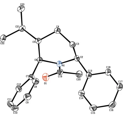
In the title compound, C22H27NO, the piperidine ring adopts a chair
conformation. The dihedral angles between the mean plane of the piperidine ring and the phenyl rings are 89.78 (7) and 48.30 (8). In the crystal, molecules
are linked into chains along theb-axis direction by C—H O hydrogen bonds. The DFT/B3LYP/6–311 G(d,p) method was used to determine the HOMO– LUMO energy levels. The molecular electrostatic potential surfaces were investigated by Hirshfeld surface analysis and two-dimensional fingerprint plots were used to analyse the intermolecular interactions in the molecule.
1. Chemical context
Piperidine is a heterocyclic six-membered ring containing nitrogen as a hetero atom and is an essential structural part of many important drugs including paroxetine, raloxifene, halo-peridol, droperidol and minoxidiln (Wagstaff et al., 2002). Piperidine derivatives exhibit a wide range of biological activities, such as antimicrobial, anti-inflammatory, antiviral, antimalarial and general anesthetic (Aridosset al., 2009). The biological properties of piperidines are highly dependent on the type and position of substituents on the heterocyclic ring. 2,6-Disubstituted piperidine derivatives have been found to possess fungicidal, bactericidal and herbicidal activities (Mobio et al., 1989). Piperidine derivatives are the inter-mediate products in agrochemicals, pharmaceuticals, rubber vulcanization accelerators and are widely used as building block molecules in many industries. Various piperidine deri-vatives are present in numerous alkaloids (Badorrey et al., 1999).
This wide range of biological activities prompted us to synthesize novel 2,6-diphenyl piperdine derivatives. Against this background, the structure of the title compound has been determined.
2. Structural commentary
The molecular structure of the title compound is shown in Fig. 1. The diphenyl-substituted piperidine compound crys-tallizes in the monoclinic space groupP21/n. The bond lengths
and angles are well within the expected limits and comparable with literature values (Allenet al., 1998).
The piperidine ring adopts a chair conformation with the puckering parameters Q2 = 0.6191 (15) A˚ and 2 =
335.12 (14) A˚ . The piperidine ring (N1/C2–C6) makes
dihed-ral angles of 89.78 (7) and 48.30 (8), respectively, with the
C7–12 and C13–C18 phenyl rings, and confirms the fact that the moieties are in an axial orientations.
The keto and methyl groups substituted at atom C19 are equatorially orientated as confirmed from the torsion angle values O1—C19—N1—C2 = 177.54 (12) and C20—C19—
N1—C6 = 172.81 (11). In the molecule, the isopropyl group
substituted at the 5-position of the piperidine ring is equato-rially oriented, as confirmed by the torsion angles of C4—C5— C21—C22 = 172.13 (14) and C6—C5—C21—C23 = 174.73 (14). The sum of the bond angles (359.87) around
atom N1 of the piperidine ring is in accordance with thesp2 -hybridization state (Beddoeset al., 1986).
3. Supramolecular features
In the crystal, molecules are linked intoC(8) chains along the
b-axis direction by C—H O hydrogen bonds (Table 1, Fig. 2). The overall crystal packing of the title compound is shown in Fig. 3.
4. DFT study
The optimized structure of the molecule in the gas phase was generated theoretically via density functional theory (DFT) using standard B3LYP functional and 6-311G(d,p) basis-set
378
Periyannanet al. C22H27NO Acta Cryst.(2020). E76, 377–381
[image:2.610.46.296.65.302.2] [image:2.610.313.565.82.125.2]research communications
Table 1
Hydrogen-bond geometry (A˚ ,).
D—H A D—H H A D A D—H A
C9—H9 O1i 0.93 2.54 3.4378 (19) 163
Symmetry code: (i)x1 2;yþ
1 2;z
[image:2.610.50.542.493.706.2]1 2.
Figure 2
A partial view along the b axis of the crystal packing of the title compound, showing the formation of a molecular chain by C—H O interactions (dotted lines).
Figure 3
The overall crystal packing of the title compound, viewed along theb-axis direction. Hydrogen bonds are shown as dashed lines, and only the H atoms involved in hydrogen bonding have been included.
Figure 1
[image:2.610.301.562.493.707.2]calculations (Becke et al., 1993), as implemented in GAUS-SIAN09(Frischet al., 2009).
The overlay diagram for the optimized structure (purple) and the structure in solid state (green) with respect to the piperidine ring is shown in Fig. 4. The piperidine rings in the two phases have an r.m.s deviation of 0.434 A˚ for the non-hydrogen atoms. The conformation of the molecules in the two phases differs with respect to the central piperidine ring, as seen in the disparity of about 38.5 in the N1—C6—C5—C4 torsion angles (39.88/1.38) and 2.25in the N1—C2—C3—C4 torsion angles (44.41/39.81) for the optimized and solid-state
molecules, respectively.
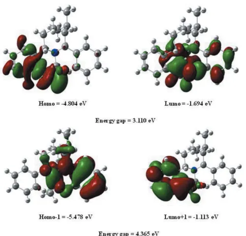
The highest-occupied molecular orbital (HOMO), acting as an electron donor, and the lowest-unoccupied molecular orbital (LUMO), acting as an electron acceptor, are known as
frontier molecular orbitals (FMOs). The FMOs play an important role in the optical and electric properties, as well as in quantum chemistry (Fleming, 1976). When the energy gap is small, the molecule is highly polarizable and has high chemical reactivity. The electron distribution of the HOMO1, HOMO, LUMO and LUMO+1 energy levels and the energy values are shown in Fig. 5. The positive and negative phases are shown in green and red, respectively.
The HOMO of the title molecule is localized on the C O group, one aromatic ring and the piperidine ring, while the LUMO is located over the whole molecule expect for the isopropyl group. The DFT study shows that the FMO energies
EHOMO andELUMO are 4.804 and1.694 eV, respectively,
and the HOMO–LUMO energy gap is 3.110 eV. The title compound has a small frontier orbital gap, hence the molecule has high chemical reactivity and low kinetic stability.
The electron affinity (I) and ionization potential (A) of the molecule were calculated using the DFT/B3LYP/6-311++G(d,p) basis set. A high value of the electrophilicity index describes a good electrophile, while a small value of electrophilicity index describes a good nucleophile. The values of the hardness (), softness (), electronegativity () and electrophilicity index (!) for the title compound are given in Table 2.
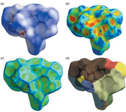
5. Hirshfeld surface analysis
CrystalExplorer17 (Turner et al., 2017) was used for the Hirshfeld surface (HS) analysis (Spackman & Jayatilaka, 2009) and to generate the associated two-dimensional finger-print plots (McKinnon et al., 2007) to quantify the various intermolecular interactions in the structure of the title compound. In the HS plotted overdnorm (Fig. 6), the white
surface indicates contacts with distances equal to the sum of the van der Waals radii, and the red and blue colours indicate distances shorter (in close contact) or longer (distinct contact) than the van der Waals radii, respectively (Venkatesanet al., 2016).
The HS mapped over curvedness and shape-index, intro-duced by Koendrink (Koenderink, 1990; Koenderink & van Doorn, 1992), give further chemical insight into molecular packing. A surface with low curvedness designates a flat region and may be indicative of–stacking in the crystal. A
research communications
Acta Cryst.(2020). E76, 377–381 Periyannanet al. C
[image:3.610.45.297.71.245.2]22H27NO
379
Figure 5 [image:3.610.314.566.93.213.2]The frontier molecular orbitals (FMOs) of the title compound. Figure 4
A structural overlay diagram (Mercury; Macrae et al., 2020) for the optimized structure (purple) and the solid-state structure (green) of the title compound.
Table 2
Calculated frontier molecular orbital analysis of the title compound.
Parameter Value
EHOMO(eV) 4.804
ELUMO(eV) 1.694
Energy gap,E(eV) 3.110
HOMO1 (eV) 5.478
LUMO+1 (eV) 1.113
Ionization potential,I(eV) 4.804
Electron affinity,A 1.694
Electrophilicity Index,! 3.394
Hardness, 1.555
Electro negativity, 3.249
[image:3.610.49.299.491.731.2]Hirshfeld surface with high curvedness is highlighted as dark-blue edges, and is indicative of the absence of –stacking (Fig. 6). The nearest neighbour coordination environment of a molecule is identified from the colour patches on the Hirshfeld surface, depending on their closeness to adjacent molecules (Mohamooda Sumayaet al., 2018).
The 2D fingerprint plots of the di and de points for the
contacts contributing to the Hirshfeld surface are shown in Fig. 7. They indicate that intermolecular H H contacts provide the largest contribution (74.2%) to the Hirshfeld surface. The percentage contributions of the other interactions are C H/H C = 18.7%, O H/H O = 7.0% and N H/ H N = 0.1%. The Hirshfeld surface analysis confirms the
importance of H-atom contacts in establishing the packing. The large number of H H, H C/C H, H O/O H and H N/N H interactions suggest that hydrogen bonding and van der Waals interactions play the major roles in the crystal packing (Hathwaret al., 2015).
6. Database survey
A search of the Cambridge Structural Database (CSD, version 5.39; Groomet al., 2016) using piperidine as the main skeleton revealed the presence of more than 30 records with different substituents on the piperidine ring. However, there are only two compounds with the same skeleton as the title compound,
viz. r-2,c-6-diphenylpiperidine (NIKYEN; Maheshwaranet al., 2013) and methyl 4-oxo-r-2,c -6-diphenylpiperidine-3-carboxylate (BIHZEY; Sampath et al., 2004). In these compounds, the piperidine ring adopts a chair conformation as the title compound. The phenyl rings substituted at the 2- and 6-positions of the piperidine ring subtend dihedral angles of 89.78 (7) and 48.30 (8), respectively, with the best plane of the
piperidine ring in the title compound and 81.04 (7) and 81.10 (7), respectively, in NIKYEN, whereas in BIHZEY
they are equatorially oriented. The C—H O interaction leads to the formation of aC(8) chain in the title compound, while it forms dimers in the other two structures.
7. Synthesis and crystallization
t-3-Isopropyl-r-2,c-6-diphenylpiperidin-4-one was reduced to the corresponding piperidine using the Wolf–Kishner reduc-tion (Ravindran & Jeyaraman, 1992). Piperidine-4-one (10 mmol) was treated with diethylene glycol (40 ml), hydra-zine hydrate (10 mmol) and KOH pellets (10 mmol) to givet -3-isopropyl-r-2,c-6-diphenylpiperidine. N-Acetyl piperidine was synthesized by the acetylation of the above piperidine. To
t-3-isopropyl-r-2,c-6-diphenylpiperidine (5 mmol) dissolved in benzene (50 ml) were added triethylamine (20 mmol) and acetyl chloride (20 mmol) to give the title compound, which was crystallized by slow evaporation from a benzene/petro-leum ether(v:v= ?:?)solution.
8. Refinement
Crystal data, data collection and structure refinement details are summarized in Table 3. H atoms were positioned geome-trically (N—H = 0.88–0.90 A˚ and C—H = 0.93–0.98 A˚) and allowed to ride on their parent atoms,with Uiso(H) =
1.5Ueq(C) for methyl H 1.2Ueq(C) for other H atoms.
Acknowledgements
The authors thank the SAIF, IIT Madras, India, for the data collection.
Funding information
KR thanks the UGC, New Delhi, for financial assistance in the form of a Minor Research Project.
380
Periyannanet al. C22H27NO Acta Cryst.(2020). E76, 377–381
[image:4.610.45.292.69.288.2]research communications
Figure 7
Two-dimensional fingerprint plot for the title compound showing the contributions of individual types of interactions: (a) all intermolecular contacts, (b) H H contacts, (c) C H/H C contacts, (d) O H/H O contacts, (e) N H/H N contacts.
Figure 6
[image:4.610.46.298.520.697.2]References
Allen, F. H., Shields, G. P., Taylor, R., Allen, F. H., Raithby, P. R., Shields, G. P. & Taylor, R. (1998).Chem. Commun.pp. 1043–1044. Aridoss, G., Parthiban, P., Ramachandran, R., Prakash, M., Kabilan,
S. & Jeong, Y. T. (2009).Eur. J. Med. Chem.44, 577–592. Badorrey, R., Cativiela, C., Dı´az-de-Villegas, M. D. & Ga´lvez, J. A.
(1999).Tetrahedron,55, 7601–7612.
Becke, A. (1993).J. Chem. Phys.98, 5648–5652.
Beddoes, R. L., Dalton, L., Joule, T. A., Mills, O. S., Street, J. D. & Watt, C. I. F. (1986).J. Chem. Soc. Perkin Trans. 2, pp. 787–797. Bruker (2008). APEX2, SAINT and SADABS. Bruker AXS Inc.,
Madison, Wisconsin, USA.
Farrugia, L. J. (2012).J. Appl. Cryst.45, 849–854.
Fleming, I. (1976).Frontier Orbitals and Organic Chemical Reactions.
London: Wiley.
Frisch, M. J., , et al. (2009). GAUSSIAN09. Gaussian Inc., Wall-ingford, CT, USA.
Groom, C. R., Bruno, I. J., Lightfoot, M. P. & Ward, S. C. (2016).Acta
Cryst.B72, 171–179.
Hathwar, V. R., Sist, M., Jørgensen, M. R. V., Mamakhel, A. H., Wang, X., Hoffmann, C. M., Sugimoto, K., Overgaard, J. & Iversen, B. B. (2015).IUCrJ,2, 563–574.
Koenderink, J. J. (1990).Solid Shape. Cambridge MA: MIT Press. Koenderink, J. J. & van Doorn, A. J. (1992).Image Vis. Comput.10,
557–564.
Macrae, C. F., Sovago, I., Cottrell, S. J., Galek, P. T. A., McCabe, P., Pidcock, E., Platings, M., Shields, G. P., Stevens, J. S., Towler, M. & Wood, P. A. (2020).J. Appl. Cryst.53, 226–235.
Maheshwaran, V., Abdul Basheer, S., Akila, A., Ponnuswamy, S. & Ponnuswamy, M. N. (2013).Acta Cryst.E69, o1371.
McKinnon, J. J., Jayatilaka, D. & Spackman, M. A. (2007).Chem.
Commun.pp. 3814–3816.
Mobio, I. G., Soldatenkov, A. T., Federov, V. O., Ageev, E. A., Sergeeva, N. D., Lin, S., Stashenku, E. E., Prostakov, N. S. & Andreeva, E. L. (1989).Khim. Farm. Zh.23, 421–427.
Mohamooda Sumaya, U., Sankar, E., Arasambattu MohanaKrishnan, K., Biruntha, K. & Usha, G. (2018).Acta Cryst.E74, 878–883. Ravindran, T. & Jeyaraman, R. (1992).Indian J. Chem.B31, 677–682. Sampath, N., Aravindhan, S., Ponnuswamy, M. N. & Nethaji, M.
(2004).Acta Cryst.E60, o2105–o2106.
Sheldrick, G. M. (2008).Acta Cryst.A64, 112–122. Sheldrick, G. M. (2015).Acta Cryst.C71, 3–8.
Spackman, M. A. & Jayatilaka, D. (2009).CrystEngComm,11, 19–32. Spek, A. L. (2020).Acta Cryst.E76, 1–11.
Turner, M. J., McKinnon, J. J., Wolff, S. K., Grimwood, D. J., Spackman, P. R., Jayatilaka, D. & Spackman, M. A. (2017).
CrystalExplorer17. University of Western Australia.
http://hirsh-feldsurface.net.
Venkatesan, P., Thamotharan, S., Ilangovan, A., Liang, H. & Sundius, T. (2016).Spectrochim. Acta, A153, 625–636.
Wagstaff, A. J., Cheer, S. M., Matheson, A. J., Ormrod, D. & Goa, K. L. (2002).Drugs,62, 655–703.
research communications
Acta Cryst.(2020). E76, 377–381 Periyannanet al. C
[image:5.610.42.296.88.366.2]22H27NO
381
Table 3Experimental details.
Crystal data
Chemical formula C22H27NO
Mr 321.44
Crystal system, space group Monoclinic,P21/n
Temperature (K) 296
a,b,c(A˚ ) 13.3077 (5), 10.3009 (4), 13.9338 (5)
(
) 104.657 (1)
V(A˚3) 1847.91 (12)
Z 4
Radiation type MoK
(mm1) 0.07
Crystal size (mm) 0.300.250.20
Data collection
Diffractometer BrukerSMARTAPEXII CCD Absorption correction Multi-scan (SADABS; Bruker,
2008)
Tmin,Tmax 0.979, 0.986 No. of measured, independent and
observed [I> 2(I)] reflections
43393, 5246, 3546
Rint 0.028
(sin/)max(A˚
1
) 0.707
Refinement
R[F2> 2(F2)],wR(F2),S 0.053, 0.169, 1.02 No. of reflections 5246
No. of parameters 221
H-atom treatment H-atom parameters constrained
max,min(e A˚
3
) 0.45,0.22
supporting information
sup-1
Acta Cryst. (2020). E76, 377-381
supporting information
Acta Cryst. (2020). E76, 377-381 [https://doi.org/10.1107/S2056989020002042]
Crystal structure, Hirshfeld surface analysis and DFT studies of 1-[
r
-2,
c
-6-di-phenyl-
t
-3-(propan-2-yl)piperidin-1-yl]ethan-1-one
P. Periyannan, M. Beemarao, K. Karthik, S. Ponnuswamy and K. Ravichandran
Computing details
Data collection: APEX2 (Bruker, 2008); cell refinement: SAINT (Bruker, 2008); data reduction: SAINT; program(s) used
to solve structure: SHELXS97 (Sheldrick, 2008); program(s) used to refine structure: SHELXL2018 (Sheldrick, 2015);
molecular graphics: ORTEP-3 for Windows (Farrugia, 2012); software used to prepare material for publication:
SHELXL97 (Sheldrick, 2008) and PLATON (Spek, 2020).
N-acetyl-3-isopropyl-2,6-diphenylpiperidine
Crystal data
C22H27NO
Mr = 321.44
Monoclinic, P21/n
a = 13.3077 (5) Å
b = 10.3009 (4) Å
c = 13.9338 (5) Å
β = 104.657 (1)°
V = 1847.91 (12) Å3
Z = 4
F(000) = 696
Dx = 1.155 Mg m−3
Mo Kα radiation, λ = 0.71073 Å
Cell parameters from 3546 reflections
θ = 1.9–30.2°
µ = 0.07 mm−1
T = 296 K
Block, white crystalline 0.30 × 0.25 × 0.20 mm
Data collection
Bruker SMART APEXII CCD diffractometer
Radiation source: fine-focus sealed tube
ω and φ scans
Absorption correction: multi-scan (SADABS; Bruker, 2008)
Tmin = 0.979, Tmax = 0.986
43393 measured reflections
5246 independent reflections 3546 reflections with I > 2σ(I)
Rint = 0.028
θmax = 30.2°, θmin = 1.9°
h = −18→18
k = −14→14
l = −19→19
Refinement
Refinement on F2
Least-squares matrix: full
R[F2 > 2σ(F2)] = 0.053
wR(F2) = 0.169
S = 1.02
5246 reflections 221 parameters 0 restraints
Hydrogen site location: inferred from neighbouring sites
H-atom parameters constrained
w = 1/[σ2(F
o2) + (0.0897P)2 + 0.2822P]
where P = (Fo2 + 2Fc2)/3
(Δ/σ)max = 0.001
Δρmax = 0.45 e Å−3
Δρmin = −0.22 e Å−3
Extinction correction: SHELXL2018 (Sheldrick, 2015),
Fc*=kFc[1+0.001xFc2λ3/sin(2θ)]-1/4
supporting information
sup-2
Acta Cryst. (2020). E76, 377-381
Special details
Geometry. All esds (except the esd in the dihedral angle between two l.s. planes) are estimated using the full covariance matrix. The cell esds are taken into account individually in the estimation of esds in distances, angles and torsion angles; correlations between esds in cell parameters are only used when they are defined by crystal symmetry. An approximate (isotropic) treatment of cell esds is used for estimating esds involving l.s. planes.
Fractional atomic coordinates and isotropic or equivalent isotropic displacement parameters (Å2)
x y z Uiso*/Ueq
C2 0.65890 (11) −0.04974 (12) 0.67960 (9) 0.0430 (3)
H2 0.710803 −0.117590 0.703575 0.052*
C3 0.55791 (12) −0.11907 (14) 0.63075 (11) 0.0535 (4)
H3A 0.509789 −0.058442 0.589793 0.064*
H3B 0.571443 −0.188289 0.588507 0.064*
C4 0.51045 (13) −0.17493 (14) 0.70987 (12) 0.0553 (4)
H4A 0.562934 −0.221549 0.758707 0.066*
H4B 0.455515 −0.235360 0.680048 0.066*
C5 0.46657 (10) −0.06466 (13) 0.75983 (10) 0.0434 (3)
H5 0.406942 −0.029180 0.710452 0.052*
C6 0.54640 (9) 0.04763 (12) 0.79099 (9) 0.0386 (3)
H6 0.561919 0.050544 0.863560 0.046*
C7 0.50324 (9) 0.18152 (12) 0.75655 (9) 0.0401 (3)
C8 0.44865 (11) 0.20754 (15) 0.65996 (11) 0.0509 (3)
H8 0.438587 0.141664 0.612841 0.061*
C9 0.40861 (12) 0.33048 (16) 0.63217 (13) 0.0608 (4)
H9 0.372408 0.346381 0.566876 0.073*
C10 0.42249 (12) 0.42814 (15) 0.70093 (15) 0.0649 (5)
H10 0.395238 0.510190 0.682456 0.078*
C11 0.47650 (13) 0.40503 (15) 0.79693 (15) 0.0639 (4)
H11 0.486050 0.471484 0.843544 0.077*
C12 0.51698 (11) 0.28257 (14) 0.82473 (11) 0.0508 (3)
H12 0.553878 0.267904 0.889987 0.061*
C13 0.69896 (10) 0.03120 (13) 0.60592 (9) 0.0435 (3)
C14 0.68956 (14) 0.16488 (15) 0.59909 (12) 0.0593 (4)
H14 0.656620 0.209142 0.640704 0.071*
C15 0.72856 (16) 0.23353 (17) 0.53114 (13) 0.0693 (5)
H15 0.722127 0.323421 0.527699 0.083*
C16 0.77685 (15) 0.16916 (19) 0.46859 (13) 0.0695 (5)
H16 0.803555 0.215281 0.423235 0.083*
C17 0.78528 (15) 0.03651 (18) 0.47370 (13) 0.0664 (5)
H17 0.816965 −0.007417 0.430922 0.080*
C18 0.74709 (12) −0.03242 (15) 0.54185 (11) 0.0528 (4)
H18 0.753688 −0.122304 0.544822 0.063*
C19 0.73417 (10) 0.06355 (13) 0.83744 (10) 0.0443 (3)
C20 0.83981 (11) 0.02750 (16) 0.82495 (13) 0.0565 (4)
H20A 0.851320 0.070404 0.767506 0.085*
H20B 0.843344 −0.064779 0.816729 0.085*
supporting information
sup-3
Acta Cryst. (2020). E76, 377-381
C21 0.42650 (12) −0.10889 (15) 0.84920 (12) 0.0546 (4)
H21 0.486985 −0.132684 0.902717 0.065*
C22 0.37021 (14) 0.00100 (17) 0.88713 (14) 0.0665 (5)
H22A 0.313140 0.030351 0.834665 0.100*
H22B 0.417520 0.071598 0.909149 0.100*
H22C 0.344502 −0.029708 0.941477 0.100*
C23 0.35598 (17) −0.22597 (19) 0.82723 (17) 0.0826 (6)
H23A 0.299342 −0.208302 0.770673 0.124*
H23B 0.329279 −0.244610 0.883608 0.124*
H23C 0.394522 −0.299376 0.813439 0.124*
N1 0.64819 (8) 0.02265 (10) 0.76798 (7) 0.0396 (2)
O1 0.72737 (8) 0.12759 (12) 0.90987 (7) 0.0591 (3)
Atomic displacement parameters (Å2)
U11 U22 U33 U12 U13 U23
C2 0.0512 (7) 0.0375 (6) 0.0451 (7) 0.0024 (5) 0.0209 (6) −0.0028 (5)
C3 0.0679 (9) 0.0462 (7) 0.0528 (8) −0.0114 (7) 0.0272 (7) −0.0134 (6)
C4 0.0683 (9) 0.0406 (7) 0.0652 (9) −0.0110 (6) 0.0322 (7) −0.0093 (6)
C5 0.0452 (7) 0.0406 (6) 0.0475 (7) −0.0036 (5) 0.0172 (5) −0.0013 (5)
C6 0.0403 (6) 0.0406 (6) 0.0370 (6) −0.0001 (5) 0.0135 (5) −0.0022 (5)
C7 0.0361 (6) 0.0390 (6) 0.0482 (7) −0.0005 (5) 0.0159 (5) −0.0026 (5)
C8 0.0510 (8) 0.0485 (7) 0.0519 (8) 0.0004 (6) 0.0102 (6) 0.0004 (6)
C9 0.0479 (8) 0.0585 (9) 0.0731 (10) 0.0054 (7) 0.0097 (7) 0.0153 (8)
C10 0.0458 (8) 0.0446 (8) 0.1057 (14) 0.0073 (6) 0.0220 (9) 0.0102 (8)
C11 0.0551 (9) 0.0444 (8) 0.0961 (13) 0.0018 (6) 0.0264 (8) −0.0164 (8)
C12 0.0490 (8) 0.0469 (7) 0.0585 (8) −0.0001 (6) 0.0171 (6) −0.0094 (6)
C13 0.0444 (7) 0.0455 (7) 0.0436 (7) −0.0016 (5) 0.0166 (5) −0.0024 (5)
C14 0.0767 (10) 0.0461 (8) 0.0656 (9) 0.0005 (7) 0.0375 (8) 0.0000 (7)
C15 0.0948 (13) 0.0505 (9) 0.0723 (10) −0.0069 (8) 0.0390 (9) 0.0065 (8)
C16 0.0826 (12) 0.0743 (11) 0.0609 (9) −0.0179 (9) 0.0357 (9) 0.0030 (8)
C17 0.0757 (11) 0.0735 (11) 0.0628 (9) −0.0068 (9) 0.0411 (8) −0.0087 (8)
C18 0.0575 (8) 0.0527 (8) 0.0548 (8) 0.0004 (6) 0.0264 (7) −0.0062 (6)
C19 0.0435 (7) 0.0426 (7) 0.0472 (7) 0.0009 (5) 0.0122 (5) 0.0026 (5)
C20 0.0419 (7) 0.0572 (9) 0.0714 (10) 0.0005 (6) 0.0162 (7) 0.0017 (7)
C21 0.0575 (8) 0.0525 (8) 0.0612 (8) −0.0021 (6) 0.0287 (7) 0.0055 (6)
C22 0.0701 (10) 0.0686 (10) 0.0743 (10) −0.0087 (8) 0.0433 (9) −0.0094 (8)
C23 0.0985 (15) 0.0604 (11) 0.1080 (15) −0.0175 (10) 0.0617 (12) −0.0009 (10)
N1 0.0411 (5) 0.0404 (5) 0.0398 (5) 0.0016 (4) 0.0149 (4) −0.0026 (4)
O1 0.0509 (6) 0.0719 (7) 0.0518 (6) −0.0013 (5) 0.0080 (4) −0.0156 (5)
Geometric parameters (Å, º)
C2—N1 1.4770 (15) C13—C14 1.384 (2)
C2—C13 1.5199 (18) C13—C18 1.3878 (18)
C2—C3 1.522 (2) C14—C15 1.384 (2)
C2—H2 0.9800 C14—H14 0.9300
supporting information
sup-4
Acta Cryst. (2020). E76, 377-381
C3—H3A 0.9700 C15—H15 0.9300
C3—H3B 0.9700 C16—C17 1.371 (3)
C4—C5 1.5238 (19) C16—H16 0.9300
C4—H4A 0.9700 C17—C18 1.381 (2)
C4—H4B 0.9700 C17—H17 0.9300
C5—C21 1.5422 (19) C18—H18 0.9300
C5—C6 1.5561 (18) C19—O1 1.2280 (16)
C5—H5 0.9800 C19—N1 1.3648 (17)
C6—N1 1.4913 (15) C19—C20 1.5061 (19)
C6—C7 1.5241 (17) C20—H20A 0.9600
C6—H6 0.9800 C20—H20B 0.9600
C7—C8 1.3843 (19) C20—H20C 0.9600
C7—C12 1.3897 (18) C21—C23 1.511 (2)
C8—C9 1.390 (2) C21—C22 1.524 (2)
C8—H8 0.9300 C21—H21 0.9800
C9—C10 1.369 (2) C22—H22A 0.9600
C9—H9 0.9300 C22—H22B 0.9600
C10—C11 1.370 (3) C22—H22C 0.9600
C10—H10 0.9300 C23—H23A 0.9600
C11—C12 1.388 (2) C23—H23B 0.9600
C11—H11 0.9300 C23—H23C 0.9600
C12—H12 0.9300
N1—C2—C13 114.24 (10) C14—C13—C18 118.30 (13)
N1—C2—C3 110.44 (10) C14—C13—C2 123.46 (12)
C13—C2—C3 112.10 (11) C18—C13—C2 118.25 (12)
N1—C2—H2 106.5 C15—C14—C13 120.84 (15)
C13—C2—H2 106.5 C15—C14—H14 119.6
C3—C2—H2 106.5 C13—C14—H14 119.6
C4—C3—C2 109.64 (12) C16—C15—C14 120.20 (16)
C4—C3—H3A 109.7 C16—C15—H15 119.9
C2—C3—H3A 109.7 C14—C15—H15 119.9
C4—C3—H3B 109.7 C17—C16—C15 119.48 (15)
C2—C3—H3B 109.7 C17—C16—H16 120.3
H3A—C3—H3B 108.2 C15—C16—H16 120.3
C3—C4—C5 109.13 (11) C16—C17—C18 120.55 (15)
C3—C4—H4A 109.9 C16—C17—H17 119.7
C5—C4—H4A 109.9 C18—C17—H17 119.7
C3—C4—H4B 109.9 C17—C18—C13 120.63 (15)
C5—C4—H4B 109.9 C17—C18—H18 119.7
H4A—C4—H4B 108.3 C13—C18—H18 119.7
C4—C5—C21 113.54 (11) O1—C19—N1 121.72 (12)
C4—C5—C6 111.60 (11) O1—C19—C20 119.50 (13)
C21—C5—C6 110.21 (11) N1—C19—C20 118.78 (12)
C4—C5—H5 107.0 C19—C20—H20A 109.5
C21—C5—H5 107.0 C19—C20—H20B 109.5
C6—C5—H5 107.0 H20A—C20—H20B 109.5
supporting information
sup-5
Acta Cryst. (2020). E76, 377-381
N1—C6—C5 113.89 (10) H20A—C20—H20C 109.5
C7—C6—C5 114.11 (10) H20B—C20—H20C 109.5
N1—C6—H6 105.2 C23—C21—C22 109.18 (14)
C7—C6—H6 105.2 C23—C21—C5 113.34 (13)
C5—C6—H6 105.2 C22—C21—C5 111.18 (13)
C8—C7—C12 117.74 (13) C23—C21—H21 107.6
C8—C7—C6 122.99 (12) C22—C21—H21 107.6
C12—C7—C6 119.25 (12) C5—C21—H21 107.6
C7—C8—C9 121.13 (14) C21—C22—H22A 109.5
C7—C8—H8 119.4 C21—C22—H22B 109.5
C9—C8—H8 119.4 H22A—C22—H22B 109.5
C10—C9—C8 120.00 (15) C21—C22—H22C 109.5
C10—C9—H9 120.0 H22A—C22—H22C 109.5
C8—C9—H9 120.0 H22B—C22—H22C 109.5
C9—C10—C11 120.01 (15) C21—C23—H23A 109.5
C9—C10—H10 120.0 C21—C23—H23B 109.5
C11—C10—H10 120.0 H23A—C23—H23B 109.5
C10—C11—C12 120.09 (15) C21—C23—H23C 109.5
C10—C11—H11 120.0 H23A—C23—H23C 109.5
C12—C11—H11 120.0 H23B—C23—H23C 109.5
C11—C12—C7 121.02 (15) C19—N1—C2 120.44 (11)
C11—C12—H12 119.5 C19—N1—C6 116.00 (10)
C7—C12—H12 119.5 C2—N1—C6 123.43 (10)
N1—C2—C3—C4 −39.81 (16) C18—C13—C14—C15 −1.0 (3)
C13—C2—C3—C4 −168.43 (12) C2—C13—C14—C15 179.32 (15)
C2—C3—C4—C5 72.53 (16) C13—C14—C15—C16 0.5 (3)
C3—C4—C5—C21 −173.93 (13) C14—C15—C16—C17 0.5 (3)
C3—C4—C5—C6 −48.63 (16) C15—C16—C17—C18 −0.9 (3)
C4—C5—C6—N1 −1.38 (15) C16—C17—C18—C13 0.4 (3)
C21—C5—C6—N1 125.74 (12) C14—C13—C18—C17 0.5 (2)
C4—C5—C6—C7 129.33 (12) C2—C13—C18—C17 −179.74 (14)
C21—C5—C6—C7 −103.55 (13) C4—C5—C21—C23 −48.70 (19)
N1—C6—C7—C8 82.58 (15) C6—C5—C21—C23 −174.73 (14)
C5—C6—C7—C8 −48.92 (16) C4—C5—C21—C22 −172.13 (14)
N1—C6—C7—C12 −98.65 (13) C6—C5—C21—C22 61.84 (16)
C5—C6—C7—C12 129.85 (12) O1—C19—N1—C2 177.54 (12)
C12—C7—C8—C9 −0.2 (2) C20—C19—N1—C2 −3.16 (18)
C6—C7—C8—C9 178.56 (13) O1—C19—N1—C6 −6.49 (18)
C7—C8—C9—C10 −0.3 (2) C20—C19—N1—C6 172.81 (11)
C8—C9—C10—C11 0.5 (3) C13—C2—N1—C19 −69.70 (15)
C9—C10—C11—C12 −0.2 (2) C3—C2—N1—C19 162.84 (12)
C10—C11—C12—C7 −0.4 (2) C13—C2—N1—C6 114.64 (13)
C8—C7—C12—C11 0.6 (2) C3—C2—N1—C6 −12.81 (17)
C6—C7—C12—C11 −178.28 (13) C7—C6—N1—C19 87.19 (13)
N1—C2—C13—C14 −23.54 (19) C5—C6—N1—C19 −141.19 (11)
C3—C2—C13—C14 103.05 (16) C7—C6—N1—C2 −96.97 (13)
supporting information
sup-6
Acta Cryst. (2020). E76, 377-381
C3—C2—C13—C18 −76.66 (16)
Hydrogen-bond geometry (Å, º)
D—H···A D—H H···A D···A D—H···A
C9—H9···O1i 0.93 2.54 3.4378 (19) 163