The wide application of technical enzymes in-creased the interest in searching the methods for their stabilisation and microbial decontami-nation. Many ways of the enzyme stabilisation were proposed using either suitable additives or chemical modifications (e.g. TORCHILIN & MARTINEK 1979; EIJSINK et al. 2004). Among the various approaches leading to microbial decon-tamination of the enzyme preparations, ionising radiation seems to be very promising because of
its simplicity and the possibility to find suitable conditions under which microorganisms are killed and the degree of an enzyme inactivation is low. It is because DNA molecules, which are essential for microbial multiplication, are more sensitive to ionising radiation than other macromolecules including proteins (KIEFER 1981). The lethal ef-fect of ionising radiation on microbial cells is very complex. Apart from the direct incidence, the free radicals formed after the adsorption of
Application of Ionising Radiation for the Stabilisation
of
Trichoderma viride
Cellulases
VÍT PLAČEK
1, KAREL VACEK
1, JAN KÁŠ
2, KATEŘINA DEMNEROVÁ
2, JARMILA ZÍDKOVÁ
2and JIŘÍ SAJDOK
21
Department of Radiation Applications, Nuclear Research Institute, Řež,
Czech Republic;
2Department of Biochemistry and Microbiology,
Faculty of Food and Biochemical Technology, Institute of Chemical Technology Prague,
Prague, Czech Republic
Abstract
PLAČEK V., VACEK K., KÁŠ J., DEMNEROVÁ K., ZÍDKOVÁ J., SAJDOK J. (2005): Application of ionising ra-diation for the stabilisation of Trichoderma viride cellulases. Czech J. Food Sci., 23: 111–115.
The solutions of cellulolytic enzymes designated as standards for the cellulase activity assay were exposed in sealed glass ampoules (containing at least 100 Cx-units per ml in 30% w/w glycerol) to gamma radiation within the dose interval of 0–18 kGy. Glycerol was found to be the best enzyme stabiliser, however, the dose for the decontamination had to be increased in comparison with the original solution because glycerol protected also the contaminating microflora. The preparation after such treatment (30% of glycerol, dose 7 kGy) retained about 95% of the initial enzymatic activity without any decrease taking place in the following 6 months. The loss of the side activities did not exceed 10.5% and no bacterial contamination was detected either after 6 months of storage following the irradiation. No difference was found in the immunoreactivity of cellulases or in protein chromatografic (FPLC) pattern between the original and the irradiated enzyme preparations.
Keywords: cellulase enzyme; stabilisation; gamma irradiation
radiation in water, either in-or outside the cell, play an important role. The final effect of ionising radiation on the biomacromolecules is strongly dependent on the environmental conditions and was recently discussed in the paper of KEMPNER and VERKMAN (1988).
The aim of the present paper was to find the most gentle and simple conditions of microbial decon-tamination of the soluble cellulase preparation by gamma radiation as well as of the simultaneous preservation of its activity.
MATERIAL AND METHODS
Chemicals. Ultrafiltrates of the fermentation broth after cultivation of Trichoderma viridea, supplied by Enzyme Plant Kolín (Czech Republic) were used as the original cellulolytic preparations. Galacturonic acid, inulin, apple pectin, and agarose were from Sigma, agar No. 3 from Oxoid, filter paper Whatman No. 1, carboxymethylcellulose from Lovosa, and the other chemicals of analytic grade purity were from Lachema Brno (Czech Republic).
Cx-activity of cellulase preparation was deter-mined using carboxymethylcellulose (CMC) as substrate and estimating the resulting reducing sugars with Somogyi-Nelson method (SOMOGYI 1950). The Cx-activity unit was defined as the amount of the enzyme releasing 1 mg of reducing sugars during the period of 30 min at pH 5 and 40°C. Very low cellulase activities were measured by plating method with Congo Red (BURIÁNOVÁ et al. 1991). FPA (filter paper activity) method was ap-plied according to MONTENECOURT and EVELEIGH (1977). The immunochemical determination of the cellulase concentration was performed using radial immunodifusion technique of MANCINI et al. (1965).
The side activities were measured in the same manner as Cx-activity except that CMC was re-placed with an appropriate substrate (apple pectin, inulin, galacturonic acid and arabinic acid).
The activity of the cellulase solutions was adjusted by dilution either to 100 or 1000 units per ml and various compounds were added as stabilisers. 10 ml of each enzyme sample was sealed in a glass ampoule and then irradiated by gamma rays from cobalt-60 facility at the room temperature. The same dose rate (0.95 kGy/h) was always used. The absorbed doses were checked by alanine dosimetry based on the Fricke dosimeter (REGULA & DEFNER 1989).
The samples of the cellulase preparation were diluted aseptically according to the presumed degree of contamination. 0.1 ml of each sample was applied on the respective cultivation medium (malt agar, complete agar, and nutrient agar) and cultivated either at 28°C (yeasts, fungi) or 37°C (bacteria); the colonies were counted after 2 and 6 days of growth (COLLINS 1964).
RESULTS AND DISCUSSION
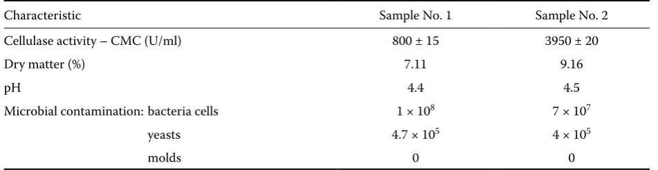
The fundamental characteristics of the original cellulase preparations are given in Table 1. The samples were contaminated with different strains of bacteria (Bacillus, Lactobacillus, gram negative non-fermentative bacteria).
[image:2.595.61.537.633.756.2]The preparation No. 1 was diluted in 0.1M ac-etate buffer, pH 5.0, to the activity of 800, 100, 10, 2 and 1 U/ml. These samples were treated with various doses of ionising radiation and then the cellulase activity was determined. Figure 1 shows that the inactivation was markedly related to the decreasing activity. Simultaneously with the activity assay, the changes in microbial contami-nation were checked. Table 2 indicates that yests are more sensitive to the ionising radiation than
Table 1. Fundamental characteristics of the cellulase preparations used
Characteristic Sample No. 1 Sample No. 2 Cellulase activity – CMC (U/ml) 800 ± 15 3950 ± 20
Dry matter (%) 7.11 9.16
pH 4.4 4.5
Microbial contamination: bacteria cells 1 × 10⁸ 7 × 10⁷
yeasts 4.7 × 10⁵ 4 × 10⁵
bacteria, and that the radiation dose of 6 kGy has a satisfactory pasteurising effect at a relatively high initial contamination level. Using this radiation dose, the loss of enzyme activity in the sample of the highest activity was only 10%. In contrast, in the highly diluted enzyme samples (2 and 1 U/ml) the inactivation of cellulase reached 74% and 92%, respectively. Consequently, some kind of the en-zyme stabilisation and also the use of samples with higher enzyme specific activities is needed for the radiation treatment. Some anticipated stabilising agents were tested in the enzyme preparations containing 1000 cellulase U/ml. Besides the
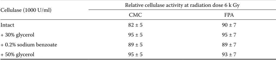
[image:3.595.68.351.84.269.2]op-timal radiation dose as determined in the previ-ous experiments (6 kGy), somewhat lower and higher radiation doses were also applied (5 and 7 kGy). The results are given in Table 3 in which the filter paper activity (FPA) is also presented for comparison. The best stabilising effect was achieved by using 30% glycerol. Further increase of the glycerol concentration did not improve the stabilisation effect and, moreover, the high viscosity of the enzyme solution makes the han-dling diffucult. Cystein, due to its free sulphydryl group, was considered as an efective scavenger of the primary products of radiolysis changing them
Table 2. Effect of radiation dose on the microbial contamination
Type of microorganism Cells/ml at radiation dose (kGy)
0 3 6 15 18
Bacteria 1 × 10⁸ 1 × 10⁴ 0 0 0
Yeasts 4.7 × 10⁵ 0 0 0 0
Molds 0 0 0 0 0
Table 3. Effect of ionising radiation on cellulase activity in the presence of various stabilisers intended
Cellulase (1000 U/ml) Relative cellulase activity at radiation dose 6 k Gy
CMC FPA
Intact 82 ± 5 90 ± 7
+ 30% glycerol 95 ± 5 95 ± 7
+ 0.2% sodium benzoate 89 ± 5 89 ± 7
+ 50% glycerol 95 ± 5 93 ± 7
CMC – activity measured by carboxymethylcellulose FPA – activity measured by filter paper
Figure 1. Cellulase activity (%) as a function of dose at various concentrations (units/ml)
-20 0 20 40 60 80 100 120
0 5 10 15 20
Dose (kGy)
Cellulase
activity
(%) 1 U/ml
[image:3.595.64.532.491.577.2] [image:3.595.63.531.620.723.2]to less reactive compounds. Unfortunately, this anticipated effect was not confirmed.
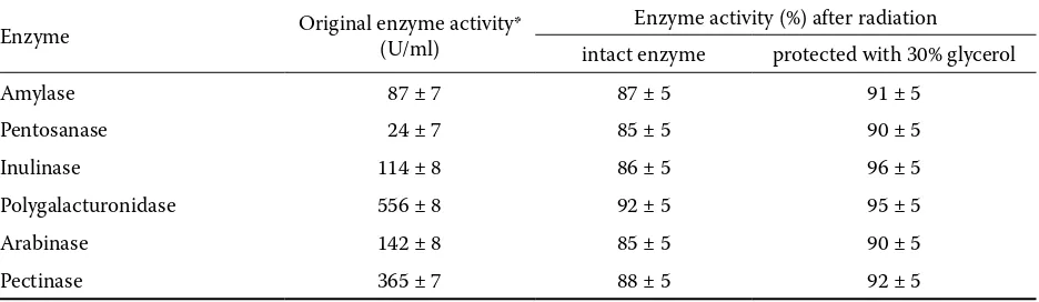
The microbial contamination (Table 2) and the changes in the levels of side activities (Table 5) of the irratiated cellulase preparations stabilised with glycerol were also tested. Table 4 demonstrates that the increase of the glycerol concentration to 50% had another unfavourable effect, i.e. the protection of bacteria against ionising radiation, and that the 30% glycerol concentration represents an accept-able compromise between the stabilisation of the enzyme and bacteria protection. Table 5 shows that also the other enzymes present are protected against radiation. Although certain small diferences between the radiation sensitivities of the individual enzymes were found, the suggested method of cel-lulase stabilisation against the inactivating effect of ionising radiation can be proposed as a general approach to the protection of other enzymes. No changes existing in the immunochemical proper-ties of the intact and irradiated cellulase, as well as the same chromatografic properties on Mono Q column of FPLC (Pharmacia, Uppsala), show
that the ionising radiation of 6 kGy is an accept-able procedure for the stabilisation of enzymes in solution when 30 w/w of glycerol is added.
References
BURIÁNOVÁ T., KOPEČNÝ J., SAJDOK J., KÁŠ J. (1991): Assay of very low cellulolytic activity in fodder supplemented with enzyme preparation. Animal Feed Science and Technology, 33: 41–48.
COLLINS C.H. (1964): Microbiological Methods Butter-worths. London: 86–109.
EIJSINK V.G.H., GASEIDNES S., SYNSTAD B., VAN DEN BURG B. (2004): Rational engineering of enzyme stabil-ity. Journal of Biotechnology, 113: 105–120.
KEMPNER E.S., VERKMAN A.S. (1988): Direct effects of ionizing-radiation unique to macromolecules. Radiation Physics and Chemistry, 32: 341–347.
KIEFER J. (1981): Biologische Strahlenwirkung. Springer Verlag, Berlin: 301–303.
[image:4.595.65.532.102.205.2]MANCINI G., CARBONARE A.O., HEREMAUS J.F. (1965): Im-munochemical quantitations of antigens by singel radial immundiffusion. Immunochemistry,2: 235–254. Table 4. Microbial contamination of cellulase preparations in dependence on radiation dose
Cellulase Bacteria counts (ml) after radiation dose (kGy)
0 3 5 6 7
Intact 7 × 10⁷ 1.3 × 10⁴ 0 0 0
+ 30% glycerol 7 × 10⁷ 2 × 10⁵ 200 0 0
+ 50% glycerol 7 × 10⁷ 5 × 10⁵ 1000 250 0
+ 30% glycerol and 0.02% sodium benzoate 7 × 10⁷ 2 × 10⁵ 500 40 0
Table 5. Changes of the side enzyme activities of cellulase preparation in intact and stabilised (30% of glycerol) forms after ionising radiation (6 kGy)
Enzyme Original enzyme activity* (U/ml) Enzyme activity (%) after radiation
intact enzyme protected with 30% glycerol
Amylase 87 ± 7 87 ± 5 91 ± 5
Pentosanase 24 ± 7 85 ± 5 90 ± 5
Inulinase 114 ± 8 86 ± 5 96 ± 5
Polygalacturonidase 556 ± 8 92 ± 5 95 ± 5
Arabinase 142 ± 8 85 ± 5 90 ± 5
Pectinase 365 ± 7 88 ± 5 92 ± 5
[image:4.595.64.532.271.407.2]Corresponding author:
Doc. Ing. JIŘÍ SAJDOK, CSc., Vysoká škola chemicko-technologická, Fakulta potravinářské a biochemické technologie, Ústav biochemie a mikrobiologie, Technická 5, 166 28 Praha 6, Česká republika
tel.: + 420 220 445 102, fax: + 420 220 445 167, e-mail: [email protected]
MONTENECOURT B.S., EVELEIGH D.E. (1977): Preparation of mutants of Trichoderma reesei with enhanced cellulase production. Applied and Environmental Microbiology,
34: 777–782.
REGULA D.F., DEFNER U. (1989): Dose estimation by ESR spectroscopy at a fatal radiation accident. Internatio-nal JourInternatio-nal of Radiation Applications and Instrumen-tation. Part A. Applied Radiation and Isotopes, 40: 1039–1043.
REESE E.T., MANDELS M. (1980): Stability of the cellulase of Trichoderma reesei under use conditions. Biotech-nology and Bioengineering, 22: 323–335.
SOMOGYI M. (1950): Studies of arteriovenous differences in blood sugar. Biological Chemistry, 186: 513–526. TORCHILIN V.P., MARTINEK K. (1979): Enzyme
stabilizati-on without cariers. Enzyme and Microbial Technology,
1: 74–82.