Crystal structure of
rac
-3-hydroxy-2-(
p
-tolyl)-2,3,3a,4,7,7a-hexahydro-1
H
-4,7-methanoisoindol-1-one
Mehmet Aslantas¸,a* Cumali C¸elik,bO¨ mer C¸elikcand Arzu Karayeld,e
aDepartment of Physics, Faculty of Sciences and Arts, University of Kahramanmaras Sutcuimam, Avsar Campus 46100, Kahramanmaras, Turkey,bYalova Community Collage, University of Yalova, 77200 Yalova, Turkey,cScience and Technology Application and Research Center, Dicle University, 21280 Diyarbakır, Turkey, dDepartment of Physics, Faculty of Sciences and Arts, Hitit University, 19030 C¸orum, Turkey, andeDepartment of Physics, Bilkent University, 06800 Ankara, Turkey. *Correspondence e-mail: aslantasmehmet@gmail.com
Received 26 January 2015; accepted 29 January 2015
Edited by H. Stoeckli-Evans, University of Neuchaˆtel, Switzerland
In the title compound, C16H17NO2, the cyclohexene ring adopts a boat conformation, and the five-membered rings have envelope conformations with the bridging atom as the flap. Their mean planes are oriented at a dihedral angle of 86.51 (7). The molecular structure is stabilized by a short
intramolecular C—H O contact. In the crystal, molecules are linked by O—H O hydrogen bonds forming chains propagating along [100]. The chains are linked by C—H
interactions, forming slabs parallel to (001).
Keywords:crystal structure; methanoisoindol-1-one; methanoisoindole-1,3-dione; O—H O hydrogen bonds; C—H interactions.
CCDC reference:1046290
1. Related literature
For medical and pharmaceutical applications of chiral tricyclic compounds, see: Abelet al.(1996); Salvatiet al.(2005). For the synthesis of the starting reagent, 2-(p-tolyl)-3a,4,7,7a-tetra-hydro-1H-4,7-methanoisoindole-1,3(2H)-dione, see: Andrade & Evilazio (2004). For the reduction reaction used to synthesise the title compound, see: Hubert et al.(1975). For the crystal structure of a similar compound, see: Takebayashi et al.(2010).
2. Experimental
2.1. Crystal data
C16H17NO2 Mr= 255.31 Monoclinic,P21=c a= 6.5067 (2) A˚ b= 9.7385 (2) A˚ c= 21.0780 (5) A˚
= 97.154 (1)
V= 1325.22 (6) A˚3 Z= 4
MoKradiation
= 0.08 mm1 T= 296 K
0.450.250.15 mm
2.2. Data collection
Bruker APEXII diffractometer Absorption correction: multi-scan
(Blessing, 1995) Tmin= 0.963,Tmax= 0.988
28760 measured reflections 5019 independent reflections 3930 reflections withI> 2(I) Rint= 0.023
2.3. Refinement
R[F2> 2(F2)] = 0.063 wR(F2) = 0.180 S= 1.09 5019 reflections 180 parameters
H atoms treated by a mixture of independent and constrained refinement
max= 0.40 e A˚
3
min=0.38 e A˚
[image:1.610.313.565.505.563.2]3
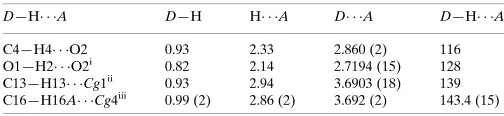
Table 1
Hydrogen-bond geometry (A˚ ,).
Cg1 and Cg4 are the centroids of the N1/C8–C11 and C2–C7 rings, respectively.
D—H A D—H H A D A D—H A
C4—H4 O2 0.93 2.33 2.860 (2) 116 O1—H2 O2i
0.82 2.14 2.7194 (15) 128 C13—H13 Cg1ii
0.93 2.94 3.6903 (18) 139 C16—H16A Cg4iii
0.99 (2) 2.86 (2) 3.692 (2) 143.4 (15)
Symmetry codes: (i)xþ1;y;z; (ii)x;y1 2;zþ
1
2; (iii)x;yþ 1 2;zþ
1 2.
Data collection: APEX2 (Bruker, 2007); cell refinement: SAINT (Bruker, 2007); data reduction:SAINT; program(s) used to solve structure:SHELXS97(Sheldrick, 2008); program(s) used to refine structure: SHELXL97 (Sheldrick, 2015); molecular graphics: ORTEP-3 for Windows(Farrugia, 2012); software used to prepare material for publication: WinGX publication routines (Farrugia, 2012).
Acknowledgements
This research was supported by Yalova University Scientific Research Projects Coordination Department (project No. 210–07). We would also like to thank DUPTAM, Dicle University, Turkey, for the use of the X-ray diffractometer.
data reports
Acta Cryst.(2015).E71, o143–o144 doi:10.1107/S2056989015001942 Aslantas¸et al.
o143
Supporting information for this paper is available from the IUCr electronic archives (Reference: SU5074).
References
Abel, M. D., Luu, H. T., Micetich, R. G., Nguyen, D. Q., Oreski, A. B., Tempest, M. L. & Daneshtalab, M. (1996).J. Heterocycl. Chem.33, 415–420. Andrade, D. S. & Evilazio, E. (2004).Synth. Commun.34, 3078–3081. Blessing, R. H. (1995).Acta Cryst.A51, 33–38.
USA.
Farrugia, L. J. (2012).J. Appl. Cryst.45, 849–854.
Hubert, J. C., Wijnberg, J. B. P. A. & Speckamp, N. W. (1975).Tetrahedron,31, 1437–1441.
Salvati, M. E., Balog, A., Wei, D. D., Pickering, D., Attar, R. M., Geng, J., Rizzo, C. A., Hunt, J. T., Gottardis, M. M., Weinmann, R. & Martinez, R. (2005).Bioorg. Med. Chem. Lett.15, 389–393.
Sheldrick, G. M. (2008).Acta Cryst.A64, 112–122. Sheldrick, G. M. (2015).Acta Cryst.C71, 3–8.
supporting information
sup-1
Acta Cryst. (2015). E71, o143–o144
supporting information
Acta Cryst. (2015). E71, o143–o144 [doi:10.1107/S2056989015001942]
Crystal structure of
rac
-3-hydroxy-2-(
p
-tolyl)-2,3,3a,4,7,7a-hexahydro-1
H
-4,7-methanoisoindol-1-one
Mehmet Aslanta
ş
, Cumali
Ç
elik,
Ö
mer
Ç
elik and Arzu Karayel
S1. Comment
Chiral tricyclic compounds in heterocyclic chemistry are important in medicinal and pharmaceutical fields (Abel et al.,
1996; Salvati et al., 2005). We report herein on the synthesis and crystal structure of the title compound, prepared by
reduction of 2-(p-tolyl)-3a,4,7,7a-tetrahydro-1H-4,7-methanoisoindole-1,3(2H)-dione, using NaBH4.
The bond lengths and angles in the title compound, Fig. 1, are close to those reported for two similar chiral structures
(Takebayashi et al., 2010). The cyclohexene ring (C9/C190/C12-C15) has a normal boat conformation [puckering
parameters: θ2 = 0.9587 (3) Å and φ2 = 169.02 (14)°]. The main bridge angle, C12—C16—C15, which connects the two
bridgeheads on the cyclohexene ring, is 93.78 (12) °. The two five-membered rings, A(C9/C10/C15/C16/C12) and
B(C12-C16) have envelope conformations with the flap atom C16 deviating from their mean planes by 0.5131 (2) and
0.4027 (2) Å, respectively. The dihedral angle between their mean planes, [A/B], is 86.51 (7)°. The whole molecule is
non-planar with the dihedral angle between the benzene (C2-C7) and imide (N1/C8-C11) rings being 26.12 (5)°. This is
much smaller than the same dihedral angle of ca.57.22 ° in the 2-phenyl derivative (Takebayashi et al., 2010) or ca. 61.37
° in the 2-(4-fluorophenyl) derivative (Takebayashi et al., 2010). In the molecule there is a strong C—H···O
intra-molecular contact present (Table 1).
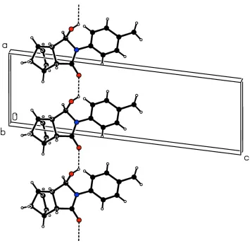
In the crystal, molecules are linked by O—H···O hydrogen bonds forming chains along [100]; see Table 1 and Fig. 2.
The chains are linked by C-H···π interactions forming slabs parallel to (001); see Table 1.
S2. Experimental
The starting reagent, 2-(p-tolyl)-3a,4,7,7a-tetrahydro-1H-4,7-methanoisoindole-1,3(2H)-dione (L), is a known compound
and was prepared from nadic anhydride and 4-toluidine (Andrade & Evilazio, 2004). The title compound was prepared by
a reduction reaction following a modification of a literature procedure (Hubert et al., 1975). NaBH4 (0.94 g) was added in
small portions at 298 K over a period of 2 h to L (0.72 g, 2.84 mmol) dissolved in ethanol (250 ml). The excess of NaBH4
was consumed in 15 min at 278 K by adding aqueous HCl (2 mol dm-3) until the pH reached 3. The mixture was stirred
for an additional 1 h at the same temperature then poured into water and extracted with dichloromethane. The organic
layer was separated, dried over Na2SO4, filtered and evaporated to yield a white solid that was purified by silica gel
chromatography [ethyl acetate/n-hexane (3:2 v/v)] which on slow evaporation of the solvent gave colourless crystals
(yield: 65%; m.p.: 475–477 K). NMR (DMSO): δ(H) 1.38–1.42 (dd, 2H, CH2), 2.24 (s, 3H, CH3), 2.59–2.60 (d, H, CH),
2.61–2.62 (d, H, CH), 3.11–3.13 (m, H, CH), 3.18–3.21 (dd, H, CH), 4.81 (s, H, CH—OH), 6.03–6.05 (dd, H, ═CH),
6.16–6.18 (dd, H, ═CH), 7.09–7.11 (d, 2H, aromatic), 7.25–7.27 (d, 2H, aromatic); δ(C) 20.96 (CH3), 45.08 (CH), 45.62
(CH), 46.56 (CH), 49.58 (CH), 51.06 (CH2), 86.17 (CH—OH), 124.07 (Cm), 129.33 (Co), 134.37 (CH═CH), 134.93 (Cq
—N), 135.87 (C—CH3), 174.39 (C═O) p.p.m.. FT—IR (ATR): 3211 (OH), 2972, 2943, 1646 (C═O), 1613 and 1515
H atoms attached to bridging atom C16 were located in a difference Fourier map and freely refined. The other H atoms
were placed in geometrically idealized positions (C—H = 0.93–0.98 Å and O—H= 0.82 Å) and treated as riding, with
[image:4.610.122.487.149.312.2]Uiso(H) = 1.5Ueq(O,C) for hydroxyl and methyl H atoms and = 1.2Ueq(C) for other H atoms.
Figure 1
The molecular structure of the title compound, with atom labelling. Displacement ellipsoids are drawn at the 30%
supporting information
sup-3
[image:5.610.128.483.66.417.2]Acta Cryst. (2015). E71, o143–o144
Figure 2
A partial view along the b axis of the crystal packing of the title compound. Dashed lines indicate the O—H···O hydrogen
bonds (see Table 1 for details).
rac-3-Hydroxy-2-(p-tolyl)-2,3,3a,4,7,7a-hexahydro-1H-4,7-methanoisoindol-1-one
Crystal data
C16H17NO2
Mr = 255.31 Monoclinic, P21/c
Hall symbol: -P 2ybc
a = 6.5067 (2) Å
b = 9.7385 (2) Å
c = 21.0780 (5) Å
β = 97.154 (1)°
V = 1325.22 (6) Å3
Z = 4
F(000) = 544
Dx = 1.280 Mg m−3
Mo Kα radiation, λ = 0.71073 Å Cell parameters from 5019 reflections
θ = 3.6–33.2°
µ = 0.08 mm−1
T = 296 K Prism, colourless 0.45 × 0.25 × 0.15 mm
Data collection
Bruker APEXII diffractometer
Radiation source: fine-focus sealed tube Graphite monochromator
φ and ω scans
Absorption correction: multi-scan (Blessing, 1995)
Tmin = 0.963, Tmax = 0.988
θmax = 33.2°, θmin = 3.6°
h = −5→9
l = −32→32
Refinement
Refinement on F2
Least-squares matrix: full
R[F2 > 2σ(F2)] = 0.063
wR(F2) = 0.180
S = 1.09 5019 reflections 180 parameters 0 restraints
Primary atom site location: structure-invariant direct methods
Secondary atom site location: difference Fourier map
Hydrogen site location: inferred from neighbouring sites
H atoms treated by a mixture of independent and constrained refinement
w = 1/[σ2(F
o2) + (0.0762P)2 + 0.4345P]
where P = (Fo2 + 2Fc2)/3
(Δ/σ)max < 0.001
Δρmax = 0.40 e Å−3
Δρmin = −0.38 e Å−3
Special details
Geometry. All e.s.d.'s (except the e.s.d. in the dihedral angle between two l.s. planes) are estimated using the full covariance matrix. The cell e.s.d.'s are taken into account individually in the estimation of e.s.d.'s in distances, angles and torsion angles; correlations between e.s.d.'s in cell parameters are only used when they are defined by crystal symmetry. An approximate (isotropic) treatment of cell e.s.d.'s is used for estimating e.s.d.'s involving l.s. planes.
Refinement. Refinement of F2 against ALL reflections. The weighted R-factor wR and goodness of fit S are based on F2,
conventional R-factors R are based on F, with F set to zero for negative F2. The threshold expression of F2 > σ(F2) is used
only for calculating R-factors(gt) etc. and is not relevant to the choice of reflections for refinement. R-factors based on F2
are statistically about twice as large as those based on F, and R- factors based on ALL data will be even larger.
Fractional atomic coordinates and isotropic or equivalent isotropic displacement parameters (Å2)
x y z Uiso*/Ueq
supporting information
sup-5
Acta Cryst. (2015). E71, o143–o144
C2 0.4079 (2) 0.82658 (16) 0.47322 (7) 0.0440 (3) C7 0.5078 (2) 0.80753 (17) 0.41974 (7) 0.0455 (3) H7 0.6327 0.7600 0.4239 0.055* C12 −0.1801 (2) 0.99380 (17) 0.15201 (8) 0.0470 (3) H12 −0.3307 1.0066 0.1470 0.056* C15 0.1436 (3) 0.98780 (17) 0.12617 (7) 0.0448 (3) H15 0.2563 0.9944 0.0995 0.054* C4 0.1392 (2) 0.94992 (18) 0.40597 (7) 0.0459 (3) H4 0.0148 0.9981 0.4021 0.055* C13 −0.1047 (3) 0.84754 (17) 0.16019 (8) 0.0530 (4) H13 −0.1791 0.7734 0.1733 0.064* C3 0.2228 (3) 0.8979 (2) 0.46493 (8) 0.0534 (4) H3 0.1517 0.9115 0.5001 0.064* C14 0.0870 (3) 0.84423 (17) 0.14533 (8) 0.0520 (4) H14 0.1720 0.7672 0.1465 0.062*
Atomic displacement parameters (Å2)
U11 U22 U33 U12 U13 U23
O1 0.0241 (4) 0.0503 (6) 0.0575 (6) −0.0066 (4) 0.0005 (4) 0.0026 (5) O2 0.0203 (4) 0.0751 (8) 0.0549 (6) 0.0010 (4) 0.0079 (4) 0.0096 (5) C16 0.0568 (9) 0.0558 (9) 0.0421 (8) −0.0040 (8) −0.0091 (7) 0.0114 (7) C1 0.0762 (14) 0.0834 (14) 0.0453 (9) −0.0020 (11) −0.0064 (9) 0.0202 (9) N1 0.0193 (4) 0.0366 (5) 0.0347 (5) 0.0025 (3) 0.0030 (3) 0.0021 (4) C11 0.0218 (4) 0.0375 (6) 0.0380 (6) 0.0015 (4) 0.0058 (4) 0.0020 (5) C5 0.0244 (5) 0.0332 (5) 0.0328 (5) 0.0007 (4) 0.0022 (4) −0.0013 (4) C8 0.0204 (4) 0.0377 (6) 0.0424 (6) 0.0021 (4) 0.0019 (4) 0.0008 (5) C9 0.0254 (5) 0.0377 (6) 0.0444 (7) 0.0034 (4) −0.0011 (4) 0.0061 (5) C10 0.0300 (5) 0.0356 (6) 0.0395 (6) −0.0011 (4) 0.0037 (4) 0.0071 (5) C6 0.0287 (5) 0.0496 (7) 0.0379 (6) 0.0092 (5) 0.0050 (5) 0.0037 (5) C2 0.0466 (7) 0.0473 (8) 0.0361 (6) −0.0043 (6) −0.0026 (5) 0.0045 (5) C7 0.0358 (6) 0.0540 (8) 0.0450 (7) 0.0090 (6) −0.0013 (5) 0.0087 (6) C12 0.0351 (6) 0.0562 (9) 0.0464 (8) −0.0055 (6) −0.0089 (6) 0.0091 (6) C15 0.0477 (8) 0.0496 (8) 0.0372 (7) 0.0010 (6) 0.0063 (6) 0.0052 (6) C4 0.0422 (7) 0.0590 (9) 0.0377 (7) 0.0157 (6) 0.0093 (5) −0.0018 (6) C13 0.0641 (10) 0.0452 (8) 0.0459 (8) −0.0178 (7) −0.0079 (7) 0.0030 (6) C3 0.0587 (9) 0.0672 (10) 0.0358 (7) 0.0103 (8) 0.0121 (6) −0.0004 (7) C14 0.0711 (11) 0.0396 (7) 0.0436 (8) 0.0024 (7) −0.0003 (7) −0.0026 (6)
Geometric parameters (Å, º)
C1—H1A 0.9600 C7—H7 0.9300 C1—H1B 0.9600 C12—C13 1.509 (3) C1—H1C 0.9600 C12—H12 0.9800 N1—C8 1.3574 (14) C15—C14 1.513 (2) N1—C5 1.4256 (16) C15—H15 0.9800 N1—C11 1.4776 (15) C4—C3 1.389 (2) C11—C10 1.5290 (18) C4—H4 0.9300 C11—H11 0.9800 C13—C14 1.324 (3) C5—C4 1.3884 (18) C13—H13 0.9300 C5—C6 1.3885 (17) C3—H3 0.9300 C8—C9 1.4984 (19) C14—H14 0.9300 C9—C10 1.5383 (18)
supporting information
sup-7
Acta Cryst. (2015). E71, o143–o144
C11—C10—C9 106.91 (10) C13—C14—H14 126.0 C11—C10—C15 116.70 (11) C15—C14—H14 126.0 C9—C10—C15 102.71 (11)
C8—N1—C11—O1 117.43 (12) C3—C2—C7—C6 −0.1 (3) C5—N1—C11—O1 −60.01 (14) C1—C2—C7—C6 179.60 (17) C8—N1—C11—C10 −2.04 (14) C5—C6—C7—C2 0.7 (2) C5—N1—C11—C10 −179.48 (11) C15—C16—C12—C13 −50.25 (14) C8—N1—C5—C4 −26.3 (2) C15—C16—C12—C9 58.64 (14) C11—N1—C5—C4 150.86 (14) C8—C9—C12—C13 −45.60 (17) C8—N1—C5—C6 155.78 (13) C10—C9—C12—C13 68.21 (15) C11—N1—C5—C6 −27.09 (17) C8—C9—C12—C16 −149.55 (12) C5—N1—C8—O2 −3.7 (2) C10—C9—C12—C16 −35.73 (14) C11—N1—C8—O2 178.96 (13) C12—C16—C15—C14 49.62 (15) C5—N1—C8—C9 176.52 (11) C12—C16—C15—C10 −60.06 (14) C11—N1—C8—C9 −0.79 (15) C11—C10—C15—C14 51.45 (17) O2—C8—C9—C10 −176.50 (14) C9—C10—C15—C14 −65.11 (15) N1—C8—C9—C10 3.26 (15) C11—C10—C15—C16 155.05 (12) O2—C8—C9—C12 −63.72 (19) C9—C10—C15—C16 38.49 (13) N1—C8—C9—C12 116.04 (13) C6—C5—C4—C3 0.1 (2) O1—C11—C10—C9 −115.65 (11) N1—C5—C4—C3 −177.82 (15) N1—C11—C10—C9 3.91 (13) C16—C12—C13—C14 33.93 (16) O1—C11—C10—C15 130.13 (12) C9—C12—C13—C14 −69.31 (17) N1—C11—C10—C15 −110.31 (12) C7—C2—C3—C4 −0.6 (3) C8—C9—C10—C11 −4.38 (14) C1—C2—C3—C4 179.80 (19) C12—C9—C10—C11 −125.16 (11) C5—C4—C3—C2 0.5 (3) C8—C9—C10—C15 118.98 (12) C12—C13—C14—C15 −0.61 (18) C12—C9—C10—C15 −1.79 (13) C16—C15—C14—C13 −32.66 (17) C4—C5—C6—C7 −0.7 (2) C10—C15—C14—C13 70.41 (17) N1—C5—C6—C7 177.28 (13)
Hydrogen-bond geometry (Å, º)
Cg1 and Cg4 are the centroids of the N1/C8–C11 and C2–C7 rings, respectively.
D—H···A D—H H···A D···A D—H···A
C4—H4···O2 0.93 2.33 2.860 (2) 116 O1—H2···O2i 0.82 2.14 2.7194 (15) 128
C13—H13···Cg1ii 0.93 2.94 3.6903 (18) 139
C16—H16A···Cg4iii 0.99 (2) 2.86 (2) 3.692 (2) 143.4 (15)