99
Varied Expression Pattern of the Small Heat Shock Protein
Gene Encoding HSP17.7 against UVA, UVB, Cu
2+and Zn
2+Stresses in Sunflower
İlker Büyük 1, Sümer ArAS 1 and Demet CAnSArAn-DumAn 2
1Department of Biology, Faculty of Science and 2Biotechnology Institute,
Ankara University, Ankara, Turkey
Abstract
Büyük İ., Aras S., Cansaran-Duman D. (2016): Varied expression pattern of the small heat shock protein gene encoding HSP17.7 against UVA, UVB, Cu2+ and Zn2+ stresses in sunflower. Plant Protect. Sci., 52: 99–106.
Today, one of the main objectives of agricultural biotechnology area is to find the responsible genes involved in stress re-sponse and engineering these genes to improve the plant rere-sponse mechanisms. Therefore the current study was conducted to gain an insight on the role of HSP17.7 gene, which is a member of sHsps family, in defence mechanism of sunflower
(Helianthus annuus L. cv. Confeta –Turkish cultivar) treated with different doses of UVA and UVB (4, 8, 12 and 20 kJ/m2)
and concentrations of copper (Cu2+) and zinc (Zn2+) (80, 160, 320, 640, and 1280 µM) heavy metals. Based on our data, it was observed that different doses of UVA and UVB irradiation resulted in increased levels of HSP17.7 mRNA in sunflower plants. The highest levels of these increases (8 and 12 kJ/m2 of UVA) were seen under UVA stress. In contrast to UV stress, only the Cu2+concentration of 1280 µM led to higher expression levels of HSP17.7 gene compared to the control. Besides this, the 1280 µM concentration of Zn2+ treatment was the peak point of increased HSP17.7 mRNA levels for all stress conditions with nearly 8 times more than in the control sample. Negative correlations were found between malondialdehyde (MDA) levels and expression levels of HSP17.7 gene in sunflower plants subjected to current abiotic stress conditions. This correlation might indicate that an effective defence mechanism was in action and it might be concluded that the HSP17.7 gene can be used for identification of cultivars tolerant to UVand high doses of Cu2+ and Zn2+ for molecular breeding studies in the near future. These findings provide evidence of the HSP17.7 gene contribution to abiotic stress response in sunflower and will be helpful for the next studies about stress tolerance improvement in sunflower plants.
Keywords: sHsps; HSP17.7 gene; qRT-PCR; abiotic stress; Helianthus annuus L.
All living organisms respond to environmental stress conditions that might be biotic or abiotic by the syn-thesis of several proteins which are referred to as ‘Stress Proteins’. These proteins protect cells against harmful effects of stress conditions. Heat shock pro-teins (HSPs), also known as molecular chaperones, are known to belong among these stress proteins while they are present in all living organisms (Kim & Hwang 2015). Besides their role in protecting cells against environmental stress conditions, HSPs are essential components of plant cells and they play an important role in protein stabilisation and cellular
functions related with plant growth (Pratt et al. 2001; Koo et al. 2015).
HSPs were first identified in the fruit fly (Drosophila melanogaster) and determined as highly conserved in all living organisms including plants. In plants, they are classified into five evolutionarily conserved families: HSP100, 90, 70, 60 (or chaperonins), and the small HSPs (sHsps) which have molecular weights of 100, 90, 70, 60, and 12–40 kDa, respectively (Al-Whaibi 2011; Koo et al. 2015). Higher plants are known to have at least 20 types of sHsps which have an α-crystallin domain consisting of 80–100 amino
acid residues in the C-terminal region (Seo et al. 2006). The amount of expressed sHsps and their types change according to the plant species. The sHsps family plays an important role in the degradation of misfolded proteins and this characteristic distin-guishes the family from other HSPs classes (Ferguson
et al. 1990; Al-Whaibi 2011). Although they have no function in the refolding of non-native proteins, there are few indications that they bind to partially folded proteins to prevent their irreversible unfold-ing and aggregation of non-native proteins (Sun et al. 2002). These characteristics of sHsps make them valuable in maintaining the functional conformation of proteins under stress conditions which is crucial for a cell to survive (Al-Whaibi 2011).
Significantly increased amounts of sHsps were observed in plants subjected to abiotic stress condi-tions such as high temperatures (Ahn & Zimmerman 2006; Volkov et al. 2006), drought and salt stress (Sato & Yokoya 2008; Zahur et al. 2009). These previous studies suggest that sHsps might play an important role in plant resistance to environmental stress conditions. In plants, they have been well characterised in some species such as maize, soy-bean (Ding et al. 2009; Wu et al. 2010), and cotton (Ronde et al. 1993; Cottee et al. 2014) and it was reported that their distribution differs within tissue, organ and plant species.
Today, one of the main objectives of agricultural biotechnology is to find the responsible genes involved in stress response and engineering these genes to improve the plant response mechanisms (Pareek
et al. 2010). Some of these genes have been well-characterised while some are suspicious in some plant species such as sunflower. Nevertheless, the analysis of these genes in many different plant species will increase the overall knowledge of their exact roles in the plant defence mechanism.
In this regard, the present study was conducted to gain an insight into the role of HSP17.7 gene, which is a member of the sHsps family, in the de-fence mechanism of sunflower (Helianthus annuus
L. cv. Confeta – Turkish cultivar)treated with differ-ent doses of UVA and UVB (4, 8, 12, and 20 kJ/m2)
and concentrations of Cu2+ and Zn2+ (80, 160, 320,
640, and 1280 µM) heavy metals. For this aim, the steady-state mRNA levels of HSP17.7 gene were determined by qRT-PCR in stressed sunflowers. All results were evaluated statistically and a probable correlation between stress conditions and HSP17.7 gene expression levels was demonstrated.
MAtEriAl And MEtHodS
Growth of plants and stress applications. Sun-flower (Helianthus annuus L.) cultivar Confeta seeds were kindly obtained from MAY Seed Company (Bursa, Turkey) and were germinated and grown hy-droponically in pots containing 0.2 l of modified 1/10 Hoagland’s solution. Hoagland’s solution included macronutrients [K2SO4, KH2PO4, MgSO4·7H2O, Ca(NO3)2·4H2O, and KCl] and micronutrients (H3BO3, MnSO4, CuSO4·5H2O, NH4Mo, and ZnSO4·7H2O) with the following final concentrations of ions: 2 mM Ca, 1 μM Mn, 4 mM NO3, 0.2 µM Cu, 1 mM Mg, 10 mM NH4, 2 mM K, 1 μM Zn, 0.2 mM P, 0.1 mM Fe, and 1 μM B. Six plants were grown in each pot in a con-trolled environmental growth chamber, under light, with photosynthetic photon flux of 250 mmol/m2·s
at 25°C and 70% relative humidity. Twenty-five-days-old plants grown in controlled media were used for the stress treatments. For the heavy metal application, Cu2+ and Zn2+ were added separately to
the hydroponic solution for 24 h at concentrations of 0 (control), 80, 160, 320, 640, and 1280 μM. For the UV application, twenty-five-days-old plants were irradiated with UVB and UVA using a BS-03 irra-diation chamber (Dr. Gobel UV-Elektronik GmbH, Ettlingen, Germany) and UVB and UVA doses were determined as 4, 8, 12 and 20 kJ/m2for the plants.
101 1.00
0.75
0.50
0.25
0
77.5 80.0 82.5 85.0 87.5 90.0 92.5 Temperature (°C)
dF/dT
RNA extraction, reverse transcription and Quan-titative Real-Time PCR. Total RNA extraction was performed using the Trizol protocol followed by the clean-up protocol using RNeasy mini clean-up kit (Cat No: 74104; Qiagen, Hilden, Germany) . RNA quantity/quality was measured with a NanoDrop 1000 ND-Spectrophotometer (Thermo Fisher Scientific, Waltham, USA). The quality of RNA was also con-firmed by gel electrophoresis which contained 1.5% agarose. cDNA synthesis which was based on reverse transcription reactions was performed with 2 µg of RNA and high fidelity cDNA synthesis kit (Roche) which contained 2.5 µM anchored-oligo (dT)18, 1X transcriptor high fidelity reverse transcriptase reac-tion buffer, 20U protector Rnase inhibitor (Roche, Mannheim, Germany), 1 mM deoxynucleotide mix, 5 mM DTT, 10U transcriptor high fidelity reverse transcriptase at final concentration. The following incubation conditions were applied; 10 min at 65°C, 30 min at 55°C, and 5 min at 85°C. cDNAs were also measured with a NanoDrop 1000 ND-Spectropho-tometer.
Quantitative Real-Time PCR (qRT-PCR) was per-formed with LightCycler® Nano System (Roche,
Mannheim, Germany), thermal cycler. The primer sequences of the target gene HSP17.7 (GenBank: U46545.1) and also housekeeping gene actin (Gen-Bank: AF282624.1) which is used for normalization were designed with Primer3 program based on the
sequences of sunflower genes available in the GenBank (http://www.ncbi.nlm.nih.gov/) (Table 1).
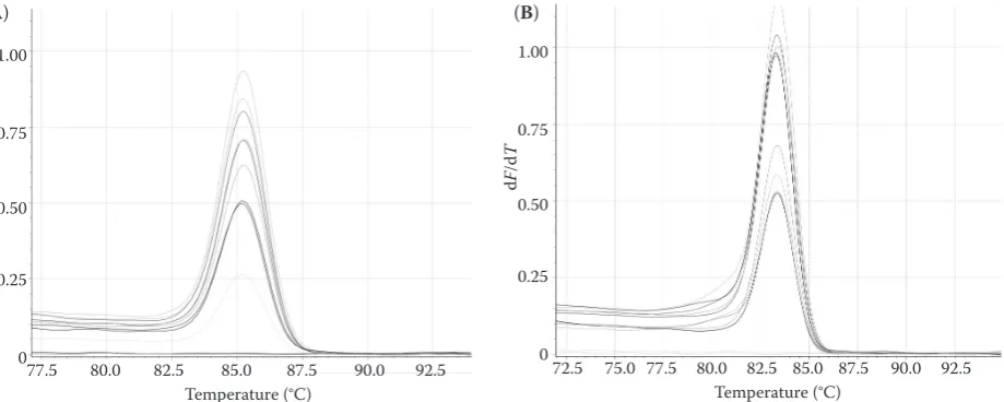
All qRT-PCR reactions were performed in three independent biological and technical triplicates with a template free control to check any contaminations. Amplifications of PCR product were monitored using an intercalator-based method including SYBR Green I dye. After pre-denaturation, followed by 10 min at 95°C, 45 cycles of 10 s at 95°C, 30 s at 60°C, and 15 s at 72°C were applied. Melting curve analysis was performed to confirm the presence of a single product and ab-sence of primer-dimers (Figure 1). Data collection for quantification was done during the annealing period. Statistical analysis. The abundance of target gene transcripts was normalised to ACT and set relative to control plants according to the 2–∆∆CT method
(Livak & Schmittgen 2001). Changes in relative expression levels (REL) of the gene were checked for statistical significance according to one-way ANOVA. Fisher’s least significant difference test at 0.05 significance levels was performed.
[(Abs 532+TBA) – (Abs 600+TBA) – (Abs 532–TBA – Abs 600–TBA)] = A (1)
[(Abs 440+TBA – Abs 600+TBA) 0.0571] = B (2)
[image:3.595.304.533.178.252.2]MDA equivalents (nmol/ml) = (A – B/157 000) × 106 (3)
Table 1.Primer sequences of HSP17.7 and ACT genes
Primer name Sequences
HSP 17.7 forwardreverse ATAAGCGGAGAGAGGAGCAGAGCATTCTCCGGCAACCTAAAC
ACT forward TGAGCAAGGAAATCACGGCTreverse TCCTCCGATCCAGACACTGT
Figure 1. Melting curve analysis of qRT-PCR reactions: (A) Single melting curve in each RT-PCR assay for ACT gene; (B) Single melting curve in each RT-PCR assay for HSP17.7 gene
(A) (B)
1.00
0.75
0.50
0.25
0
72.5 75.0 77.5 80.0 82.5 85.0 87.5 90.0 92.5 Temperature (°C)
d
F
/d
[image:3.595.72.532.544.728.2]rESUltS
Lipid peroxidation analysis. In the present study, primarily we evaluated malondialdehyde (MDA) levels as lipid peroxidation can be considered as a marker under stress conditions. MDA contents of sunflower plants were changed at almost all concentrations and doses of all stress conditions and these changes seemed consistent with mRNA levels of HSP17.7 gene (Figure 2). According to the analysis, all doses of UVA and UVB stresses led to statistically signifi-cantly higher levels of MDA compared to the non-stressed control sample. As documented in Table 2, the highest MDA level was seen in sunflower plants subjected to 20 kJ/m2 UVA stress among UVA and
UVB stressed plants while the lowest MDA level was found out in 8 kJ/m2 UVA stressed plants (P < 0.05).
Statistically non-significant increases were observed between 4–8 kJ/m2 and 12–20 kJ/m2 of UVA stress
treatment (Table 2).
Table 2 shows that all concentrations of Cu2+
con-tamination except for 1280 µM led to statistically significantly higher levels of MDA compared to the non-stressed control plants (Table 2). When the
ef-fects of Zn2+ stress on MDA levels were evaluated,
it was observed that statistically significantly higher levels of MDA compared to the non-stressed control only were seen at 320 µM of stress treatment. After 320 µM concentration of Zn2+ treatment, gradual
decreases were observed in MDA levels compared to this concentration (Table 2).
Gene expression analysis. Although the expression
profile of many genes of sHSPs have been studied in different plants, there are no available data on the effect of different doses and concentrations of UVA, UVB, Cu2+, and Zn2+ stresses on HSP17.7 gene expression
in the sunflower species. Therefore, qRT-PCR analysis of this gene in the sunflower species is one of the important issues of the present study.
The expression level of HSPS17.7 gene was altered in sunflower plants subjected to almost all concentrations of heavy metals and doses of UV stress conditions. Higher mRNA levels of HSP17.7 compared to the non-stressed control were seen in sunflower plants subjected to all doses of UVA and UVB stress treatments except for 20 kJ/m2 of UVA (Figure 3A and Table 3). Based
on the qRT-PCR results, it can be correctly concluded that 8 kJ/m2 of both UVA and UVB treatments led to
0 1 2 3 4 5
C
ontrol Cu
80
C
u
160
C
u
320
C
u
640
C
u
12
80
Zn
80
Zn 16
0
Zn 32
0
Zn 64
0
Zn
1
280
MDA e
equivalents
(nmol/ml)
[image:4.595.57.525.129.240.2]Concentrations of Cu2+ and Zn2+
Figure 2.Effects of (A) UVA and UVB (4, 8, 12, and 20 kJ/m2) and (B) Cu2+ and Zn2+ (80, 160, 320, 640, and 1280 μM) treatment on malondialdehyd (MDA) (lipid peroxidation) levels in sunflower plants
0 1 2 3 4 5
Control UVB
4
UVB
8
UVB 12 UVB 20 UVA 4 UVA 8 UVA 12 UVA 20
MDA
e
qui
vale
nt
s
(nmol/ml)
Doses of UVA and UVB
Table 2. Malondialdehyde (MDA) content in sunflower plants subjected to different doses of UV and different con-centrations of heavy metal treatments
UV doses
(kJ/m2) UVB-MDA (nM/g) UVA-MDA (nM/g) Heavy metal concentrations (μM) Cu
+2-MDA
(nM/g) Zn
+2-MDA (nM/g)
Control 0.834a 0.834a Control 0.75a 0.75a
4 1.53b 2.112b 80 2.04b 0.92a
8 1.545b 0.43c 160 3.12c 0.89a
12 2.099c 0.64d 320 3.5c 1.197b
20 1.873c 3.72e 640 3.8c 0.8ac
1280 0.6c 0.185d
a–dmeans within each column followed by the same letter are not significantly different at the P = 0.05 level
[image:4.595.57.525.609.721.2]103 the highest HSP17.7 mRNA expression compared to
their own control. In addition, comparative evaluation of UVA and UVB effects on sunflower plants revealed that UVA stress in that dose increased the mRNA levels of HSP17.7 nearly 5 times more than in the control sample and twice more than in UVB affected sunflower plants (Figure3A and Table 3).
The heavy metal contamination also resulted in altered mRNA levels of HSP17.7 gene as it was ob-served in UV stressed sunflowers. In contrast to UV stress, Cu2+ contaminations at concentrations of 80,
160, 320, and 640 µM reduced HSP17.7 mRNA ex-pression. On the other hand, the gene in sunflower plants subjected to 1280 µM Cu2+ was expressed nearly
3 times more than in the control sample (Table 3B). According to the results illustrated in Figure 3B, it is obvious that all concentrations of Zn2+ contamination
except for 320 µM led to increased mRNA levels of HSP17.7 compared to the control sample (Figure 3B and Table 3). Zn2+ contamination at 1280 µM revealed
a statistically significant increase and this was the peak point of increased HSP17.7 mRNA levels among all stress conditions with nearly 8 times more than in the control sample (Figure 3B and Table 3).
diSCUSSion
Plants are often subjected to many abiotic stresses such as salinity, drought, freezing, heavy metals, and ultraviolet radiation which affect yields of some important crops like sunflower in the world. Due to their sessile nature, plants have some adaptation and defence strategies to survive under rapidly changing environmental conditions (Boyer 1982; Soydam et al. 2013). In addition, the stress specific regulation of transcription is seen in plants under environmental stress conditions and some of these regulated genes and their proteins play an important role in general stress response (Rizhsky et al. 2004; Larkindale & Vierling 2008). Although in the past few decades, a large number of candidate genes have been iden-tified by several researchers, several stress related molecular mechanisms still seem veiled.
Heat shock proteins which are known as chaperons are key components for cellular homeostasis in plant cells by assisting protein refolding under environmen-tal stress conditions (Sairam & Tyagi 2004). Heat shock proteins which are also called stress proteins exist in all living organisms and the synthesis of these
0 1 2 3 4 5 6
Control UVB
4
UVB
8
UVB 12 UVB 20 UVA 4 UVA 8 UVA 12 UVA 20
H
SP 17.7 gene
ex
pression
fold change
Doses of UVA and UVB
0 2 4 6 8 10
Control Cu 80 Cu 160 Cu 320 Cu 640 Cu 1280 Zn 80 Zn 160 Zn 320 Zn 640 Zn 1280
H
SP 17.7 gene
ex
pression
fold chan
ge
[image:5.595.66.529.94.216.2]Concentrations of Cu2+ and Zn2+
Figure 3.Effects of (A) UVA and UVB (4, 8, 12, and 20 kJ/m2) and (B) Cu2+ and Zn2+ (80, 160, 320, 640, and 1280 μM) treatment on mRNA levels of HSP17.7 in sunflower
Table 3. Transcription changes of HSP17.7 gene in sunflower plants subjected to different doses of UV and different concentrations of heavy metal treatments
UV doses
(kJ/m2) UVB-HSP17.7 (nM/g) UVA-HSP17.7 (nM/g) Heavy metal concentrations (μM) Cu
+2-HSP17.7
(nM/g) Zn
+2-HSP17.7 (nM/g)
Control 1a 1a Control 1a 1a
4 2.09b 1.42b 80 0.417b 1.37b
8 2.23b 4.72c 160 0.3c 1.46bc
12 1.151ac 4.35c 320 0.2cd 0.48d
20 2.099bd 0.63d 640 0.122e 1.56bce
1280 3.36f 7.77f
a–fmeans within each column followed by the same letter are not significantly different at the P = 0.05 level
[image:5.595.66.532.626.738.2]proteins increases under some acute environmental stress factors such as high temperature, cold, and oxygen deficiency (Henle et al. 1998; Wang et al. 2004). sHsps are among the important HSP classes and their exact roles have been investigated recently. Hence, in the present study, we aimed to investigate the possible role of HSP17.7 gene which encodes a sHSP in a sunflower plant against UVA, UVB, and heavy metal stress response.
Ultraviolet (UV) radiation is an environmental factor which has important roles in the regulation of many physiological and ecological processes in plant ecosystems and which is spectrally divided into three types: UV-A, UV-B, and UV-C (Müller-Xing et al. 2014). Among these UV types, UV-C (200–280 nm) is completely absorbed by the ozone layer and thus cannot reach the Earth’s surface; while UV-A (320–400 nm) and UV-B (280–320 nm) can reach it and are involved in cellular processes. Hence, the present study was conducted with UVA and UVB stresses which can pose environmental adverse ef-fects in excess quantities (Müller-Xing et al. 2014). To our knowledge no studies have been published revealing the interactions between sHSPs and UV stress in a sunflower plant. There are some studies in other crop plants including maize (Ferreyra et al. 2010; Casati et al. 2011), barley (Kravets et al. 2012), cucumber (Shinkle et al. 2010), and grapevine (Pontin et al. 2010; Martinez-Luscher et al. 2013). In the present study, different doses of UVA and UVB irradiation resulted in increased levels of HSP17.7 mRNA in sunflower plants. The highest levels of these increases (8 and 12 kJ/m2 of UVA)
were found out in UVA stressed sunflower plants for the two types of UV treatment. However, the MDA level, which gives information about damage to the cell membranes caused by stress conditions, was lower than in the non-stressed control sample at 8 and 12 kJ/m2 of UVA treatment, indicating that
an effective defence mechanism was in action (Rao
et al. 2005). The negative correlation between MDA levels and expression levels of stress responsive genes in many organisms subjected to abiotic stress condi-tions was also shown by previous researches (Cho & Seo 2005; Li et al. 2012). All these results showed that HSP17.7 may play an important role in the de-fence system against UV stress in sunflower plants.
Copper and zinc are important micronutrients and they participate in many enzyme systems and structure of photosynthetic proteins. They induce toxicity above their optimal levels in plants as well
as in nearly all living organisms and cause a problem of both agricultural and environmental significance (Chamseddine et al. 2009; Millaleo et al. 2010). They are accumulated until the toxic levels in plants are reached, which leads to adverse effects on plant growth and morphology (Marschner 1995). In addition, excess concentrations of these metals are also hazardous to genetic stability by leading to genotoxicity in many plants (Conte et al. 1998; Savva 1998; Piraino et al. 2006; Liu et al. 2007).
To identify the possible role of HSP17.7 gene in defence against stress caused by different concen-trations of Cu2+ and Zn2+, qRT-PCR and MDA data
were also evaluated together. The data showed that only 1280 µM of Cu2+ treatment resulted in increased
levels of HSP17.7 mRNA in sunflower plants com-pared to the control sample and the MDA level was lower than in the non-stressed control sample at this concentration of Cu2+, indicating that an
effec-tive defence mechanism was in action. At the other concentrations of Cu2+ treatment, the MDA level was
found higher compared to the non-stressed control sample while HSP17.7 expression levels were lower than in the control (Tables 2 and 3).
Zn2+ treatment led to statistically significant
in-creases in the mRNA levels of HSP17.7 gene except for the application of 320 µM. Above this concentra-tion, the expression of HSP17.7 gene was gradually increased and reached the peak level at 1280 µM and a negative correlation which reflects the possible role of this gene in defence against Zn2+ stress was
again observed between MDA levels and HSP17.7 gene expression levels (Table 3). In previous studies, HSP18.1 and MsHsp18.2 genes in medicago sativa
and AtHsp17.7 gene in Arabidopsis thaliana were identified as the key components whose role in the protection of plants against heavy metal stresses is similar to the results of the present study (Sun et al. 2002).
As a result, changes in the mRNA levels of HSP17.7 gene seem more sensitive to UV stress rather than to heavy metal stress except for the Zn2+ treatment of
1280 µM. The results obtained in the present study about the more pronounced gene expression levels induced by UVA irradiation are in accordance with the previous studies suggesting that 8 and 12 kJ/m2
105 studies in the near future. At the same time, these
findings can be used for improving stress tolerance in sunflower plants by cloning and functional analyses.
references
Ahn Y.J., Zimmerman J.L. (2006): Introduction of the carrot HSP17.7 into potato (Solanum tuberosum L.) enhances cellular membrane stability and tuberization in vitro. Plant, Cell and Environment, 29: 95–104.
Al-Whaibi M.H. (2011): Plant heat-shock proteins: a mini review. Journal of King Saud University – Science, 23: 139–150.
Boyer J.S. (1982): Plant productivity and environment. Sci-ence, 218: 443–448.
Casati P., Campi M., Morrow D.J., Fernandes J.F., Walbot V. (2011): Transcriptomic, proteomic and metabolomic analysis of UV-B signaling in maize. BMC Genomics, 12: 321.
Chamseddine M., Wided B.A., Guy H., Marie-Edith C., Fatma J. (2009): Cadmium and copper induction of oxida-tive stress and antioxidaoxida-tive response in tomato (Solanum
lycopersicon) leaves. Plant Growth Regulation, 57: 89–99.
Cho N., Seo N. (2005): Oxidative stress in Arabidopsis thali-ana exposed to cadmium is due to hydrogen peroxide accumulation. Plant Science, 168: 113–120.
Conte C., Mutti I., Puglisi P., Ferrarini A., Regina G., Maestri E., Marmiroli N. (1998): DNA fingerprinting analysis by a PCR based method for monitoring the genotoxic effects of heavy metals pollution. Chemosphere, 37: 2739–2749. Cottee N.S., Wilson I.W., Tan D.K.Y., Bange M.P. (2014): Understanding the molecular events underpinning culti-var differences in the physiological performance and heat tolerance of cotton (Gossypium hirsutum). Functional Plant Biology, 41: 56–67.
Ding D., Zhang L., Wang H., Liu Z., Zhang Z., Zheng Y. (2009): Differential expression of miRNAs in response to salt stress in maize roots. Annals of Botany, 103: 29–38. Ferguson D.L., Guikema J.A., Paulsen G.M. (1990):
Ubiq-uitin pool modulation and protein degradation in wheat roots during high temperature stress. Plant Physiology, 92: 740–746.
Ferreyra M.L.F., Rius S., Emiliani J., Pourcel L., Feller A., Morohashi K., Casati P., Grotewold E. (2010): Cloning and characterization of a UV-B-inducible maize flavonol synthase. The Plant Journal, 62: 77–91.
Henle K.J., Jethmalani S.M., Nagle W.A. (1998): Stress pro-teins and glycopropro-teins (review). International Journal of Molecular Medicine, 1: 25–32.
Hodges D.M., Delong J.M., Forney C.F., Prange R.K. (1999): Improving the thiobarbituric acid-reactive-substances
as-say for estimating lipid peroxidation in plant tissues con-taining anthocyanin and other interfering compounds. Planta, 207: 604–611.
Kim N.H., Hwang B.K. (2015): pepper heat shock protein 70a interacts with the type III effector AvrBsT and trig-gers plant cell death and immunity. Plant Physiology, 167: 307–322.
Koo H.J., Park S.M., Kim K.P., Suh M.C., Lee M.O., Lee S.L., Xinli X., Hong C.B. (2015): Small heat shock proteins can release light dependence of tobacco seed during germina-tion. Plant Physiology, 167: 1030–1038.
Kravets E., Zelena L., Zabara E., Blume Y. (2012): Adapta-tion strategy of barley plants to UV-B radiaAdapta-tion. Emirates Journal of Food and Agriculture, 24: 632–645.
Larkindale J., Vierling E. (2008): Core genome responses involved in acclimation to high temperature. Plant Physi-ology, 146: 748–761.
Li H., Luo H., Li D., Hu T., Fu J. (2012): Antioxidant enzyme activity and gene expression in response to lead stress in perennial ryegrass. Journal of the American Society for Horticultural Science, 137: 80–85.
Liu Y., Wang X., Zeng G., Qu D., Gu J., Zhou M., Chai L. (2007): Cadmium-induced oxidative stress and response of the ascorbate-glutathione cycle in Bechmeria nivea (L.) Gaud. Chemosphere, 69: 99–107.
Livak K.J., Schmittgen T.D. (2001): Analysis of relative gene expression data using real-time quantitative PCR and the 2–∆∆CT method. Methods, 25: 402–408.
Marschner H. (1995): Mineral Nutrition of Higher Plants. 2nd Ed. San Diego, Academic Press.
Martinez-Luscher J., Morales F., Delrot S., Sanchez-Diaz M., Gomes E., Aguirreolea J. (2013): Short- and long-term physiological responses of grapevine leaves to UV-B radiation. Plant Science, 213: 114–122.
Millaleo R., Reyes-Diaz M., Ivanov A.G., Mora M.L., Alber-di M. (2010): Manganese as essential and toxic element for plants: transport, accumulation and resistance mecha-nisms. Soil Science and Plant Nutrition, 10: 476–494. Müller-Xing R., Xing Q., Goodrich J. (2014): Footprints of the
sun: memory of UV and light stress in plants. Frontiers in Plant Science, 5: Art. No. 474. doi: 10.3389/fpls.2014.00474 Pareek A., Sopory S.K., Bohnert H.J., Govindjee. (2010):
Abiotic Stress Adaptation in Plants. Physiological, Mo-lecular and Genomic Foundation. 1st Ed. Dordrecht, Springer.
Pontin M.A., Piccoli P.N., Francisco R., Bottini R., Mar-tinez-Zapater J.M., Lijavetzky D. (2010): Transcriptome changes in grapevine (Vitis vinifera L.) cv. Malbec leaves induced by ultraviolet-B radiation. BMC Plant Biology, 10: 224.
Pratt W.B., Krishna P., Olsen L.J. (2001): Hsp90-binding immunophilins in plants: the protein movers. Trends in Plant Science, 6: 54–58.
Rao G.M., Sumita P., Roshni M., Ashtagimatt M.N. (2005): Plasma antioxidant vitamins and lipid peroxidation prod-ucts in pregnancy induced hypertension. Indian Journal of Clinical Biochemistry. 20: 198–200.
Rizhsky L., Davletova S., Liang H., Mittler R. (2004): The zinc finger protein Zat12 Is required for cytosolic ascor-bate peroxidase 1 expression during oxidative stress in
Arabidopsis. The Journal of Biology Chemistry, 279:
11736–11743.
Ronde J.A. de, Mescht A., van der Cress W.A. (1993): Heat-shock protein synthesis in cotton is cultivar dependent. South African Journal of Plant and Soil, 10: 95–97. Sairam R.K., Tyagi A. (2004): Physiology and molecular
biology of salinity stress tolerance in plants. Current Science, 86: 407–421.
Sato Y., Yokoya S. (2008): Enhanced tolerance to drought stress in transgenic rice plants overexpressing a small heat-shock protein, sHSP17.7. Plant Cell Reports, 27: 329–334.
Savva D. (1998): Use of DNA fingerprinting to detect geno-toxic effects. Ecogeno-toxicology and Environmental Safety, 41: 103–106.
Seo J.S., Lee Y.M., Park H.G., Lee J.S. (2006): The intertidal copepod Tigriopus japonicus small heat shock protein 20 gene (Hsp20) enhances thermotolerance of transformed
Escherichia coli. Biochemical and Biophysical Research
Communications, 340: 901–908.
Shinkle J.R., Edwards M.C., Koenig A., Shaltz A., Barnes P.W. (2010): Photomorphogenic regulation of increases in UV-absorbing pigments in cucumber (Cucumis sativus)
and Arabidopsis thaliana seedlings induced by different
UV-B and UV-C wavebands. Physiologia Plantarum, 138: 113–121.
Soydam Aydin S., Büyük I., Aras S. (2013): Relationships among lipid peroxidation, SOD enzyme activity, and SOD gene expression profile in Lycopersicum esculentum L. exposed to cold stress. Genetics and Molecular Research, 12: 3220–3229.
Sun W., Van Montagu M., Verbruggen N. (2002): Small heat shock proteins and stress tolerance in plants. Biochimica et Biophysica Acta, 1577: 1–9.
Volkov R.A., Panchuk I.I., Mullineaux P.M., Schöffl F. (2006): Heat stress-induced H2O2 is required for effec-tive expression of heat shock genes in Arabidopsis. Plant Molecular Biology, 61: 733–746.
Wang L., Renault G., Garreau H., Jacquet M. (2004): Stress induces depletion of Cdc25p and decreases the cAMP producing capability in Saccharomyces cerevisiae. Mi-crobiology, 150: 3383–3391.
Wu H.C., Hsu S.F., Luo D.L., Chen S.J., Huang W.D., Lur H.S., Jinn T.L. (2010): Recovery of heat shock-triggered released apoplastic Ca2+ accompanied by pectin methylesterase ac-tivity is required for thermotolerance in soybean seedlings. Journal Experimental Botany, 61: 2843–2852.
Zahur M., Maqbool A., Irfan M., Barozai M.Y.K., Qaiser U., Rashid B., Husnain T., Riazuddin S. (2009): Functional analysis of cotton small heat shock protein promoter region in response to abiotic stresses in tobacco using Agrobacterium-mediated transient assay. Plant Molecu-lar Biology Reporter, 36: 1915–1921.
Received: 2015–10–09 Accepted after corrections: 2016–01–06
Corresponding author: