4-[(1-Benzyl-1
H
-1,2,3-triazol-4-yl)meth-yl]-2
H
-1,4-benzothiazin-3(4
H
)-one
Nada Kheira Sebbar,a* Abdelfettah Zerzouf,b El Mokhtar Essassi,aMohamed Saadicand Lahcen El Ammaric
aLaboratoire de Chimie Organique He´te´rocyclique URAC 21, Pharmacochimie, Avenue Ibn Battouta, BP 1014, Faculte´ des Sciences, Universite´ Mohammed V-Agdal, Rabat, Morocco,bLaboratoire de Chimie Organique et Etudes Physico-chimique, ENS Takaddoum, Rabat, Morocco, andcLaboratoire de Chimie du Solide Applique´e, Faculte´ des Sciences, Universite´ Mohammed V-Agdal, Avenue Ibn Battouta, BP 1014, Rabat, Morocco
Correspondence e-mail: nk_sebbar@yahoo.fr
Received 11 January 2014; accepted 13 January 2014
Key indicators: single-crystal X-ray study;T= 296 K; mean(C–C) = 0.003 A˚; Rfactor = 0.046;wRfactor = 0.149; data-to-parameter ratio = 16.2.
In the title compound, C18H16N4OS, the six-membered heterocycle of the benzothiazine fragment exhibits a screw-boat conformation. The dihedral angles between the plane through the triazole ring and those through the fused and terminal benzene rings are 76.68 (11) and 71.0 (1),
respec-tively; the benzene rings are nearly perpendicular [dihedral angle = 79.6 (1)]. In the crystal, molecules are linked by C—
H N and C—H O interactions, forming a three-dimen-sional network.
Related literature
For the biological activity of 1,4-benzothiazin-3-one deriva-tives, see: Rathore & Kumar (2006); Barazarte et al.(2008); Chiaet al. (2008). For related structures, see: Ouzidan et al.
(2011); Sebbar et al.(2014). For puckering parameters, see: Cremer & Pople (1975).
Experimental
Crystal data
C18H16N4OS Mr= 336.41 Monoclinic,P21=c a= 13.283 (2) A˚
b= 5.3661 (10) A˚
c= 23.281 (4) A˚ = 96.633 (10)
V= 1648.3 (5) A˚3 Z= 4
MoKradiation = 0.21 mm1
T= 296 K
0.390.350.28 mm
Data collection
Bruker X8 APEX diffractometer Absorption correction: multi-scan
(SADABS; Bruker, 2009)
Tmin= 0.649,Tmax= 0.747
15366 measured reflections 3526 independent reflections 2540 reflections withI> 2(I)
Rint= 0.044
Refinement
R[F2> 2(F2)] = 0.046 wR(F2) = 0.149
S= 1.03 3526 reflections
217 parameters
H-atom parameters constrained
max= 0.22 e A˚
3
min=0.33 e A˚
3
Table 1
Hydrogen-bond geometry (A˚ ,).
D—H A D—H H A D A D—H A
C8—H8 N2i
0.93 2.44 3.344 (3) 165
C8—H8 N3i
0.93 2.44 3.339 (3) 164
C5—H5 O1ii 0.93 2.58 3.477 (2) 163
Symmetry codes: (i)x;yþ1;z; (ii)xþ1;y1 2;zþ12.
Data collection:APEX2(Bruker, 2009); cell refinement: SAINT-Plus(Bruker, 2009); data reduction:SAINT-Plus; program(s) used to solve structure: SHELXS97(Sheldrick, 2008); program(s) used to refine structure:SHELXL97(Sheldrick, 2008); molecular graphics: ORTEP-3 for Windows(Farrugia, 2012); software used to prepare material for publication: PLATON (Spek, 2009) and publCIF (Westrip, 2010).
The authors thank the Unit of Support for Technical and Scientific Research (UATRS, CNRST) for the X-ray data.
Supporting information for this paper is available from the IUCr electronic archives (Reference: TK5287).
organic compounds
o160
Sebbaret al. doi:10.1107/S1600536814000786 Acta Cryst.(2014). E70, o160–o161 Acta Crystallographica Section EStructure Reports
Online
References
Barazarte, A., Camacho, J., Dominguez, J., Lobo, G., Gamboa, N., Rodrigues, J., Capparelli, M. V., Alvarez-Larena, A., Andujar, S., Enriz, D. & Charris, J. (2008).Bioorg. Med. Chem.16, 3661–3674.
Bruker (2009). APEX2, SAINT-Plus and SADABS. Bruker AXS Inc., Madison, Wisconsin, USA.
Chia, E. W., Pearce, A. N., Berridge, M. V., Larsen, L., Perry, N. B., Sansom, C. E., Godfrey, C. A., Hanton, L. R., Lu, G. L., Walton, M., Denny, W. A., Webb, V. L., Copp, B. R. & Harper, J. L. (2008).Bioorg. Med. Chem.16, 9432–9442.
Cremer, D. & Pople, J. A. (1975).J. Am. Chem. Soc.97, 1354–1358. Farrugia, L. J. (2012).J. Appl. Cryst.45, 849–854.
Ouzidan, Y., Kandri Rodi, Y., Fronczek, F. R., Venkatraman, R., Essassi, E. M. & El Ammari, L. (2011).Acta Cryst.E67, o890–o891.
Rathore, B. S. & Kumar, M. (2006).Bioorg. Med. Chem.14, 5678–5682. Sebbar, N. K., Zerzouf, A., Essassi, E. M., Saadi, M. & El Ammari, L. (2014).
Acta Cryst.E70, o116.
supporting information
sup-1 Acta Cryst. (2014). E70, o160–o161
supporting information
Acta Cryst. (2014). E70, o160–o161 [doi:10.1107/S1600536814000786]
4-[(1-Benzyl-1
H
-1,2,3-triazol-4-yl)methyl]-2
H
-1,4-benzothiazin-3(4
H
)-one
Nada Kheira Sebbar, Abdelfettah Zerzouf, El Mokhtar Essassi, Mohamed Saadi and Lahcen El
Ammari
S1. Experimental
S1.1. Synthesis and crystallization
A mixture of 4-(prop-2-yn-1-yl)-3,4-dihydro-2H-1,4-benzothiazin-3-one (0.27 g, 0.35 mmol) and benzylazide (1.34 ml,
0.35 mmol) in ethanol (5 ml) was stirred at room temperature for 24 h. After cooling, the solid obtained was purified by
column chromatography on silica gel with ethyl acetate-hexane (1/2) as eluent. Crystals were isolated when the solvent
was allowed to evaporate.
S1.2. Refinement
The H atoms were located in a difference map and treated as riding with C—H = 0.93 Å (aromatic) and C—H = 0.97 Å
(methylene), and with Uiso(H) = 1.2 Ueq(C).
S2. Results and discussion
1,4-Benzothiazine derivatives have a wide spectrum of pharmaceutical and biological activities, such as anti-microbial
(Rathore & Kumar, 2006); anti-malarial (Barazarte et al., 2008) and anti-inflammatory (Chia et al., 2008). The present
work is a continuation of the investigation of the benzothiazine derivatives published recently by our team (Ouzidan et
al., 2011; Sebbar et al., 2014). The aim of the present paper was to study the crystal structure of the recently synthesized
4-[(1-benzyl-1H-1,2,3-triazol-4-yl)methyl]-3,4-dihydro-2H-1,4-benzothiazin-3-one compound.
The molecule of the title compound is built up from two fused six-membered rings linked to a triazole ring, via the
heterocyclic, which in turn is attached to benzene ring as shown in Fig. 1. The 1,4-thiazine ring adopts a screw boat
conformation as indicated by the puckering amplitude Q = 0.6197 (17) Å, spherical polar angle θ = 64.42 (16)° and with
φ = 329.6 (2)° (Cremer & Pople, 1975). The triazole ring (N1N2N3C8C9) makes dihedral angles of 76.68 (11) and 71.0
(1)° with the benzene fused to the 1,4-thiazine ring (C11 to C16) and the other benzene ring (C1 to C6), respectively.
Moreover, the two benzene rings are nearly perpendicular as indicated by the dihedral angle between them of 79.6 (1)°.
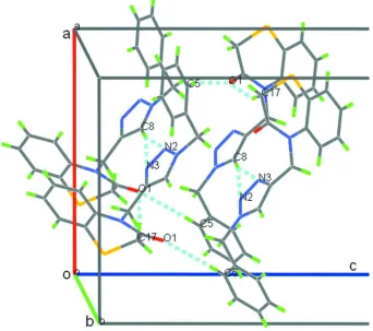
In the crystal, the molecules are linked together by a weak intermolecular C8–H8···N2, C8–H8···N3 and C5–H5···O1
Figure 1
Molecular structure of the title compound with the atom-labelling scheme. Displacement ellipsoids are drawn at the 50%
probability level. H atoms are represented as small circles.
Figure 2
Partial three dimensional plot of the title compound, showing molecules linked through C8–H8···N2, C8–H8···N3 and
[image:4.610.136.478.285.588.2]supporting information
sup-3 Acta Cryst. (2014). E70, o160–o161
4-[(1-Benzyl-1H-1,2,3-triazol-4-yl)methyl]-2H-1,4-benzothiazin-3(4H)-one
Crystal data
C18H16N4OS
Mr = 336.41 Monoclinic, P21/c Hall symbol: -P 2ybc
a = 13.283 (2) Å
b = 5.3661 (10) Å
c = 23.281 (4) Å
β = 96.633 (10)°
V = 1648.3 (5) Å3
Z = 4
F(000) = 704
Dx = 1.356 Mg m−3
Mo Kα radiation, λ = 0.71073 Å Cell parameters from 3526 reflections
θ = 1.8–27.1°
µ = 0.21 mm−1
T = 296 K Block, colourless 0.39 × 0.35 × 0.28 mm
Data collection
Bruker X8 APEX diffractometer
Radiation source: fine-focus sealed tube Graphite monochromator
φ and ω scans
Absorption correction: multi-scan (SADABS; Bruker, 2009)
Tmin = 0.649, Tmax = 0.747
15366 measured reflections 3526 independent reflections 2540 reflections with I > 2σ(I)
Rint = 0.044
θmax = 27.1°, θmin = 1.8°
h = −16→17
k = −6→5
l = −29→29
Refinement
Refinement on F2 Least-squares matrix: full
R[F2 > 2σ(F2)] = 0.046
wR(F2) = 0.149
S = 1.03 3526 reflections 217 parameters 0 restraints
Primary atom site location: structure-invariant direct methods
Secondary atom site location: difference Fourier map
Hydrogen site location: inferred from neighbouring sites
H-atom parameters constrained
w = 1/[σ2(F
o2) + (0.087P)2 + 0.1602P] where P = (Fo2 + 2Fc2)/3
(Δ/σ)max < 0.001 Δρmax = 0.22 e Å−3 Δρmin = −0.33 e Å−3
Special details
Geometry. All e.s.d.'s (except the e.s.d. in the dihedral angle between two l.s. planes) are estimated using the full covariance matrix. The cell e.s.d.'s are taken into account individually in the estimation of e.s.d.'s in distances, angles and torsion angles; correlations between e.s.d.'s in cell parameters are only used when they are defined by crystal symmetry. An approximate (isotropic) treatment of cell e.s.d.'s is used for estimating e.s.d.'s involving l.s. planes.
Refinement. Refinement of F2 against all reflections. The weighted R-factor wR and goodness of fit S are based on F2, conventional R-factors R are based on F, with F set to zero for negative F2. The threshold expression of F2 > σ(F2) is used only for calculating R-factors(gt) etc. and is not relevant to the choice of reflections for refinement. R-factors based on F2 are statistically about twice as large as those based on F, and R- factors based on all data will be even larger.
Fractional atomic coordinates and isotropic or equivalent isotropic displacement parameters (Å2)
x y z Uiso*/Ueq
C1 0.22144 (17) 1.2104 (4) 0.32721 (9) 0.0608 (6)
H1 0.2703 1.3328 0.3361 0.073*
C2 0.13131 (19) 1.2201 (4) 0.35176 (10) 0.0663 (6)
C3 0.05772 (18) 1.0436 (4) 0.33828 (10) 0.0620 (6)
H3 −0.0028 1.0501 0.3547 0.074*
C4 0.07467 (19) 0.8572 (5) 0.30010 (9) 0.0675 (7)
H4 0.0246 0.7387 0.2901 0.081*
C5 0.16506 (19) 0.8438 (4) 0.27650 (8) 0.0577 (6)
H5 0.1759 0.7140 0.2515 0.069*
C6 0.23928 (15) 1.0199 (4) 0.28950 (7) 0.0457 (4)
C7 0.33713 (17) 1.0077 (4) 0.26263 (8) 0.0597 (6)
H7A 0.3309 0.8829 0.2323 0.072*
H7B 0.3490 1.1673 0.2450 0.072*
C8 0.48825 (17) 1.0970 (4) 0.33613 (9) 0.0545 (5)
H8 0.4885 1.2703 0.3354 0.065*
C9 0.55342 (15) 0.9457 (3) 0.36961 (7) 0.0424 (4)
C10 0.64140 (18) 1.0099 (4) 0.41219 (9) 0.0555 (5)
H10A 0.6711 1.1646 0.4006 0.067*
H10B 0.6181 1.0363 0.4497 0.067*
C11 0.72308 (14) 0.6346 (4) 0.46251 (7) 0.0438 (4)
C12 0.64100 (17) 0.6069 (5) 0.49446 (8) 0.0581 (6)
H12 0.5823 0.6994 0.4847 0.070*
C13 0.6465 (2) 0.4417 (5) 0.54082 (8) 0.0727 (8)
H13 0.5923 0.4296 0.5627 0.087*
C14 0.7301 (2) 0.2979 (6) 0.55454 (8) 0.0780 (8)
H14 0.7327 0.1873 0.5854 0.094*
C15 0.81136 (19) 0.3164 (5) 0.52239 (8) 0.0683 (7)
H15 0.8680 0.2154 0.5311 0.082*
C16 0.80820 (15) 0.4867 (4) 0.47686 (7) 0.0499 (5)
C17 0.84430 (15) 0.5783 (4) 0.36944 (7) 0.0526 (5)
H17A 0.8910 0.6067 0.3410 0.063*
H17B 0.8032 0.4341 0.3572 0.063*
C18 0.77735 (15) 0.8007 (4) 0.37190 (7) 0.0480 (5)
N1 0.42420 (12) 0.9464 (3) 0.30471 (6) 0.0442 (4)
N2 0.44742 (13) 0.7078 (3) 0.31697 (6) 0.0482 (4)
N3 0.52629 (12) 0.7072 (3) 0.35664 (6) 0.0461 (4)
N4 0.71926 (12) 0.8149 (3) 0.41721 (6) 0.0451 (4)
O1 0.77092 (14) 0.9600 (3) 0.33403 (6) 0.0689 (5)
S1 0.91484 (4) 0.51598 (14) 0.43858 (2) 0.0692 (2)
Atomic displacement parameters (Å2)
U11 U22 U33 U12 U13 U23
C1 0.0529 (13) 0.0505 (13) 0.0794 (14) −0.0036 (10) 0.0088 (11) −0.0073 (10)
C2 0.0640 (15) 0.0553 (13) 0.0817 (14) 0.0067 (11) 0.0168 (12) −0.0186 (11)
C3 0.0530 (13) 0.0659 (14) 0.0694 (13) 0.0021 (11) 0.0169 (11) 0.0023 (11)
C4 0.0738 (16) 0.0651 (15) 0.0658 (12) −0.0245 (12) 0.0177 (12) −0.0075 (11)
C5 0.0784 (16) 0.0501 (12) 0.0461 (9) −0.0030 (11) 0.0137 (10) −0.0047 (9)
C6 0.0480 (11) 0.0503 (11) 0.0385 (8) 0.0108 (9) 0.0041 (7) 0.0125 (8)
C7 0.0553 (13) 0.0785 (16) 0.0465 (10) 0.0182 (11) 0.0108 (9) 0.0225 (10)
supporting information
sup-5 Acta Cryst. (2014). E70, o160–o161
C9 0.0480 (11) 0.0325 (9) 0.0483 (9) −0.0044 (8) 0.0125 (8) −0.0029 (7)
C10 0.0621 (13) 0.0426 (11) 0.0615 (11) −0.0112 (10) 0.0064 (10) −0.0110 (9)
C11 0.0433 (10) 0.0563 (11) 0.0312 (7) −0.0180 (9) 0.0015 (7) −0.0067 (7)
C12 0.0483 (12) 0.0824 (15) 0.0449 (10) −0.0193 (11) 0.0101 (9) −0.0094 (10)
C13 0.0673 (16) 0.114 (2) 0.0379 (9) −0.0397 (15) 0.0129 (10) −0.0035 (11)
C14 0.0828 (19) 0.110 (2) 0.0378 (9) −0.0344 (16) −0.0084 (10) 0.0178 (11)
C15 0.0655 (15) 0.0909 (18) 0.0437 (10) −0.0127 (13) −0.0145 (9) 0.0138 (11)
C16 0.0410 (10) 0.0736 (14) 0.0332 (8) −0.0165 (10) −0.0039 (7) −0.0012 (8)
C17 0.0461 (11) 0.0736 (14) 0.0391 (8) −0.0139 (10) 0.0094 (8) −0.0017 (9)
C18 0.0469 (11) 0.0581 (12) 0.0387 (8) −0.0221 (9) 0.0036 (7) −0.0004 (8)
N1 0.0450 (9) 0.0419 (9) 0.0469 (8) 0.0054 (7) 0.0102 (7) 0.0093 (6)
N2 0.0531 (10) 0.0375 (9) 0.0513 (8) 0.0015 (7) −0.0049 (7) 0.0012 (7)
N3 0.0538 (10) 0.0318 (8) 0.0503 (8) −0.0042 (7) −0.0035 (7) −0.0009 (6)
N4 0.0435 (9) 0.0497 (9) 0.0419 (7) −0.0123 (7) 0.0044 (6) −0.0033 (7)
O1 0.0803 (12) 0.0684 (10) 0.0588 (8) −0.0190 (8) 0.0122 (8) 0.0172 (7)
S1 0.0364 (3) 0.1170 (6) 0.0528 (3) −0.0050 (3) −0.0005 (2) 0.0036 (3)
Geometric parameters (Å, º)
C1—C6 1.385 (3) C10—H10B 0.9700
C1—C2 1.386 (3) C11—C16 1.390 (3)
C1—H1 0.9300 C11—C12 1.397 (3)
C2—C3 1.371 (3) C11—N4 1.428 (2)
C2—H2 0.9300 C12—C13 1.392 (3)
C3—C4 1.374 (3) C12—H12 0.9300
C3—H3 0.9300 C13—C14 1.360 (4)
C4—C5 1.379 (3) C13—H13 0.9300
C4—H4 0.9300 C14—C15 1.387 (4)
C5—C6 1.374 (3) C14—H14 0.9300
C5—H5 0.9300 C15—C16 1.396 (3)
C6—C7 1.508 (3) C15—H15 0.9300
C7—N1 1.464 (3) C16—S1 1.766 (2)
C7—H7A 0.9700 C17—C18 1.493 (3)
C7—H7B 0.9700 C17—S1 1.798 (2)
C8—N1 1.330 (3) C17—H17A 0.9700
C8—C9 1.364 (3) C17—H17B 0.9700
C8—H8 0.9300 C18—O1 1.224 (2)
C9—N3 1.354 (2) C18—N4 1.379 (2)
C9—C10 1.483 (3) N1—N2 1.340 (2)
C10—N4 1.466 (3) N2—N3 1.314 (2)
C10—H10A 0.9700
C6—C1—C2 120.5 (2) C16—C11—C12 118.32 (18)
C6—C1—H1 119.7 C16—C11—N4 121.53 (16)
C2—C1—H1 119.7 C12—C11—N4 120.14 (19)
C3—C2—C1 120.5 (2) C13—C12—C11 120.4 (2)
C3—C2—H2 119.8 C13—C12—H12 119.8
C2—C3—C4 119.0 (2) C14—C13—C12 120.8 (2)
C2—C3—H3 120.5 C14—C13—H13 119.6
C4—C3—H3 120.5 C12—C13—H13 119.6
C3—C4—C5 120.7 (2) C13—C14—C15 119.9 (2)
C3—C4—H4 119.6 C13—C14—H14 120.1
C5—C4—H4 119.6 C15—C14—H14 120.1
C6—C5—C4 120.80 (19) C14—C15—C16 120.0 (2)
C6—C5—H5 119.6 C14—C15—H15 120.0
C4—C5—H5 119.6 C16—C15—H15 120.0
C5—C6—C1 118.43 (18) C11—C16—C15 120.55 (19)
C5—C6—C7 120.64 (18) C11—C16—S1 120.37 (14)
C1—C6—C7 120.92 (19) C15—C16—S1 119.07 (18)
N1—C7—C6 112.60 (14) C18—C17—S1 111.42 (13)
N1—C7—H7A 109.1 C18—C17—H17A 109.3
C6—C7—H7A 109.1 S1—C17—H17A 109.3
N1—C7—H7B 109.1 C18—C17—H17B 109.3
C6—C7—H7B 109.1 S1—C17—H17B 109.3
H7A—C7—H7B 107.8 H17A—C17—H17B 108.0
N1—C8—C9 106.02 (17) O1—C18—N4 120.9 (2)
N1—C8—H8 127.0 O1—C18—C17 121.52 (17)
C9—C8—H8 127.0 N4—C18—C17 117.51 (16)
N3—C9—C8 107.47 (18) C8—N1—N2 110.29 (16)
N3—C9—C10 122.51 (17) C8—N1—C7 129.57 (18)
C8—C9—C10 130.00 (18) N2—N1—C7 120.13 (17)
N4—C10—C9 112.43 (15) N3—N2—N1 107.31 (15)
N4—C10—H10A 109.1 N2—N3—C9 108.92 (15)
C9—C10—H10A 109.1 C18—N4—C11 123.52 (17)
N4—C10—H10B 109.1 C18—N4—C10 115.48 (16)
C9—C10—H10B 109.1 C11—N4—C10 120.46 (15)
H10A—C10—H10B 107.8 C16—S1—C17 95.91 (9)
Hydrogen-bond geometry (Å, º)
D—H···A D—H H···A D···A D—H···A
C8—H8···N2i 0.93 2.44 3.344 (3) 165
C8—H8···N3i 0.93 2.44 3.339 (3) 164
C5—H5···O1ii 0.93 2.58 3.477 (2) 163