research communications
Acta Cryst.(2018). E74, 349–351 https://doi.org/10.1107/S2056989018002360
349
Received 1 January 2018 Accepted 9 February 2018
Edited by H. Ishida, Okayama University, Japan
Keywords:crystal structure; 1,2-dihydro-naphthalene derivative; hydrogen bonding.
CCDC reference:1823056
Supporting information:this article has supporting information at journals.iucr.org/e
Crystal structure of dimethyl
1-oxo-2,4-diphenyl-1,2-dihydronaphthalene-2,3-dicarboxylate
Gajendran Jagadeesan,a* Immanuel Monica Chandramalar,aJayachandran Karunakaran,b Solaiappan Gopinathcand Arasambattu K. Mohanakrishnanb
aDepartment of Physics, Jeppiaar Engineering College, Jeppiaar Nagar, OMR, Chennai 600 119, India,bDepartment of
Organic Chemistry, University of Madras, Guindy Campus, Chennai 600 025, India, andcDepartment of Physics, RKM
Vivekananda College (Autonomous), Chennai 600 004, India. *Correspondence e-mail: g.jagan85@gmail.com
In the title compound, C26H20O5, a 1,2-dihydronaphthalene derivative, the cyclohexa-1,3-diene ring of the 1,2-dihydronaphthalene ring system adopts a half-chair conformation. The mean plane of the 1,2-dihydronapthalene ring system makes dihedral angles of 86.23 (6) and 64.80 (7)with two phenyl rings.
The carbonyl O atom attached to the dihydronaphthalene ring system deviates from the mean plane of the 1,2-dihydronaphthalene ring system by 0.618 (1) A˚ . In the crystal, the molecules are linked into layers parallel to thebcplane via
two kinds of C—H O interactions, one of which forms aC(10) chain motif running along the c-axis direction and the other forms an R22(6) ring motif. Adjacent layers are further connected by C—H and offset–interactions [centroid–centroid distance = 3.6318 (9) A˚ ].
1. Chemical context
Naphthalene derivatives have manifested applications in many fields, for example, as colorants, explosives, disinfectants, insecticides and the plant hormone auxin. Naphthalene is believed to play a role in the chemical defence against biological enemies (Wiltz et al., 1998; Wright et al., 2000). Naphthalene derivatives have been identified as a new range of potent anti-microbials that are effective against a wide range of human pathogens and have diverse and interesting antibiotic properties with minimum toxicity (Rokade & Sayyed, 2009; Upadhayayaet al., 2010). Compounds with non-coplanarly accumulated aromatic rings have received atten-tion from organic chemists and materials chemists as unique structural building blocks, because they provide characteristic optical and electronic properties originating from their structural features. For example, biphenyl and binaphthyl are applied to optically active molecular catalysts and polymer materials on the basis of their axial chiralities (Deria et al., 2013). The structures of similar 1-oxo-1,2-dihydronaphtalene derivatives, namely, dimethyl 4-(4-methoxyphenyl)-2-(4- methylphenyl)-1-oxo-1,2-dihydronaphthalene-2,3-dicarboxyl-ate, dimethyl 1-oxo-2-(pyren-4-yl)-4-(thiophen-2-yl)-1,2-di-hydronaphthalene-2,3-dicarboxylate and ethyl 1-oxo-2- phenyl-2,4-bis(thiophen-2-yl)-1,2-dihydronaphthalene-3-carboxylate, have been reported by Gopinathet al.(2017).
2. Structural commentary
1,3-diene C5–C10 ring adopts a half-chair conformation with puckering and smallest displacement parameters q2 = 0.3091 (14) A˚ , q3 = 0.1461 (14) A˚ , ’2 = 155.9 (3) and = 64.7 (2)andC
s= 4.41 (19). The dihedral angle between the C1–C6 and C5–C10 rings is 10.15 (6). The C11–C16 phenyl
ring is almost perpendicular to the 1,2-dihydronaphthalene C1–C10 ring system with a dihedral angle of 83.83 (7) between them. The other phenyl ring (C21–C26) makes dihedral angles of 64.80 (7) and 29.06 (8)with the mean plane
of C1–C10 ring system and the C11–C16 phenyl ring, respec-tively. Atom O1 of the carbonyl group deviates from the mean plane of the 1,2-dihydronaphthalene ring system by 0.647 (1) A˚ .
3. Supramolecular features
In the crystal, the molecules are linked via C—H O hydrogen bonds (C24—H24 O2i; symmetry code as in
Table 1), which generatesC(10) zigzag chains running along thec-axis direction (Fig. 2). In addition, the chains are linked
via pairs of C—H O interactions (C20—H20B O5ii; Table 2) with an R2
2(6) ring motif (Fig. 3), forming layers
350
Jagadeesanet al. C26H20O5 Acta Cryst.(2018). E74, 349–351 [image:2.610.102.242.232.408.2] [image:2.610.313.566.260.368.2]research communications
Table 1
Hydrogen-bond geometry (A˚ ,).
Cg3 is the centroid of the phenyl C11–C16 ring.
D—H A D—H H A D A D—H A
C24—H24 O2i 0.93 2.59 3.449 (3) 155
C20—H20B O5ii 0.96 2.59 3.430 (2) 146
C3—H3 Cg3iii 0.93 2.77 3.6338 (16) 154
Symmetry codes: (i)x;yþ3 2;z
1
2; (ii)xþ1;yþ1;z; (iii)x;yþ1;z.
Figure 2
A packing diagram of the title compound, showing aC(10) zigzag chain along to thecaxis formedviaC—H O hydrogen bonds (dashed lines). The H atoms not involved in the hydrogen bonding have been excluded for clarity. [Symmetry code: (i)x,3
2y,
1 2+z.]
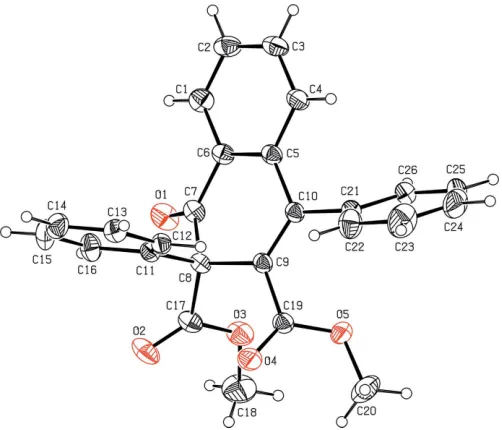
Figure 1
The molecular structure of the title compound with the atom-numbering scheme. The displacement ellipsoids are drawn at the 30% probability level. H atoms are shown as spheres of arbitrary radii.
Figure 3
[image:2.610.318.564.435.700.2] [image:2.610.46.297.494.709.2]parallel to thebcplane. Between the layers there are also C— H (C3—H3 Cg3iii; Table 1) and – stacking inter-actions (Fig. 4) [Cg1 Cg1iii = 3.6318 (9) A˚ , interplanar distance = 3.343 (1) A˚ and offset distance = 1.419 (1) A˚ ; symmetry code: (iii) x, 1 y, z; Cg1 and Cg3 are the centroids of the C1–C6 and C11–C16 rings, respectively].
4. Synthesis and crystallization
To a solution of 1,3-diphenylisobenzofuran (1 g, 3.70 mmol) in dry dichloromethane, dimethyl acetylenedicarboxylate (0.58 g, 4.07 mmol) was added and the reaction mixture was stirred at room temperature for 1 h. Removal of solvent followed by column chromatographic purification (silica gel; 15% ethyl acetate in hexane) afforded isobenzofurandimethyl acetylenedicarboxylate adduct as a colourless solid (1.10 g, 72%). To a solution of the adduct (0.50 g, 1.21 mmol) in dry dichloromethane, BF3OEt2 (0.075 g, 0.52 mmol) was added and the reaction mixture was stirred at room temperature for 5 min. Removal of solvent followed by column chromato-graphic purification (silica gel; 15% ethyl acetate in hexane) gave the title compound as a colourless solid (0.45 g, 94%). Single crystals suitable for X-ray diffraction were prepared by slow evaporation of an ethyl acetate solution of the title compound at room temperature (m.p. = 454–456 K).
5. Refinement
Crystal data, data collection and structure refinement details are summarized in Table 2. H atoms were localized in a difference-Fourier map and then were treated as riding atoms, with C—H = 0.93 and 0.96 A˚ for aryl and methyl groups, respectively, and with Uiso(H) = 1.2Ueq(aryl C) and 1.5Ueq(methyl C), allowing for free rotation of the methyl groups.
Acknowledgements
The authors thank Dr P. K. Sudhadevi Antharjanam, Tech-nical Officer, SAIF, IIT Madras, Chennai, India, for the data collection. GJ thanks Jeppiaar Engineering College, Rajiv Gandhi Salai, Chennai, India for their support.
References
Bruker (2008). APEX2, SAINT and SADABS. Bruker AXS Inc., Madison, Wisconsin, USA.
Deria, P., Von Bargen, C. D., Olivier, J. H., Kumbhar, A. S., Saven, J. G. & Therien, M. J. (2013).J. Am. Chem. Soc.135, 16220–16234. Farrugia, L. J. (2012).J. Appl. Cryst.45, 849–854.
Gopinath, S., Narayanan, P., Sethusankar, K., Karunakaran, J., Nandakumar, M. & Mohanakrishnan, A. K. (2017). Acta Cryst.
E73, 177–182.
Macrae, C. F., Bruno, I. J., Chisholm, J. A., Edgington, P. R., McCabe, P., Pidcock, E., Rodriguez-Monge, L., Taylor, R., van de Streek, J. & Wood, P. A. (2008).J. Appl. Cryst.41, 466–470.
Rokade, Y. B. & Sayyed, R. Z. (2009).Rasayan J. Chem.2, 972–980. Sheldrick, G. M. (2008).Acta Cryst.A64, 112–122.
Spek, A. L. (2009).Acta Cryst.D65, 148–155.
Upadhayaya, R. S., Vandavasi, J. K., Kardile, R. A., Lahore, S. V., Dixit, S. S., Deokar, H. S., Shinde, P. D., Sarmah, M. P. & Chattopadhyaya, J. (2010).Eur. J. Med. Chem.45, 1854–1867. Wiltz, B. A., Henderson, G. & Chen, J. (1998).Environ. Entomol.27,
936–940.
Wright, M. S., Lax, A. R., Henderson, G. & Chen, J. A. (2000).
Mycologia,92, 42–45.
research communications
[image:3.610.313.561.92.358.2]Acta Cryst.(2018). E74, 349–351 Jagadeesanet al. C26H20O5
351
Figure 4
[image:3.610.47.297.550.694.2]A packing diagram of the title compound, showing C—H and– interactions (dashed lines), whereCg1 andCg3 are the centroids of the phenyl C1–C6 and C11–C16 rings, respectively. [Symmetry code: (iii)x, 1y,z.]
Table 2
Experimental details.
Crystal data
Chemical formula C26H20O5
Mr 412.42
Crystal system, space group Monoclinic,P21/c
Temperature (K) 296
a,b,c(A˚ ) 15.8021 (8), 7.4706 (4), 17.8599 (9)
() 96.581 (2)
V(A˚3) 2094.49 (19)
Z 4
Radiation type MoK
(mm1) 0.09
Crystal size (mm) 0.350.300.25
Data collection
Diffractometer Bruker Kappa APEXII
Absorption correction Multi-scan (SADABS; Bruker, 2008)
Tmin,Tmax 0.969, 0.978
No. of measured, independent and observed [I> 2(I)] reflections
21819, 4614, 3375
Rint 0.028
(sin/)max(A˚
1) 0.641
Refinement
R[F2> 2(F2)],wR(F2),S 0.039, 0.108, 1.03
No. of reflections 4614
No. of parameters 283
H-atom treatment H-atom parameters constrained
max, min(e A˚3) 0.22,0.15
Computer programs:APEX2andSAINT(Bruker, 2008),SHELXS97andSHELXL97
supporting information
sup-1
Acta Cryst. (2018). E74, 349-351
supporting information
Acta Cryst. (2018). E74, 349-351 [https://doi.org/10.1107/S2056989018002360]
Crystal structure of dimethyl
1-oxo-2,4-diphenyl-1,2-dihydronaphthalene-2,3-dicarboxylate
Gajendran Jagadeesan, Immanuel Monica Chandramalar, Jayachandran Karunakaran,
Solaiappan Gopinath and Arasambattu K. Mohanakrishnan
Computing details
Data collection: APEX2 (Bruker, 2008); cell refinement: SAINT (Bruker, 2008); data reduction: SAINT (Bruker, 2008);
program(s) used to solve structure: SHELXS97 (Sheldrick, 2008); program(s) used to refine structure: SHELXL97
(Sheldrick, 2008); molecular graphics: ORTEP-3 for Windows (Farrugia, 2012) and Mercury (Macrae et al., 2008);
software used to prepare material for publication: SHELXL97 (Sheldrick, 2008) and PLATON (Spek, 2009).
Dimethyl 1-oxo-2,4-diphenyl-1,2-dihydronaphthalene-2,3-dicarboxylate
Crystal data
C26H20O5
Mr = 412.42 Monoclinic, P21/c
Hall symbol: -P 2ybc
a = 15.8021 (8) Å
b = 7.4706 (4) Å
c = 17.8599 (9) Å
β = 96.581 (2)°
V = 2094.49 (19) Å3
Z = 4
F(000) = 864
Dx = 1.308 Mg m−3
Mo Kα radiation, λ = 0.71073 Å Cell parameters from 3375 reflections
θ = 2.3–27.1°
µ = 0.09 mm−1
T = 296 K Block, colourless 0.35 × 0.30 × 0.25 mm
Data collection
Bruker Kappa APEXII diffractometer
Radiation source: fine-focus sealed tube Graphite monochromator
ω & φ scans
Absorption correction: multi-scan (SADABS; Bruker, 2008)
Tmin = 0.969, Tmax = 0.978
21819 measured reflections 4614 independent reflections 3375 reflections with I > 2σ(I)
Rint = 0.028
θmax = 27.1°, θmin = 2.3°
h = −20→13
k = −9→8
l = −22→21
Refinement
Refinement on F2
Least-squares matrix: full
R[F2 > 2σ(F2)] = 0.039
wR(F2) = 0.108
S = 1.03 4614 reflections 283 parameters 0 restraints
Primary atom site location: structure-invariant direct methods
Secondary atom site location: difference Fourier map
Hydrogen site location: inferred from neighbouring sites
supporting information
sup-2
Acta Cryst. (2018). E74, 349-351
w = 1/[σ2(F
o2) + (0.0478P)2 + 0.4036P]
where P = (Fo2 + 2Fc2)/3
(Δ/σ)max = 0.017
Δρmax = 0.22 e Å−3
Δρmin = −0.14 e Å−3
Extinction correction: SHELXL, Fc*=kFc[1+0.001xFc2λ3/sin(2θ)]-1/4
Extinction coefficient: 0.0033 (8)
Special details
Geometry. All esds (except the esd in the dihedral angle between two l.s. planes) are estimated using the full covariance matrix. The cell esds are taken into account individually in the estimation of esds in distances, angles and torsion angles; correlations between esds in cell parameters are only used when they are defined by crystal symmetry. An approximate (isotropic) treatment of cell esds is used for estimating esds involving l.s. planes.
Refinement. Refinement of F2 against ALL reflections. The weighted R-factor wR and goodness of fit S are based on F2,
conventional R-factors R are based on F, with F set to zero for negative F2. The threshold expression of F2 > 2sigma(F2) is
used only for calculating R-factors(gt) etc. and is not relevant to the choice of reflections for refinement. R-factors based on F2 are statistically about twice as large as those based on F, and R- factors based on ALL data will be even larger.
Fractional atomic coordinates and isotropic or equivalent isotropic displacement parameters (Å2)
x y z Uiso*/Ueq
supporting information
sup-3
Acta Cryst. (2018). E74, 349-351
C20 0.50781 (11) 0.6419 (3) 0.08805 (11) 0.0783 (6) H20A 0.5019 0.7671 0.0983 0.117* H20B 0.5429 0.6268 0.0480 0.117* H20C 0.5338 0.5825 0.1325 0.117* C21 0.27693 (8) 0.6601 (2) −0.02840 (7) 0.0398 (3) C22 0.28407 (11) 0.8357 (2) −0.00451 (10) 0.0593 (4) H22 0.2708 0.8659 0.0433 0.071* C23 0.31073 (13) 0.9673 (3) −0.05105 (14) 0.0824 (6) H23 0.3156 1.0851 −0.0344 0.099* C24 0.33002 (12) 0.9242 (4) −0.12164 (13) 0.0846 (7) H24 0.3474 1.0127 −0.1531 0.102* C25 0.32355 (11) 0.7507 (4) −0.14553 (9) 0.0723 (6) H25 0.3375 0.7214 −0.1932 0.087* C26 0.29671 (9) 0.6184 (3) −0.10003 (8) 0.0531 (4) H26 0.2918 0.5010 −0.1173 0.064* O1 0.18036 (7) 0.07330 (14) 0.12689 (6) 0.0561 (3) O2 0.32467 (8) 0.21704 (16) 0.25637 (6) 0.0629 (3) O3 0.37245 (7) 0.18860 (15) 0.14387 (6) 0.0548 (3) O4 0.38995 (7) 0.60087 (17) 0.18244 (5) 0.0600 (3) O5 0.42502 (6) 0.56566 (15) 0.06595 (5) 0.0508 (3)
Atomic displacement parameters (Å2)
U11 U22 U33 U12 U13 U23
supporting information
sup-4
Acta Cryst. (2018). E74, 349-351
C25 0.0470 (9) 0.128 (2) 0.0429 (9) 0.0034 (11) 0.0090 (7) 0.0312 (11) C26 0.0438 (8) 0.0809 (12) 0.0347 (7) 0.0026 (8) 0.0045 (6) 0.0059 (7) O1 0.0767 (8) 0.0338 (6) 0.0554 (6) −0.0059 (5) −0.0029 (5) 0.0052 (5) O2 0.0806 (8) 0.0670 (8) 0.0383 (6) 0.0193 (6) −0.0058 (5) 0.0116 (5) O3 0.0531 (6) 0.0581 (7) 0.0521 (6) 0.0213 (5) 0.0011 (5) −0.0005 (5) O4 0.0472 (6) 0.0932 (9) 0.0387 (5) −0.0121 (6) 0.0013 (4) −0.0186 (6) O5 0.0362 (5) 0.0741 (8) 0.0430 (5) −0.0041 (5) 0.0081 (4) −0.0076 (5)
Geometric parameters (Å, º)
C1—C2 1.379 (2) C14—H14 0.9300 C1—C6 1.3873 (19) C15—C16 1.376 (2) C1—H1 0.9300 C15—H15 0.9300 C2—C3 1.370 (2) C16—H16 0.9300 C2—H2 0.9300 C17—O2 1.1905 (16) C3—C4 1.381 (2) C17—O3 1.3328 (18) C3—H3 0.9300 C18—O3 1.4456 (19) C4—C5 1.3942 (17) C18—H18A 0.9600 C4—H4 0.9300 C18—H18B 0.9600 C5—C6 1.3940 (19) C18—H18C 0.9600 C5—C10 1.4796 (18) C19—O4 1.1985 (15) C6—C7 1.4777 (18) C19—O5 1.3256 (17) C7—O1 1.2090 (17) C20—O5 1.4399 (19) C7—C8 1.5397 (18) C20—H20A 0.9600 C8—C9 1.5212 (18) C20—H20B 0.9600 C8—C17 1.5392 (17) C20—H20C 0.9600 C8—C11 1.5408 (18) C21—C22 1.380 (2) C9—C10 1.3457 (16) C21—C26 1.3867 (19) C9—C19 1.4897 (18) C22—C23 1.384 (2) C10—C21 1.4852 (18) C22—H22 0.9300 C11—C12 1.3844 (19) C23—C24 1.369 (3) C11—C16 1.3866 (19) C23—H23 0.9300 C12—C13 1.3774 (19) C24—C25 1.365 (3) C12—H12 0.9300 C24—H24 0.9300 C13—C14 1.371 (2) C25—C26 1.377 (3) C13—H13 0.9300 C25—H25 0.9300 C14—C15 1.369 (2) C26—H26 0.9300
supporting information
sup-5
Acta Cryst. (2018). E74, 349-351
C3—C4—H4 119.6 O3—C18—H18B 109.5 C5—C4—H4 119.6 H18A—C18—H18B 109.5 C6—C5—C4 117.90 (12) O3—C18—H18C 109.5 C6—C5—C10 120.27 (11) H18A—C18—H18C 109.5 C4—C5—C10 121.82 (12) H18B—C18—H18C 109.5 C1—C6—C5 120.89 (12) O4—C19—O5 123.97 (13) C1—C6—C7 119.35 (13) O4—C19—C9 122.85 (13) C5—C6—C7 119.76 (12) O5—C19—C9 113.11 (11) O1—C7—C6 122.90 (12) O5—C20—H20A 109.5 O1—C7—C8 121.30 (11) O5—C20—H20B 109.5 C6—C7—C8 115.69 (11) H20A—C20—H20B 109.5 C9—C8—C17 110.48 (11) O5—C20—H20C 109.5 C9—C8—C7 110.63 (10) H20A—C20—H20C 109.5 C17—C8—C7 106.22 (10) H20B—C20—H20C 109.5 C9—C8—C11 112.92 (10) C22—C21—C26 118.68 (14) C17—C8—C11 111.96 (10) C22—C21—C10 119.79 (12) C7—C8—C11 104.24 (10) C26—C21—C10 121.49 (14) C10—C9—C19 123.16 (12) C21—C22—C23 120.60 (17) C10—C9—C8 122.34 (11) C21—C22—H22 119.7 C19—C9—C8 114.40 (10) C23—C22—H22 119.7 C9—C10—C5 119.96 (12) C24—C23—C22 120.1 (2) C9—C10—C21 122.49 (12) C24—C23—H23 119.9 C5—C10—C21 117.40 (10) C22—C23—H23 119.9 C12—C11—C16 117.97 (13) C25—C24—C23 119.64 (18) C12—C11—C8 122.16 (11) C25—C24—H24 120.2 C16—C11—C8 119.81 (12) C23—C24—H24 120.2 C13—C12—C11 120.92 (13) C24—C25—C26 120.94 (18) C13—C12—H12 119.5 C24—C25—H25 119.5 C11—C12—H12 119.5 C26—C25—H25 119.5 C14—C13—C12 120.45 (14) C25—C26—C21 120.02 (18) C14—C13—H13 119.8 C25—C26—H26 120.0 C12—C13—H13 119.8 C21—C26—H26 120.0 C15—C14—C13 119.25 (14) C17—O3—C18 115.77 (13) C15—C14—H14 120.4 C19—O5—C20 117.16 (12) C13—C14—H14 120.4
supporting information
sup-6
Acta Cryst. (2018). E74, 349-351
C5—C6—C7—O1 −158.09 (14) C7—C8—C17—O2 −101.40 (17) C1—C6—C7—C8 −154.54 (12) C11—C8—C17—O2 11.8 (2) C5—C6—C7—C8 25.65 (18) C9—C8—C17—O3 −42.97 (14) O1—C7—C8—C9 145.22 (13) C7—C8—C17—O3 77.06 (14) C6—C7—C8—C9 −38.46 (15) C11—C8—C17—O3 −169.76 (11) O1—C7—C8—C17 25.29 (17) C10—C9—C19—O4 140.82 (15) C6—C7—C8—C17 −158.39 (11) C8—C9—C19—O4 −35.58 (19) O1—C7—C8—C11 −93.12 (15) C10—C9—C19—O5 −42.04 (18) C6—C7—C8—C11 83.21 (13) C8—C9—C19—O5 141.56 (12) C17—C8—C9—C10 146.53 (12) C9—C10—C21—C22 −62.17 (19) C7—C8—C9—C10 29.18 (17) C5—C10—C21—C22 113.49 (15) C11—C8—C9—C10 −87.22 (15) C9—C10—C21—C26 120.02 (15) C17—C8—C9—C19 −37.04 (15) C5—C10—C21—C26 −64.33 (17) C7—C8—C9—C19 −154.38 (11) C26—C21—C22—C23 −0.2 (2) C11—C8—C9—C19 89.22 (13) C10—C21—C22—C23 −178.11 (15) C19—C9—C10—C5 179.15 (12) C21—C22—C23—C24 0.3 (3) C8—C9—C10—C5 −4.73 (19) C22—C23—C24—C25 −0.6 (3) C19—C9—C10—C21 −5.3 (2) C23—C24—C25—C26 0.9 (3) C8—C9—C10—C21 170.82 (12) C24—C25—C26—C21 −0.9 (2) C6—C5—C10—C9 −11.87 (19) C22—C21—C26—C25 0.6 (2) C4—C5—C10—C9 168.95 (13) C10—C21—C26—C25 178.39 (13) C6—C5—C10—C21 172.36 (12) O2—C17—O3—C18 −3.7 (2) C4—C5—C10—C21 −6.82 (18) C8—C17—O3—C18 177.83 (14) C9—C8—C11—C12 −0.86 (16) O4—C19—O5—C20 −4.2 (2) C17—C8—C11—C12 124.60 (13) C9—C19—O5—C20 178.73 (14) C7—C8—C11—C12 −121.00 (12)
Hydrogen-bond geometry (Å, º)
Cg3 is the centroid of the phenyl C11–C16 ring.
D—H···A D—H H···A D···A D—H···A
C24—H24···O2i 0.93 2.59 3.449 (3) 155
C20—H20B···O5ii 0.96 2.59 3.430 (2) 146
C3—H3···Cg3iii 0.93 2.77 3.6338 (16) 154