Original Article
Different expression of NOD2 in decidual stromal cells
between normal and unexplained recurrent
spontaneous abortion women during first
trimester gestation
Yuanyuan Zhang1, Chunfeng Yang1, Shuai Fu1, Xin Chen1, Shining Zhang2, Yiyang Li3, Meirong Du4, Jianping
Zhang1
1Department of Obstetrics and Gynecology, Sun Yatsen Memorial Hospital of Sun Yatsen University, Guangzhou, China; 2Department of Interventional Radiology, Second People Hospital of Guangdong, Guangzhou, China; 3Department of Obstetrics and Gynecology, The first Hospital of JiLin University, Changchun, China; 4Laboratory for Reproductive Immunology, Hospital and Institute of Obstetrics and Gynecology, Fudan University Shanghai Medical College, Shanghai, China
Received October 9, 2014; Accepted December 1, 2014; Epub December 1, 2014; Published December 15, 2014
Abstract: The NOD2 gene, encoding intracellular paternal recognition receptor (PRR) also called caspase activation and recruitment domain 15 (CARD15), is mutated in Crohn’s disease, an autoimmune-disorder. Unexplained recur-rent spontaneous abortion (URSA) involved in complex auto-immune disorder. However, little is known about the expression of NOD2 protein at maternal-fetal interface with URSA patients. Our aim was to compare the expression levels of NOD2 in the decidual stromal cells (DSCs) from patients with normal pregnancy to those with unexplained recurrent spontaneous abortion (URSA) during first trimester pregnancy. Tissues and DSCs were collected from 12 patients with URSA and 26 patients with normal pregnancies that required abortion. DSCs in the normal pregnancy group showed significantly higher mRNA and protein levels of NOD2 than those isolated from the URSA group using real time PCR and in cell western. The appropriate expression of NOD2 in normal DSCs suggests that this protein may be required to sustain normal pregnancy.
Keywords: NOD2, decidual stromal cells (DSCs), unexplained spontaneous recurrent abortion
Introduction
Spontaneous miscarriages (abortions) occur in approximately 14% to 16% of naturally con-ceived pregnancies [1]. Abortions may arise from an abnormal uterine cavity, parental karyotypes, endocrine factors, infection, auto-immunity [2, 3], and unknown mechanisms [4]. A loss of three or more consecutive pregnan-cies before the 20th week of gestation with unknown etiology is called unexplained recur-rent spontaneous abortion (URSA) and occurs in approximately 1-3% of fertile women [5]. Some of these abortions may arise from the loss of maternal immuno-tolerance or abnor-mal maternal responses to the fetus.
In full term pregnancies, the maternal immune responses remain tolerant from early
[16], asthma [17] and NOD2-associated autoin-flammatory disease (NAID) [18].
DSC as one of the most important elements at maternal-fetal interface plays essential role for immunologic protection of the fetus, but the expression of NOD2 in DSCs (normal and URSA) have not been elucidated. In this study, we compared the expression levels of NOD2 in DSCs during normal pregnancy with URSA patients. Our study suggests that loss of func-tion of NOD2 may be responsible for the URSA.
Materials and methods
Samples
Human first trimester pregnancy decidual sam-ples (26 cases normal pregnancy women and 12 cases URSA patients) were obtained with informed consent from first trimester voluntary termination of gestation in Sun Yat-Sen Memorial Hospital and Red-House hospital of Fudan University from January 21st 2012 to
October 6th 2012. The inclusion criteria for
URSA group: genetics, anatomy, endocrine, hor-mone, certain coagulation and serum levels of immune regulatory proteins were normal. The patients with infection, smoking and alcohol consumption, environmental factors, psycho-logical trauma, and stressful life event were excluded. All patients had signed consent forms of the tissue collection, and the study was approved by Human Research Ethics Committee of Obstetrics and Gynecology Hospital, Fudan University and Sun Yat-Sen University’s Human Investigations Committee.
Immunohistochemistry
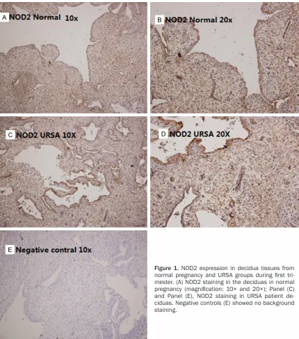
Immunohistochemistry was performed essen-tially as described previously [19]. Briefly, all sections were fixed in 4% paraformaldehyde over night, then deparaffinized, rehydrated through a graded alcohol series, and subjected
NOD2 mAb (Abcam, Cambridge, UK) or PBS, and then stained by haematoxylin (Sigma), dehydrated with ethanol and mounted from xylene.
DSCs isolation
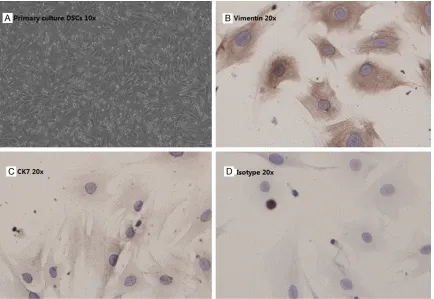
Decidual stromal cells were isolated from fresh tissue which collected in ice-cold DMEM/F12 (Gibco, Grand Island, NY, USA). The individual patient tissues were washed in calcium- and magnesium-free Hanks balanced salt solution (HBSS), then subjected to dissociate by collage-nase IV/DNase-I digestion (Sigma, Saint Louis, Missouri, USA) and isolated by discontinuous Percoll gradient centrifugation, as described [20]. DSCs of density between 1.042 and 1.062 g/ml were collected and cultured in DMEM/F12 (Gibco, Grand Island, NY, USA) complete medium (10% heat-inactivated fetal bovine serum (FBS, Gibco), 100 U/ml penicillin, and 100 μg/ml streptomycin) in 5% CO2 at 37°C. After 30 min, non-adherent hematopoi-etic and immune cells were washed away, leav-ing 98% pure DSCs (confirmed by stainleav-ing of mouse Cytokeratin7 mAb and mouse anti-vimentin mAb (ZSGB-BIO, Beijing, China).
RNA extraction and QPCR
The purified DSCs were seeded in 12-well plates at a density of 5 × 10⁵ cells/well. After
as follows: 5’-TGCGGACTCTACTCTTTGAGC-3’ (forward) and 5’-CCGTGAACCTGAACTTGAACT-3’ (reverse); for human glyceraldehyde-3-phos-phatedehydrogenase (GAPDH): 5’-GCACCGTC- AAGGCTGAGAAC-3’ (forward) and 5’-TGGTGAA- GACGCCAGTGGA-3’ (reverse). 3 μl cDNA was diluted into 12 μl, and 2 μl dilution was added for each PCR reaction. PCRs were run on an ABI 7900HT (Perkin-Elmer Applied Biosystems, USA). Each cDNA was amplified in triplicate and the corresponding sample without reverse tran-scriptase (ddH2O) was included as the negative
control. The expression of the NOD2 was nor-malized to that of GAPDH. The replicates were then averaged, and fold induction was deter-mined in a ΔΔCt-based fold-change calcula- tions.
In-cell Western
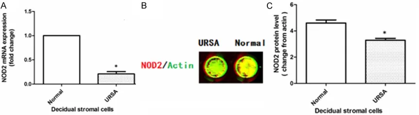
[image:3.612.91.521.69.556.2]In-cell Western was carried out to determine the protein level of NOD2 in the DSCs, as described [19]. Purified isolated DSCs were seeded (1.5 × 10⁴/well) in 96-well plate and
cultured 24 hrs, then Cells were fixed with 4% paraformaldehyde for 20 min at room tempera-ture. After washing with 0.1% Triton 3 times, cells were blocked by adding 100 μl of LI-COR Odyssey Blocking Buffer (LI-COR Biosciences, Lincoln, Nebraska, USA) for 90 min at room temperature. Cells were incubated with mouse anti-human mAb NOD2 (25 μg/ml, Abcam, Cambridge, UK) and the rabbit anti-human β-actin (1:80, Santa Cruz, CA, USA) which detected the control reference protein over-night at 4°C. Cells were washed 3 times with 1 × PBS, and incubated with the corresponding secondary antibodies: anti-mouse IRDyeTM- 700DX conjugated (1:60, affinity purified, Red fluorescence) and anti-rabbit IRDyeTM800DX conjugated (1:80, affinity purified, green fluo-rescence) in darkness (secondary antibodies: Rockland, Inc, Gilbertsville, PA, USA). Images of NOD2 were analyzed by Odyssey Infrared Imaging System (LI-COR Biosciences GmbH). The protein level of NOD2 was analyzed by the ratio of the intensity of NOD2 (green fluores-cence) to that of β-actin (red fluoresfluores-cence). Each experiment was repeated three times.
Statistics
All values were expressed as the mean ± SEM and differences were compared by Student
t-test with GraphPad Prism 5.0. The differences were accepted as significant at P < 0.05.
Results
[image:4.612.91.525.71.370.2](Figure 2). Second, the expression of NOD2 in DSCs from the primary cultures was evaluated by real time PCR and In-cell Western blot analy-sis. Consistent with the up regulation of the mRNA of NOD2, the DSCs from each woman in the normal pregnancy group expressed higher NOD2 protein than DSCs in each individual URSA patient (Figure 3).
Discussion
Till now, in human gestation system, NOD2 expression has been observed in first trimester placenta, specifically in the trophoblast cell [15], and in endometrial epithelial cell [8]. NOD2 acts by regulating chemokins/cytokines secretions, at least in part by protecting mater-nal-fetal interface from pathogens evading, For example, MDP stimulation of endometrial epi-thelial cells significantly increased expression of IL-8 and TNF-α [8], MDP stimulation of tro-phoblasts from the first trimester up-regulated secretion of GRO-α, IL-6, IL-8, and MCP-1 [15, 20]. This data suggested that NOD2 is function-al at maternfunction-al-fetfunction-al interface. Defective NOD2 expression affects the cytokines secretion baseline, for example ectopic expression of NOD2 in cell line H8 (a first trimester tropho-blast cell line), produced higher levels of cyto-kines than that with vehicle treatment alone [20]. The appropriate level of cytokines aids the tight regulation of cytokines and chemokines necessary to maintain the maternal tolerance towards the fetus. Interestingly, pretreatment of a murine macrophage cell line by NOD2 ligand: MDP, the cell releases its tight chaper-one, heat shock protein 90, and degrades the activated Nod2 protein, which blocks Nod2’s continual activation [21]. Based on this data, if
MDP stimulation also down regulates expres-sion of NOD2 in human DSCs, the short burst of NOD2 activation can contribute to the tightly regulated temporal and spatial expression of chemokines/cytokines at maternal-fetal inter- face.
In this study, we demonstrated for the first time that different expression of NOD2, an impor-tant cytosolic pattern recognition receptor (PRR), was lower in decidual stromal cells from patients with unexplained recurrent spontane-ous abortion in first trimester than those from patients with normal pregnancies. The physio-logical roles of NOD2 in DSCs during normal pregnancy are not well understood but appear to be broader than only surveillance for intra-cellular pathogens.
[image:5.612.92.523.71.190.2]thwart the pathogenic invader in sufficient time. Growing evidence suggests that obligate intracellular bacteria, such as Chlamydia, may be responsible for reproductive tract infections, and Chlamydia infection is associated with an increased risk of miscarriage [27]. Fifth, activa-tion of NOD2 requires sufficient intracellular NOD2 levels [15]. Since many URSA patients show an inflammatory cytokine milieu rather than the finely tuned cytokine orchestra observed during normal pregnancy [4], this may help explain why URSA patients have lower NOD2 expression.
Limitations of this study were the lack of data on the effects of deficiency of Nod2 in animal model may induce recurrent spontaneous abor-tion. Next, we will set up the model of Nod2-dificiency mouse to observe the affection dur-ing mouse pregnancy.
In summary, to our knowledge this is the first report demonstrating a difference in the expression of NOD2 in DSCs between partici-pants with normal pregnancy and those with URSA. These findings suggest that an appropri-ate level of NOD2 is necessary for pregnancy maintenance.
Acknowledgements
This study was supported partially by National Nature Science Foundation of China (Nos. 81170625 and 81270754), Guangdong Natural Science Foundation (NO. 2012B031800352). The funders had no role in study design, data collection and analysis, decision to publish, or preparation of the manuscript.
Disclosure of conflict of interest
None.
Address correspondence to: Dr. Jianping Zhang, Department of Obstetrics and Gynecology, Sun
Yat-[1] Wang JX, Norman RJ and Wilcox AJ. Incidence of spontaneous abortion among pregnancies produced by assisted reproductive technology. Hum Reprod 2004; 19: 272-277.
[2] Pandey MK, Rani R and Agrawal S. An update in recurrent spontaneous abortion. Arch Gynecol Obstet 2005; 272: 95-108.
[3] Meng LL, Chen H, Tan JP, Wang ZH, Zhang R, Fu S and Zhang JP. Evaluation of etiological characteristics of Chinese women with recur-rent spontaneous abortions: a single-centre study. Chin Med J (Engl) 2011; 124: 1310-1315.
[4] Saini V, Arora S, Yadav A and Bhattacharjee J. Cytokines in recurrent pregnancy loss. Clin Chim Acta 2011; 412: 702-708.
[5] Rull K, Tomberg K, Koks S, Mannik J, Mols M, Sirotkina M, Varv S and Laan M. Increased pla-cental expression and maternal serum levels of apoptosis-inducing TRAIL in recurrent mis-carriage. Placenta 2013; 34: 141-148.
[6] Ren L, Liu YQ, Zhou WH and Zhang YZ. Trophoblast-derived chemokine CXCL12 pro-motes CXCR4 expression and invasion of hu-man first-trimester decidual stromal cells. Hum Reprod 2012; 27: 366-374.
[7] Negishi Y, Wakabayashi A, Shimizu M, Ichikawa T, Kumagai Y, Takeshita T and Takahashi H. Disruption of maternal immune balance main-tained by innate DC subsets results in sponta-neous pregnancy loss in mice. Immunobiology 2012; 217: 951-961.
[8] King AE, Horne AW, Hombach-Klonisch S, Mason JI and Critchley HO. Differential expres-sion and regulation of nuclear oligomerization domain proteins NOD1 and NOD2 in human endometrium: a potential role in innate im-mune protection and menstruation. Mol Hum Reprod 2009; 15: 311-319.
[9] Teles A, Zenclussen AC and Schumacher A. Regulatory T cells are baby’s best friends. Am J Reprod Immunol 2013; 69: 331-339.
[11] Nakashima A, Shima T, Inada K, Ito M and Saito S. The balance of the immune system be-tween T cells and NK cells in miscarriage. Am J Reprod Immunol 2012; 67: 304-310.
[12] Sayama S, Nagamatsu T, Schust DJ, Itaoka N, Ichikawa M, Kawana K, Yamashita T, Kozuma S and Fujii T. Human decidual macrophages suppress IFN-gamma production by T cells through costimulatory B7-H1: PD-1 signaling in early pregnancy. J Reprod Immunol 2013; 100: 109-117.
[13] Trowsdale J and Betz AG. Mother’s little help-ers: mechanisms of maternal-fetal tolerance. Nat Immunol 2006; 7: 241-246.
[14] Lin Y, Li C, Shan B, Wang W, Saito S, Xu J, Di J, Zhong Y and Li DJ. Reduced stathmin-1 expres-sion in natural killer cells associated with spontaneous abortion. Am J Pathol 2011; 178: 506-514.
[15] Costello MJ, Joyce SK and Abrahams VM. NOD protein expression and function in first trimes-ter trophoblast cells. Am J Reprod Immunol 2007; 57: 67-80.
[16] Hugot JP, Chamaillard M, Zouali H, Lesage S, Cezard JP, Belaiche J, Almer S, Tysk C, O’Morain CA, Gassull M, Binder V, Finkel Y, Cortot A, Modigliani R, Laurent-Puig P, Gower-Rousseau C, Macry J, Colombel JF, Sahbatou M and Thomas G. Association of NOD2 leucine-rich repeat variants with susceptibility to Crohn’s disease. Nature 2001; 411: 599-603.
[17] Kabesch M, Peters W, Carr D, Leupold W, Weiland SK and von Mutius E. Association be-tween polymorphisms in caspase recruitment domain containing protein 15 and allergy in two German populations. J Allergy Clin Imm- unol 2003; 111: 813-817.
[18] Yao Q, Zhou L, Cusumano P, Bose N, Piliang M, Jayakar B, Su LC and Shen B. A new category of autoinflammatory disease associated with NOD2 gene mutations. Arthritis Res Ther 2011; 13: R148.
[19] Zhang YY, Chen H, Sun C, Wang HZ, Liu ML, Li YY, Nie XL, Du MR, Li DJ and Zhang JP. Expression and functional characterization of NOD2 in decidual stromal cells isolated during the first trimester of pregnancy. PLoS One 2014; 9: e99612.
[20] Mulla MJ, Yu AG, Cardenas I, Guller S, Panda B and Abrahams VM. Regulation of Nod1 and Nod2 in first trimester trophoblast cells. Am J Reprod Immunol 2009; 61: 294-302.
[21] Lee KH, Biswas A, Liu YJ and Kobayashi KS. Proteasomal degradation of Nod2 protein me-diates tolerance to bacterial cell wall compo-nents. J Biol Chem 2012; 287: 39800-39811. [22] Daher S, de Arruda GDK, Blotta MH, Mamoni
RL, Reck AP, Camano L and Mattar R. Cytokines in recurrent pregnancy loss. J Reprod Immunol 2004; 62: 151-157.
[23] Chaouat G, Zourbas S, Ostojic S, Lappree-Delage G, Dubanchet S, Ledee N and Martal J. A brief review of recent data on some cytokine expressions at the materno-foetal interface which might challenge the classical Th1/Th2 dichotomy. J Reprod Immunol 2002; 53: 241-256.
[24] Hess AP, Hamilton AE, Talbi S, Dosiou C, Nyegaard M, Nayak N, Genbecev-Krtolica O, Mavrogianis P, Ferrer K, Kruessel J, Fazleabas AT, Fisher SJ and Giudice LC. Decidual stromal cell response to paracrine signals from the tro-phoblast: amplification of immune and angio-genic modulators. Biol Reprod 2007; 76: 102-117.
[25] Penack O, Holler E and van den Brink MR. Graft-versus-host disease: regulation by mi-crobe-associated molecules and innate im-mune receptors. Blood 2010; 115: 1865-1872.
[26] Erlebacher A. Mechanisms of T cell tolerance towards the allogeneic fetus. Nat Rev Immunol 2013; 13: 23-33.
![8 Chloro 5 (4 phenethylpiperazin 1 yl)pyrido[2,3 b][1,5]benzoxazepine](data:image/gif;base64,R0lGODlhAQABAIAAAP///wAAACH5BAEAAAAALAAAAAABAAEAAAICRAEAOw==)