Introduction
Neurofibroma are common benign peripheral
nerve sheath tumors that occur sporadically as
solitary nodules, or as multiple lesions in
pa-tients with NF1. Neurofibroma most commonly
occur as cutaneous nodules, however, they may
be found along any peripheral nerve, plexus or
trunk. Malignant transformation occurs in
ap-proximately 5% of large plexiform lesions, but is
rare in cutaneous and soft tissue neurofibroma.
Malignant transformation is also more likely in
patients with NF1 [1]. Malignant peripheral
nerve sheath tumors (MPNST) are uncommon,
aggressive malignancies that arise within
pe-ripheral nerves and account for approximately
5% of soft tissue tumors. They may arise
spon-taneously; however, around 60% arise from
neurofibroma, often of the plexiform type.
Al-though rare in the general clinical population
(incidence of 0.001%), MPNST arise in 8-13% of
patients with neurofibromatosis 1[2-4]. These
tumors can be highly metastatic and often recur
after resection and radiation therapy, often
leading to death within 10 years of diagnosis.
Due to this fact, MPNST are a major factor
con-Original Article
CD44 and p53 immunoexpression patterns in NF1
neoplasms - indicators of malignancy and infiltration
Nicole D. Riddle
1, Lemuel Gorden
2, Mumtaz V. Rojiani
1, Ardeshir Hakam
1,3, Amyn M. Rojiani
1,31Department of Pathology and Cell Biology, University of South Florida, Tampa, FL, USA; 2Department of Pathology,
University of Florida, Gainesville, FL, USA; 3Department of Anatomic Pathology, Moffitt Cancer Center, Tampa, FL,
USA.
Received April 21, 2010, accepted May 20, 2010, available online June 12, 2010
tributing to NF1 patient mortality [5, 6].
The putative tumor suppressor gene, p53, has
been shown to play an important role in many
malignancies [7, 8]. The p53 gene is located on
the short arm of chromosome 17 and contains
393 amino acids. The wild type p53 represses
abnormal cell proliferation and growth by acting
at various cellular pathways. In recent years
there has been additional evidence to suggest
that genetic alterations at sites other than the
NF1 gene may be important in the malignant
transformation of NF1 neoplasms [9-13]. A
pos-sible candidate in this process is p53, which
has clearly been shown to play a role in various
other cancers [14-16].
CD44 is a major cell surface receptor for
hyalu-ronic acid that is included in the group of cell
adhesion molecules and is a membrane
glyco-protein that is found in a wide variety of cell
types. In lymphocytes, it functions as a homing
receptor, but more importantly it is recognized
for its interactions with various extracellular
matrix components including hyaluronic acid.
CD44 has multiple isoforms that are generated
by alternative splicing of the mRNA, including or
excluding certain exons. These variant exons
(v3-v10) contribute to the molecule’s ability to
mediate significantly different functions, playing
roles in cell-cell and cell-matrix adhesion and
activation of high-affinity growth factor
recep-tors. The extracellular matrix interactions of
CD44 and its isoforms have also been shown to
play a role in the growth of and infiltrative and
metastatic behavior of various tumors [17-20].
The standard form of CD44 is present on the
surface of most human cells. Altered levels of
CD44 expression have been detected in many
types of human neoplasms [21-23]. Thus
detec-tion of abnormal reguladetec-tion of CD44 splicing
could be helpful in diagnosis and prognostic
evaluation of certain tumors as well as a
possi-ble diagnostic marker for theses neoplasms.
We have examined the expression of CD44 in
benign and malignant neoplasms in NF1
pa-tients to determine whether the differential
ex-pression of CD44, if any, correlates with
infiltra-tion or malignancy in these lesions. We have
also undertaken an immunohistochemical
as-sessment of the presence of functional wild
type and mutant p53 protein in these tumors to
elucidate whether or not p53 is also involved in
the malignant transformation of neurofibroma.
Materials and methods
Twenty-eight benign and 16 malignant
periph-eral nerve sheath tumors were identified from
the surgical pathology archives. Only specimens
resected from patients with a known and
docu-mented clinical diagnosis of Neurofibromatosis
Type I were selected for this study. Hematoxylin
and eosin (H & E) stained sections along with
any available special studies (S-100, etc.) were
also reviewed to confirm the diagnosis.
Forty-four tumors from 33 patients were selected and
evaluated for immunoexpression of CD44,
CD44 v6 and p53.
Antibodies
p53: Mouse monoclonal antibody, DO-7
(Novocastra Laboratories Ltd) is specific for
hu-man p53 protein, wild type and mutant forms.
Dilution used: 1:100. Immunopositivity with this
antibody has been correlated with altered
ex-pression of the p53 gene and is an accepted
method of detecting p53 abnormalities.
P53 immuno expression was semi-quantitated
using Image ProPlus Image Analysis System
(Media Cybernetics, Silver Spring, MD). Three
areas with maximal immunoreactivity were
sam-pled from each case. A medium power (20X)
field was video-captured and analyzed for
nu-clear staining. Data is expressed as the number
of positive nuclei/20X field and categorized as
follows: 0-10%, 10-30%, 30-60% and greater
than 60% immunopositive nuclei.
CD44H: Mouse, anti-human CD44 monoclonal
antibody that recognizes human standard CD44
and all protein isoforms. It is unable to
distin-guish splice variants. Dilution used: 1:100. (R
and D Systems).
CD44v6: Anti-human CD44 variant 6, mouse
monoclonal antibody that recognizes any
pro-tein containing the variant 6 exon. Dilution
used: 1:100. (R and D Systems).
(Zymed Laboratories, Inc., South San Francisco,
CA). Slides were rinsed and incubated with the
chromogen DAB (3,3’-diaminobenzidine) and
counterstained with methyl green or
hematoxy-lin. Lymphoid tissue, normal epithelium (skin)
and a known adenocarcinoma of the colon
served as the positive controls for CD44, while
the same tumor also served as a positive
con-trol for p53. Negative concon-trols included either
no primary antibody or nonspecific IgG applied
to the sections.
Statistical analysis was performed with
compari-son between percentages of the individual
groups using nonparametric t-test as well as
chi-squared analysis. Statistical significance was
defined as a p-value of less than 0.05.
Results
p53
Immunoreactivity for p53 was seen as discrete
nuclear staining. Twenty six of the 28 benign
neurofibroma (93%) showed limited (< 30%)
p53 immunoreactivity. Two cases (7%) did show
between 30-60% p53 staining. In contrast, 12
of 16 (75%) of MPNST showed moderate to
strong positivity (>30% positive nuclei). Of the
30 lesions that had < 30% positivity 26 (87%)
were neurofibroma and 4 (13%) were MPNST.
Twelve of 14 (86%) of the cases with >30%
im-munoreactivity were MPNST and 2 (14%) were
neurofibroma. This data shows a strong positive
correlation between p53 immunoreactivity and
MPNST (p<0.001) (Figure 1a-c).
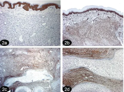
[image:3.612.113.497.81.413.2]CD44
CD44v6 splice variant did not stain any of the
tumors examined although epidermal positivity
was seen (Figure 2a).
Eight of 11 (73%) of locally infiltrative
cutane-ous neurofibroma were diffusely positive for
CD44, both in the tumor cells and the overlying
epidermis (the latter providing an internal
posi-tive control). The remaining 3 cases (27%) were
only focally positive within the tumor cells.
Wide-spread positivity was seen in 13 of 16 (81%) of
MPNST examined. The remaining 3 cases (19%)
were either only focally positive or negative.
Thus CD44 expression could not be correlated
with the benign or malignant status of the
tu-mors.
The 17 (non-cutaneous) benign tumors
exam-ined, in general, showed minimal or only focal
immunoreactivity for CD44. However 3 of 5
plexiform neurofibroma and all cutaneous
tu-mors with a tumor nodule (n=5) showed an
un-usual pattern of immunoreactivity. Tumors that
were well-defined and nodular or appeared
“well encapsulated,” were minimally positive for
CD44. Subcutaneous tumors that were poorly
defined (Figure 2b) and even with a small focus
of tumor were strongly immunoreactive.
Plexi-form neurofibroma demonstrated a similar
pat-tern. The ‘nodular’ portions of the tumor were
often negative while the more cellular, loose,
infiltrative, “non-confined” portions of the
tu-mors tumor surrounding these nodules were
markedly positive (Figure 2c and d).
[image:4.612.99.515.82.391.2]Discussion
Malignant peripheral nerve sheath tumors
(MPNST) are aggressive soft tissue tumors that
occur either sporadically or in patients with
neu-rofibromatosis type 1. The malignant
transfor-mation of the benign neurofibroma to MPNST is
not completely understood at the molecular
level.
Not surprisingly, patients with distant
me-tastasis have a significantly worse outcome. In
several series, the estimated 10-year
disease-specific survival for patients presenting with
metastatic disease was approximately 8%
ver-sus 30% for patients presenting with primary
disease and 25 % for patients presenting with
recurrent disease, despite the use of
conven-tional therapy [24, 25]. This dismal prognosis
highlights the importance of identifying patients
with
neurofibroma
that may progress to MPNST
as well as those with localized MPNST who are
at high risk for metastatic development.
We
therefore investigated CD44 and p53
expres-sion in benign and malignant peripheral nerve
sheath tumors and found that malignant
trans-formation from neurofibroma to MPNST occurs
in part to the inactivation of p53 tumor
suppres-sor gene and upregulation of CD44 adhesion
molecule.
Mutations in the p53 tumor suppressor gene
appear to result in changes in the molecular
structure of the protein, such that there is
in-creased stability and higher steady-state level of
the protein. Thus overexpression of the protein
as detected by Immunohistochemistry,
corre-lates with mutations of the p53 gene. This
cor-relation has been demonstrated for a number of
tumors including breast, lung, ovary,
endo-metrium and gastrointestinal neoplasms
[26-39]. There is increasing evidence that
muta-tions in genes other than the NF1 gene
contrib-ute to the development of malignancies in
pa-tients with NF1 and p53 has been suggested to
participate in the malignant transformation of
tumors these patients [9-11, 13, 40]. There is
also evidence that increased p53
immunoreac-tivity may correlate adversely with prognosis
[41].
CD44 is a cell adhesion molecule that promotes
cell-extracellular matrix protein interactions.
Both CD44 and its splice variants have been
shown to play a role in both infiltration and
me-tastasis in various malignancies, including
those of the central nervous system [17, 18, 21
-23, 42-46]. The marked positivity for CD44
seen in most MPNST is in favor of a role for this
molecule in infiltration and possibly metastasis.
In our series we had a single MPNST metastatic
to the regional lymph nodes. This tumor was
intensely CD44 immunoreactive, with positive
staining of almost every tumor cell. Additionally,
the pattern of immunoreactivity in locally
infiltra-tive cutaneous neurofibroma which
histologi-cally have very poorly defined margins,
com-bined with the immunoreactivity seen in
“nonencapsulated” areas of plexiform and
cuta-neous tumors further supports this role in
infil-tration and progression. In summary CD44
im-munoreactivity is most intense in infiltrative and
nonencapsulated tumors, irrespective of benign
or malignant status.
In conclusion the present study defines a
corre-lation between p53 overexpression and
malig-nancy in NF1. A marked increase in the nuclear
expression of mutant p53 in MPNST was seen
in 12/16 cases (75%). Additionally, p53 positive
nuclei were also seen in some benign
neurofi-broma, however, in significantly lower numbers.
These findings support a role for p53 mutations
in the malignant transformation of benign NF1
tumors, although other molecular factors are
also involved in this process.
Additionally the study describes the
immunoex-pression pattern of CD44, in that within the
con-fines of encapsulated tumors expression is
lim-ited, while in poorly circumscribed neurofibroma
CD44 expression is upregulated. CD44 was also
strongly positive in invasive MPNST. This is
thought to reflect the interaction of CD44+
tu-mor cells with extracellular matrix, hence
facili-tating infiltrative behavior.
Please address correspondence to: Amyn M. Rojiani MD, Department of Pathology and Cell Biology, 12901 Bruce B Downs Blvd. MDC 11, Tampa, FL 33610, USA. Tel: (813) 8750, Fax: (813) 974-5536, E-mail: [email protected]
References
[1] Louis DN, Ohgaki H, Wiestler OD and Cavenee WK. WHO Classification of Tumours of the Cen-tral Nervous System. 2007;
[2] Baser ME, Friedman JM, Wallace AJ, Ramsden RT, Joe H and Evans DG. Evaluation of clinical diagnostic criteria for neurofibromatosis 2. Neu-rology 2002; 59: 1759-1765.
nerve sheath tumours in neurofibromatosis 1. J Med Genet 2002; 39: 311-314.
[4] Szudek J, Evans DG and Friedman JM. Patterns of associations of clinical features in neurofibro-matosis 1 (NF1). Hum Genet 2003; 112: 289-297.
[5] Ducatman BS, Scheithauer BW, Piepgras DG, Reiman HM and Ilstrup DM. Malignant periph-eral nerve sheath tumors. A clinicopathologic study of 120 cases. Cancer 1986; 57: 2006-2021.
[6] Wong WW, Hirose T, Scheithauer BW, Schild SE and Gunderson LL. Malignant peripheral nerve sheath tumor: analysis of treatment outcome. Int J Radiat Oncol Biol Phys 1998; 42: 351-360. [7] Nigro JM, Baker SJ, Preisinger AC, Jessup JM,
Hostetter R, Cleary K, Bigner SH, Davidson N, Baylin S, Devilee P and et al. Mutations in the p53 gene occur in diverse human tumour types. Nature 1989; 342: 705-708.
[8] Porter PL, Gown AM, Kramp SG and Coltrera MD. Widespread p53 overexpression in human malig-nant tumors. An immunohistochemical study using methacarn-fixed, embedded tissue. Am J Pathol 1992; 140: 145-153.
[9] Agesen TH, Florenes VA, Molenaar WM, Lind GE, Berner JM, Plaat BE, Komdeur R, Myklebost O, van den Berg E and Lothe RA. Expression pat-terns of cell cycle components in sporadic and neurofibromatosis type 1-related malignant pe-ripheral nerve sheath tumors. J Neuropathol Exp Neurol 2005; 64: 74-81.
[10] Birindelli S, Perrone F, Oggionni M, Lavarino C, Pasini B, Vergani B, Ranzani GN, Pierotti MA and Pilotti S. Rb and TP53 pathway alterations in sporadic and NF1-related malignant peripheral nerve sheath tumors. Lab Invest 2001; 81: 833-844.
[11] Carroll SL and Stonecypher MS. Tumor suppres-sor mutations and growth factor signaling in the pathogenesis of NF1-associated peripheral nerve sheath tumors: II. The role of dysregulated growth factor signaling. J Neuropathol Exp Neu-rol 2005; 64: 1-9.
[12] Halling KC, Scheithauer BW, Halling AC, Nasci-mento AG, Ziesmer SC, Roche PC and Wollan PC. p53 expression in neurofibroma and malignant peripheral nerve sheath tumor. An immunohisto-chemical study of sporadic and NF1-associated tumors. Am J Clin Pathol 1996; 106: 282-288. [13] Subramanian S, Thayanithy V, West RB, Lee CH,
Beck AH, Zhu S, Downs-Kelly E, Montgomery K, Goldblum JR, Hogendoorn PC, Corless CL, Oliveira AM, Dry SM, Nielsen TO, Rubin BP, Fletcher JA, Fletcher CD and van de Rijn M. Ge-nome-wide transcriptome analyses reveal p53 inactivation mediated loss of miR-34a expres-sion in malignant peripheral nerve sheath tu-mours. J Pathol 220: 58-70.
[14] Koga T, Iwasaki H, Ishiguro M, Matsuzaki A and Kikuchi M. Frequent genomic imbalances in chromosomes 17, 19, and 22q in peripheral
nerve sheath tumours detected by comparative genomic hybridization analysis. J Pathol 2002; 197: 98-107.
[15] Legius E, Dierick H, Wu R, Hall BK, Marynen P, Cassiman JJ and Glover TW. TP53 mutations are frequent in malignant NF1 tumors. Genes Chro-mosomes Cancer 1994; 10: 250-255.
[16] Menon AG, Anderson KM, Riccardi VM, Chung RY, Whaley JM, Yandell DW, Farmer GE, Freiman RN, Lee JK, Li FP and et al. Chromosome 17p deletions and p53 gene mutations associated with the formation of malignant neurofibrosarco-mas in von Recklinghausen neurofibromatosis. Proc Natl Acad Sci U S A 1990; 87: 5435-5439. [17] Su W, Gutmann DH, Perry A, Abounader R,
Laterra J and Sherman LS. CD44-independent hepatocyte growth factor/c-Met autocrine loop promotes malignant peripheral nerve sheath tumor cell invasion in vitro. Glia 2004; 45: 297-306.
[18] Wiranowska M, Ladd S, Smith SR and Gottschall PE. CD44 adhesion molecule and neuro-glial proteoglycan NG2 as invasive markers of glioma. Brain Cell Biol 2006; 35: 159-172.
[19] Su W, Sin M, Darrow A and Sherman LS. Malig-nant peripheral nerve sheath tumor cell invasion is facilitated by Src and aberrant CD44 expres-sion. Glia 2003; 42: 350-358.
[20] Ponta H, Sherman L and Herrlich PA. CD44: from adhesion molecules to signalling regulators. Nat Rev Mol Cell Biol 2003; 4: 33-45.
[21] Baltuch GH, de Tribolet N and Van Meir EG. Ex-pression of the CD44 adhesion molecule in tu-mours of the central and peripheral nervous system. J Neurooncol 1995; 26: 191-198. [22] Li H, Liu J and Hofmann M. [CD44 expression
patterns in primary and secondary brain tumors]. Zhonghua Yi Xue Za Zhi 1995; 75: 525-528, 573.
[23] Sherman L, Jacoby LB, Lampe J, Pelton P, Aguzzi A, Herrlich P and Ponta H. CD44 expression is aberrant in benign Schwann cell tumors pos-sessing mutations in the neurofibromatosis type 2, but not type 1, gene. Cancer Res 1997; 57: 4889-4897.
[24] Zou C, Smith KD, Liu J, Lahat G, Myers S, Wang WL, Zhang W, McCutcheon IE, Slopis JM, Lazar AJ, Pollock RE and Lev D. Clinical, pathological, and molecular variables predictive of malignant peripheral nerve sheath tumor outcome. Ann Surg 2009; 249: 1014-1022.
[25] Carli M, Ferrari A, Mattke A, Zanetti I, Casanova M, Bisogno G, Cecchetto G, Alaggio R, De Sio L, Koscielniak E, Sotti G and Treuner J. Pediatric malignant peripheral nerve sheath tumor: the Italian and German soft tissue sarcoma coopera-tive group. J Clin Oncol 2005; 23: 8422-8430. [26] Ahmed AA, Etemadmoghadam D, Temple J,
Pathol
[27] Cai KQ, Wu H, Klein-Szanto AJ and Xu XX. Acqui-sition of a second mutation of the Tp53 alleles immediately precedes epithelial morphological transformation in ovarian tumorigenicity. Gyne-col OnGyne-col 2009; 114: 18-25.
[28] Chiba I, Takahashi T, Nau MM, D'Amico D, Curiel DT, Mitsudomi T, Buchhagen DL, Carbone D, Piantadosi S, Koga H and et al. Mutations in the p53 gene are frequent in primary, resected non-small cell lung cancer. Lung Cancer Study Group. Oncogene 1990; 5: 1603-1610.
[29] Geyer JT, Lopez-Garcia MA, Sanchez-Estevez C, Sarrio D, Moreno-Bueno G, Franceschetti I, Palacios J and Oliva E. Pathogenetic pathways in ovarian endometrioid adenocarcinoma: a mo-lecular study of 29 cases. Am J Surg Pathol 2009; 33: 1157-1163.
[30] Hirshfield KM, Rebbeck TR and Levine AJ. Germ-line mutations and polymorphisms in the origins of cancers in women. J Oncol 2010: 297671. [31] Hollstein M, Sidransky D, Vogelstein B and Harris
CC. p53 mutations in human cancers. Science 1991; 253: 49-53.
[32] Hollstein MC, Metcalf RA, Welsh JA, Montesano R and Harris CC. Frequent mutation of the p53 gene in human esophageal cancer. Proc Natl Acad Sci U S A 1990; 87: 9958-9961.
[33] Iggo R, Gatter K, Bartek J, Lane D and Harris AL. Increased expression of mutant forms of p53 oncogene in primary lung cancer. Lancet 1990; 335: 675-679.
[34] Kovach JS, McGovern RM, Cassady JD, Swanson SK, Wold LE, Vogelstein B and Sommer SS. Di-rect sequencing from touch preparations of hu-man carcinomas: analysis of p53 mutations in breast carcinomas. J Natl Cancer Inst 1991; 83: 1004-1009.
[35] Rodrigues NR, Rowan A, Smith ME, Kerr IB, Bod-mer WF, Gannon JV and Lane DP. p53 mutations in colorectal cancer. Proc Natl Acad Sci U S A 1990; 87: 7555-7559.
[36] Stanojevic Z, Djordjevic B, Pajovic SB, Zivanov-Curlis J and Najman S. Molecular pathogenesis of borderline and invasive ovarian tumors. J BUON 2009; 14: 7-18.
[37] Takahashi T, D'Amico D, Chiba I, Buchhagen DL and Minna JD. Identification of intronic point mutations as an alternative mechanism for p53 inactivation in lung cancer. J Clin Invest 1990; 86: 363-369.
[38] Thompson AM and Lane DP. p53 transcriptional pathways in breast cancer: the good, the bad and the complex. J Pathol 220: 401-403. [39] Varley JM, Brammar WJ, Lane DP, Swallow JE,
Dolan C and Walker RA. Loss of chromosome 17p13 sequences and mutation of p53 in hu-man breast carcinomas. Oncogene 1991; 6: 413-421.
[40] Holtkamp N, Atallah I, Okuducu AF, Mucha J, Hartmann C, Mautner VF, Friedrich RE, Mawrin C and von Deimling A. MMP-13 and p53 in the progression of malignant peripheral nerve sheath tumors. Neoplasia 2007; 9: 671-677. [41] Brekke HR, Kolberg M, Skotheim RI, Hall KS,
Bjerkehagen B, Risberg B, Domanski HA, Man-dahl N, Liestol K, Smeland S, Danielsen HE, Mertens F and Lothe RA. Identification of p53 as a strong predictor of survival for patients with malignant peripheral nerve sheath tumors. Neuro Oncol 2009; 11: 514-528.
[42] Heyse TJ, Malcherczyk D, Moll R, Timmesfeld N, Wapelhorst J, Fuchs-Winkelmann S, Paletta JR and Schofer MD. CD44: survival and metastasis in chondrosarcoma. Osteoarthritis Cartilage [43] Kunimura T, Yoshida T, Sugiyama T and
Moroho-shi T. The RelationMoroho-ships Between Loss of Stan-dard CD44 Expression and Lymph Node, Liver Metastasis in T3 Colorectal Carcinoma. J Gastro-intest Cancer 2009; 40: 115-118.
[44] Toole BP. Hyaluronan-CD44 Interactions in Can-cer: Paradoxes and Possibilities. Clin Cancer Res 2009; 15: 7462-7468.
[45] Wang SJ, Wong G, de Heer AM, Xia W and Bour-guignon LY. CD44 variant isoforms in head and neck squamous cell carcinoma progression. Laryngoscope 2009; 119: 1518-1530.