Int J Clin Exp Pathol 2013;6(12):2757-2764 www.ijcep.com /ISSN:1936-2625/IJCEP1310028
Original Article
Development of neural stem cells at different sites of
fetus brain of different gestational age
Xiaojuan Yin*, Lihua Li*, Xiaoying Zhang*, Yao Yang, Yannan Chai, Xiao Han, Zhichun Feng
Affiliated Bayi Children’s Hospital, Beijing Military Region General Hospital, No 5, Nan Mencang, Dongcheng Dis
-trict, Beijing 100700, China. *Equal contributors.
Received October 11, 2013; Accepted November 7, 2013; Epub November 15, 2013; Published December 1, 2013
Abstract: Objective: This study aimed to investigate the development of neural stem cells (NSCs) in fetal brain, which may provide experimental evidence for the clinical treatment of brain injury in children. Methods: A total of 60 fetus-es were collected after labor induction and divided into 6 groups according to the gfetus-estational age (16 w, 20 w, 24 w, 28 w, 32 w and 36 w; n=10 per group). The hippocampus, striatum, subventricular zone, frontal lobe, temporal lobe, occipital lobe and parietal lobe were harvested. In situ hybridization, immunohistochemistry and light microscopy
were done to determine the morphology and quantity of NSCs. Results: NSCs were identified in the brain of fetuses
with different gestational age. NSCs were round, oval, spindle-shaped, starlike, triangular or polygonal. NSC colony was also observed with symmetrical or asymmetrical division. Single NSC, group-like NSCs and cluster-like NSCs were found in the different sites of fetal brain, and NCSs interacted with each other via synapses. However, the dis-tribution, morphology, growth and quantity of NSCs were different in the brain of fetuses with different gestational age. The number of NSCs reduced with the increase in gestational age, but they were always observed. Conclusion: The morphology of NSCs in fetal brain is variable and they are widely distributed in the hippocampus, subventricular zone, striatum and cortex. The number of NSCs reduced with the increase of gestational age.
Keywords: Human, fetal brain, nestin
Introduction
Neural stem cells (NSCs) are multipotent cells that can give rise to various cell types in the central nervous system (CNS), including neu-rons, astrocytes, and oligodendrocytes [1]. NSCs can be generated from either the fetus, the adult, embryonic stem cells (ESCs) or induced pluripotent stem cells (iPSCs) [2]. In adult mammals, neurogenesis is limited to two brain regions; the subventricular zone (SVZ) by the lateral ventricles and the subgranular zone (SGZ) of the dentate gyrus in the hippocampus. The microenvironment of SVZ and SGZ is ben-eficial for the survival of NSCs [3-6]. To date, NSCs have been identified in the cortex, hippo-campus, striatum, olfactory bulb, and tissues along the cerebral ventricle (lateral ventricle, third ventricle and fourth ventricle), diencepha-lon, midbrain, cerebellum, spinal cord and reti-na [7, 8].
NSCs from specific brain region and those from
confounding cells in transplanted NSCs and the tumorigenesis and immune rejection after NSC transplantation. In addition, in diseases with diffused lesions in nervous system, cell trans-plantation is usually performed repeatedly, and thus, the therapeutic efficacy is often poor. On the basis of above findings, auto-repair of ner-vous system seems to be more feasible [10, 11]. Hypoxic-ischemic brain damage (HIBD) is a disease of nervous system with multiple lesions. Neonates are a continuation of fetal life. We speculate that NSCs are more widely distributed in neonates than in adults. Thus, to induce the proliferation and differentiation of endogenous NSCs has better prospective in the treatment of HIBD in neonates.
This study aimed to investigate the distribution, morphology, growth and quantity of NSCs in fetal brain. Our findings may provide an experi-mental and theoretical basis for in vivo and in vitro regulation of proliferation and differentia-tion of NSCs and the clinical treatment of dege- nerative disease of nervous system with NSCs.
Materials and methods
Samples
A total of 60 fetuses were collected after labor induction and divided into 6 groups according
to the gestational ages (16 w, 20 w, 24 w, 28 w, 32 w and 36 w; n=10 per group). The hippo-campus, striatum, subventricular zone, frontal lobe, temporal lobe, occipital lobe and parietal lobe were harvested from each fetus. Prenatal ultrasonography showed refractory congenital heart diseases in these fetuses and induced labor was required by the mother and her rela-tives. These refractory congenital heart diseas-es included persistent truncus arteriosus (PTA), total abnormal pulmonary venous drainage (TAPVD), critical tetralogy of Fallot (TOF), critical aortic stenosis and critical pulmonary stenosis. Physical examination showed the mothers were healthy. Informed consent was obtained from the mothers and this study protocol was approved by the Ethics Committee of General Hospital of Beijing Military Region.
Main reagents
[image:2.612.90.526.72.290.2]Development of neural stem cells in fetus brain
3’) were used in the present study. Other reagents were domestic and analytically pure. Immunohistochemistry for Nestin at 7 sites of fetal brain
Immunohistochemistry was done with SABC method. Tissue was harvested from embryos, fixed in 4% paraformaldehyde (PFA), equilibrat-ed in 30% sucrose at 4°C and frozen in Tissue-Tek OCT. Cryosections (20 μm) were blocked with 10% goat serum in 0.1% Triton X-100 at room temperature and primary antibodies (rab-bit anti-human nestin polyclonal antibody) was applied overnight at 4°C. Secondary antibodies included biotin conjugated goat anti-rabbit IgG. Incubation was done at 37°C for 30 min. After addition of SABC, incubation was done at 37°C for 30 min, and visualization was done with DAB for 5 min, followed by counterstaining with hematoxylin. After washing in water for 1 min, observation was done under a light microscope and representative photographs were cap-tured. In negative control group, the primary antibody was replaced with 0.01 mol/L KPBS and the secondary antibody with normal goat serum.
In situ hybridization for Nestin mRNA at 7 sites of fetal brain
Tissue was harvested from embryos, fixed in 4% paraformaldehyde (PFA), equilibrated in
30% sucrose at 4°C and frozen in Tissue-Tek OCT. Cryosections (15 μm) were blocked with 10% goat serum in 0.1% Triton X-100 at room temperature. Digestion was done with pepsin in 3% citric acid at 37°C for 100 s, followed by fixation in 1% paraformaldehyde in /0.1 M PBS (pH 7.2-7.6) at room temperature for 10 min. After addition of prehybridization solution, pre-hybridization was done at 42°C for 4 h. The digoxin labeled probe in in situ hybridization solution was added followed by hybridization at 42°C for 20 h. After washing in series of SSC solution (2×, 0.5×, 0.2×) at 37°C, blocking solu-tion was added followed by addisolu-tion of biotinyl-ated anti-digoxin antibody and subsequent incubation at 37°C for 90 min. Incubation was performed with SABC at 37°C for 30 min and then with biotinylated peroxidase at 37°C for 30 min. Visualization was done with DAB, fol-lowed by counterstaining with hematoxylin for 1 min. After washing in water for 1 min, observa-tion was done under a microscope and repre-sentative photographs were captured. In nega-tive control group, probes or antibody were not used.
Quantification and qualification of NSCs in fetal brain
[image:3.612.93.523.71.264.2]were included in one field. At different sites, observation was done in 3 sections, and 10 fields were randomly selected from each sec-tion. Thus, a total of 300 fields were used for the detection of total cells and positive cells in each group. The proportion of positive cells to total cells was calculated at different sites in each group, which was used as the proportion of Nestin positive cells at different sites. Statistical analysis
Statistical analysis was done with SPSS version 13.0. The proportion of NSCs positive for Nestin protein and mRNA at different sites was com-pared with chi square test. A value of P<0.05 was considered statistically significant.
Results
NSCs morphology from fetus brain
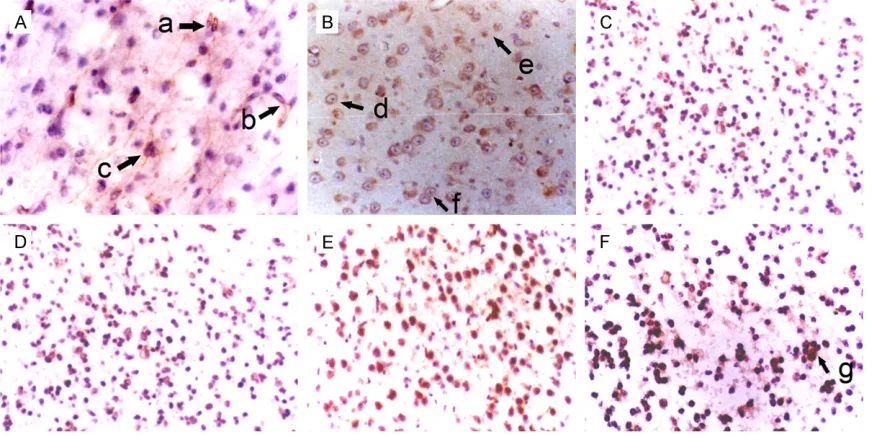
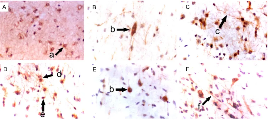
In the hippocampus, NSCs were round, oval, tri-angular or star-like. Most of NSCs were round and single cell was observed. Symmetrical divi-sion, colony formed by 2-8 cells and cluster-like distribution were found. One NSC usually had 0-3 processes. Some NSCs interact with other neurons via synapses (Figure 1). In the subven-tricular zone, NSCs were star-like, round, oval or spindle-shaped, and most of them were star-like. NSCs had scattered distributed in astro-cytes and single cell was frequently observed.
Most NSCs had 1-4 nucleoli. Symmetrical and asymmetrical division was found in these NSCs which had 0-2 processes. Some NSCs interact with other neurons via synapses (Figure 2). In the striatum, NSCs were round, oval, polygonal or pleomorphic, and most cells were oval. The nucleus was vacuole-like and had 1-4 nucleoli. One NSC had 0-3 processes and single NSC was found among neurons. Symmetrical divi-sion was occadivi-sionally observed in NSCs with colony-like distribution (Figure 3). In the cortex, NSCs were round or oval, or occasionally polyg-onal. Most NSCs were round and had 1-5 nucle-oli. NSCs were group-like, had colony-like growth and presented with regional distribution (Figure 4).
Proportion of nestin positive NSCs at different sites of fetal brain
In the brain of fetuses with specific gestational age, the proportion of nestin positive cells reduced in the following order: hippocampus, subventricular zone, striatum, frontal lobe, tem-poral lobe, parietal lobe and occipital lobe (P<0.01). At the same site, the proportion of nestin positive cells reduced with the increase in gestational age (P<0.01) (Table 1, Figure 5). Nestin mRNA positive NSCs in fetal brain
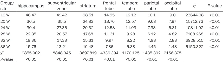
[image:4.612.91.523.74.274.2]Development of neural stem cells in fetus brain
NSCs reduced in the following order: hippocam-pus, subventricular zone, striatum, frontal lobe, temporal lobe, parietal lobe and occipital lobe (P<0.01). At the same site, the proportion of nestin mRNA positive cells reduced with the increase in gestational age (P<0.01) (Table 2,
Figure 6).
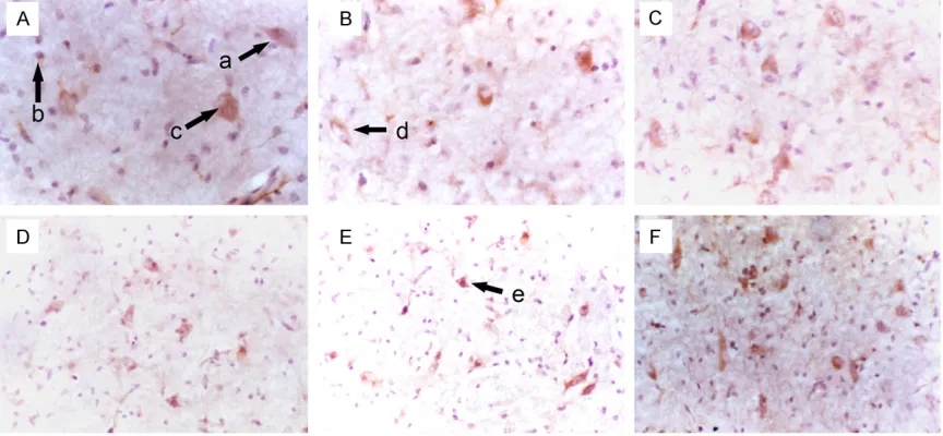
[image:5.612.90.526.72.201.2] [image:5.612.91.379.279.598.2]neurons completes, the nestin expression stops. In the CNS of mammalians, change in intermediate filament protein occurs at the key stage of cell differentiation. In the CNS, NSCs express nestin, and the nestin expression reduces rapidly in the order from proliferating NSCs to neurons. In the nestin gene, an Figure 4. Morphology of NSCs in the cortex of fetuses with different gestational age (SABC, DAB staining, ×200). A: Frontal lobe; B: Temporal lobe; C: Parietal lobe; D: Occipital lobe; a: Group-like growth of NSCs; b: Round NSCs; c: Oval NSCs; d: Colony-like growth of NSCs.
Table 1. Proportion of Nestin positive cells at different sites of fetal brain (%) Group/
site hippocampus subventricular zone striatum frontal lobe temporal lobe parietal lobe occipital lobe χ2 P-value
16 w 37.5 31.35 26.93 15.59 12.25 10.09 8.31 13219.62 <0.01
20 w 31.4 28.31 23.37 13.48 10.54 7.73 6.52 10245.49 <0.01
24 w 24.8 22.46 18.70 11.62 9.57 5.42 3.87 9210.018 <0.01
28 w 20.2 18.92 15.98 10.52 8.73 4.13 2.58 8014.862 <0.01
32 w 17.09 14.97 12.87 9.87 7.53 3.46 1.65 6830.853 <0.01
36 w 12.38 10.88 8.57 6.68 4.16 1.98 0.96 5693.014 <0.01
χ2 6253.974 4644.602 4050.503 1265.152 1448.848 2275.611 2869.747
P-value <0.01 <0.01 <0.01 <0.01 <0.01 <0.01 <0.01
Discussion
enhancer is found in the intron 2 and can act on NSCs of CNS. This suggests that there is a transcriptional regulation of NSCs in the CNS. Of note, this enhancer in the intron has no influ-ence on peripheral nervous system [12, 13]. The introns of human nestin gene are geneti-cally protected, and 2 of 3 introns are shared with neurofilaments. Before the reclassification of neurofilaments, nestin and neurofilament share a precursor [14]. To date, nestin has been widely used as a marker for NSCs in in
vivo and in vitro studies and employed to iden-tify NSCs from cells in nervous system [15]. In the present study, in situ hybridization and immunohistochemistry were employed to detect the nestin mRNA and protein, respec-tively, at 7 sites of human fetal brain, and NSCs positive for nestin protein and mRNA were observed in the fetal brain. In addition, the pro-tein and mRNA expression of nestin showed similar trend in the fetal brain. These findings indicate that nestin is a favorable antigen used to identify NSCs.
gyrus into specific layer, and these nerve fibers are also NSCs [16]. NSCs are a group of cells with self-renewal and differentiation potential, and in the non-terminal state of differentiation. NSCs can symmetrically and asymmetrically divide into new NSCs or differentiate into small daughter cells which finally differentiate into three types of cells: neurons, astrocytes and oligodendrocytes [17]. In previous studies, NSCs are regarded to origin from the subven-tricular zone and dentate gyrus [18]. In depth studies reveal that NSCs also exist in the olfac-tory bulb, ependyma, and subependymal zone of rats, and the amount of NSCs in the brain reduces over gestational age [8]. Subsequently, NSCs are also identified in the hippocampus, cortex, striatum and subependymal zone of humans [19]. However, the alteration of NSCs during the development of embryonic brain has never comprehensively studied.
To date, no studies have been conducted to investigate the changes in the distribution,
[image:6.612.92.521.86.209.2]P-value <0.01 <0.01 <0.01 <0.01 <0.01 <0.01 <0.01
Figure 6. Proportion of Nestin mRNA positive NSCs at different sites of fetal brain.
[image:6.612.97.389.101.410.2]Development of neural stem cells in fetus brain
morphology, growth and amount of NSCs over gestational age. In the present study, in situ hybridization and immunohistochemistry were employed to detect the mRNA and protein expression of nestin (a marker of NSCs) at 7 sites of the brain of fetuses with different ges-tational ages. Results demonstrated NSCs were found in the brain of fetuses with different gestational age, but the distribution, morphol-ogy, growth and amount of NSCs varied among different sites and among fetuses with differ-ent gestational ages. Our results showed NSCs were round, oval, spindle-shaped, star-like, tri-angular or polygonal and had small or large size. NSCs with symmetrical or asymmetrical division were also observed, and 2-8 NSCs could form a colony and presented with group-like or cluster-group-like growth. The group-group-like or cluster-like NSCs formed NSC colonies or the center for the generation of new NSCs. Some NSCs had processes extending to surrounding cells and interacted with other cells via synaps-es. The amount of NSCs reduced with the increase in gestational age. The above charac-teristics of NSCs have never been reported pre-viously. One may imagine that any thing has its special morphology and functions, and the morphology and structure of cells determine their biological functions. The morphology and growth of NSCs in fetal brain indicate that NSCs with different morphologies, at different sites of the brain or at different gestational age may have distinct biological functions. Our results also revealed that the amount of NSCs at a spe-cific site reduced with the increase in gestation-al age; in the brain of fetus with a specific ges-tational age, the amount of NSCs reduced in the following order: hippocampus, subventricu-lar zone, striatum, frontal lobe, temporal lobe, parietal lobe, and occipital lobe, and significant difference was observed between any two sites. These findings indicate that the amount of NSCs in fetal brain is larger than that in adult brain. Neonates are a continuation of fetal life and may develop further into children and final-ly into adults. Once focal ischemia/hypoxia is present in the brain, NSCs at other sites may proliferate and differentiate to repair this injury. Thus, our results not only uncovered the devel-opment of NSCs at different sites of the brain of fetuses with different gestational age, but vali-dated our hypothesis that to induce the prolif-eration and differentiation of endogenous NSCs is better for the treatment of HIBD in neonates.
Although great progresses have been achieved in studies on NSCs, some problems still exist in the NSC transplantation, such as immune rejection, reconstruction of morphology and cell circuit, functional integration and evalua-tion of short-term and long-term prognosis. Thus, it has a long way before the transplanta-tion of NSCs is used in clinical practice. Directional induction of NSC differentiation is crucial for the clinical application of NSCs. Different types of neural cells might be required for the repair of lesions of different types or at different sites in nervous system. Although in vitro experiments have confirmed that some substances may induce the directional differ-entiation of NSCs, the differdiffer-entiation of NSCs might vary in distinct environments. Thus, it is difficult to collect NSCs with the same ancestry, homogeneity and identical differentiation level. In addition, the brain is an integrant that can auto-regulate according to the pathophysiologi-cal conditions, which can not be mimicked in vitro due to its complexity, and the direction of NSC differentiation can not be accurately deter-mined. In addition, whether transplanted NSCs induce immune rejection is still controversial. Thus, the in vivo survival and proliferation of NSCs are required to further investigated. NSCs exist in the whole life of mammalians. They may generate precursor cells and directionally dif-ferentiate into cells to maintain the homeosta-sis. Thus, NSCs may self-renew and stay in a quiescent state. Transplantation of NSCs is a new strategy for the treatment of brain injury, and exert therapeutic effects via multiple mechanisms, such as neural replacement, neu-roprotection, inflammation and apoptosis [20]. It is accepted that NSCs can be used to treat nervous system injury and degenerative dis-eases. However, exogenous NSCs have limita-tions, and to induce the endogenous NSCs to repair brain injury may be a better strategy. In HIBD of neonates, to induce the proliferation and differentiation of endogenous NSCs may be a preferred strategy. Of note, the time win-dow for the induction of NSC proliferation and differentiation should be paid attention to.
Conclusion
Address correspondence to: Dr. Zhichun Feng or
Xiaojuan Yin, Affiliated Bayi Children’s Hospital,
Beijing Military Region General Hospital, No 5, Nan Mencang, Dongcheng District, Beijing 100700, P. R. China. E-mail: [email protected] (Zhichun Feng); [email protected] (Xiaojuan Yin)
References
[1] Alvarez-Buylla A, Seri B and Doetsch F.
Identification of neural stem cells in the adult
vertebrate brain. Brain Res Bull 2002; 57: 751-758.
[2] Kim HJ and Jin CY. Stem Cells in Drug Screen-ing for Neurodegenerative Disease. Korean J Physiol Pharmacol 2012; 16: 1-9.
[3] Zhao C, Deng W and Gage FH. Mechanisms and functional implications of adult neurogen-esis. Cell 2008; 132: 645-660.
[4] Carreira BP, Carvalho CM and Araújo IM. Regu-lation of injury-induced neurogenesis by nitric oxide. Stem Cells Int 2012; 2012: 895659. [5] Jin K, Minami M, Lan JQ, Mao XO, Batteur S,
Simon RP and Greenberg DA. Neurogenesis in dentate subgranular zone and rostral subven-tricular zone after focal cerebral ischemia in the rat. Proc Natl Acad Sci U S A 2001; 98: 4710-4715.
[6] Akerblom M, Sachdeva R and Jakobsson J. Functional Studies of microRNAs in Neural Stem Cells: Problems and Perspectives. Front Neurosci 2012; 6: 14.
[7] Doetsch F, Caille I, Lim DA, Garcia-Verdugo JM and Alvarez-Buylla A. Subventricular zone as-trocytes are neural stem cells in the adult mammalian brain. Cell 1999; 97: 703-716. [8] Tropepe V, Coles BL, Chiasson BJ, Horsford DJ,
Elia AJ, McInnes RR and van der Kooy D. Reti-nal stem cells in the adult mammalian eye. Sci-ence 2000; 287: 2032-2036.
[9] Gincberg G, Arien-Zakay H, Lazarovici P and Le-lkes PI. Neural stem cells: therapeutic poten-tial for neurodegenerative diseases. Br Med Bull 2012; 104: 7-19.
transfected cells. Biochim Biophys Acta 2001; 1520: 251-254.
[13] Zimmerman L, Parr B, Lendahl U, Cunningham M, McKay R, Gavin B, Mann J, Vassileva G and McMahon A. Independent regulatory elements in the nestin gene direct transgene expression to neural stem cells or muscle precursors. Neuron 1994; 12: 11-24.
[14] Dahlstrand J, Zimmerman LB, McKay RD and Lendahl U. Characterization of the human nes-tin gene reveals a close evolutionary
relation-ship to neurofilaments. J Cell Sci 1992; 103:
589-597.
[15] Messam CA, Hou J and Major EO. Coexpres-sion of nestin in neural and glial cells in the
developing human CNS defined by a human-specific anti-nestin antibody. Exp Neurol 2000;
161: 585-596.
[16] Pincus DW, Goodman RR, Fraser RA, Neder-gaard M and Goldman SA. Neural stem and progenitor cells: a strategy for gene therapy and brain repair. Neurosurgery 1998; 42: 858-867; discussion 867-858.
[17] Cataudella T, Conti L and Cattaneo E. Neural stem and progenitor cells: choosing the right Shc. Prog Brain Res 2004; 146: 127-133. [18] Galli R, Gritti A, Bonfanti L and Vescovi AL.
Neu-ral stem cells: an overview. Circ Res 2003; 92: 598-608.
[19] Poltavtseva RA, Revishchin AV, Aleksandrova MA, Korochkin LI, Viktorov IV and Sukhikh GT. Neural stem and progenitor cells of human embryos and fetuses as a basis of biomedical new technologies. Ontogenez 2003; 34: 211-215.