INTRODUCTION
Electric fish detect, discriminate and perhaps recognize objects using active electroreception. These animals create an electric field by activating an electric organ (EO) (Bennett, 1971; Caputi, 2011). The characteristics of the field created by this discharge (EOD) depend on the degree of polarization of the elements of the scene – which in turn depend on objects’ impedance in relation to the conductivity of the water and the fish – as well as on the relative position of the elements, and the field geometry (Lissmann and Machin, 1958; Nelson and MacIver, 2006; Caputi et al., 1998; Budelli et al., 2002; Nelson and MacIver, 1999; Nelson, 2005; Chen et al., 2005; Caputi and Budelli, 2006; Snyder et al., 2007; Pereira and Caputi, 2010). Theoretical analyses suggest that objects with a conductance different from water are polarized by electric fields in such a way that they behave as a virtual electric source (Lissmann and Machin, 1958). Because of this theoretical argument they can be replaced by an electric source having an electromotive force whose time course is a function of the local field in the absence of the object. This source has been called the ‘object’s stamp in a given scene’ (Caputi et al., 2008; Pereira and Caputi, 2010). The corollary of the concept of ‘stamp’ is that the presence of every polarized object may also polarize the rest of the objects in the scene with a ponderance inversely dependent on a power function of the distance between the objects. This concept of electric image formation provides some suggestions about how object images might be modified by the presence of other objects in the scene (Sicardi et al., 2000; Rother et al., 2003; Chen et al., 2005; Caputi and Budelli, 2006; Nelson and MacIver, 2006) and how clues that objects offer to an active electrolocating agent vary
with context (Pereira et al., 2007; Pereira and Caputi, 2010; Sanguinetti et al., 2011).
The stamp of one object depends on the sum of fields of two different kinds: primary fields directly created by the fish and secondary fields resulting from the polarization of the elements of the scene.
A minimal scene consists of the fish (i.e. the first object affecting the field) and a second object. Analysis of this type of scene show important effects of geometry and conductance of the fish’s body on the spatial profile and amplitude of the image (Migliaro et al., 2005). Theoretical predictions and experimental results indicate that the image of that second object is modified by the presence of a third object (besides the object generating the image and the fish’s body). Modeling analysis of this type of scene predicted that: (a) the presence of a third object in a scene may cause a change in the clues that another object offers to the agent (Rother et al., 2003; Chen et al., 2005) and (b) contextual objects may contribute with parallax references for object active electrosensing (Babineau et al., 2006; Babineau et al., 2007). However, these studies focused mainly on the side of the fish and not on the foveal region, where the electroreceptor density and central representation are more important (Castelló et al., 2000). In this region, experimental results indicate that a third object may affect the fish’s body stamp and also the stamp of the second object besides the fish’s body (as is the case of a fish inside a tube) (Pereira et al., 2005).
In this study, we examined the effects of the presence of a third object on the electric image of another placed at the foveal region inGymnotus omarorum (Richer de Forges et al., 2009), a pulse-emitting gymnotiform fish. The sensory meaning of The Journal of Experimental Biology 215, 1533-1541
© 2012. Published by The Company of Biologists Ltd doi:10.1242/jeb.067223
RESEARCH ARTICLE
Active electrolocation in pulse gymnotids: sensory consequences of objects
ʼ
mutual
polarization
Pedro A. Aguilera, Ana Carolina Pereira and Ángel A. Caputi*
Departamento de Neurociencias Integrativas y Computacionales, Instituto de Investigaciones Biológicas Clemente Estable, Av. Italia 3318, Montevideo, Uruguay
*Author for correspondence ([email protected])
Accepted 9 January 2012
SUMMARY
We examined non-linear effects of the presence of one object on the electric image of another placed at the foveal region in Gymnotus omarorum. The sensory consequences of object mutual polarization on electric images were also depicted using behavioral procedures. Image measurements show that objects whose electric image is not detectable may modify the electric image of another placed closer to the fish and suggest that detection range and discrimination parameters used for one object may be affected when the presence of others enriches the scene. Behavioral experiments confirm that these changes in object images resulting from mutual polarization may be exploited for improving perception. While conductive objects close to the skin allow the fish to detect other objects placed out of the active electrodetection range, non-conductive objects may hide objects that otherwise show clear electric images. This suggests that fish movements may orient the self-generated field to exploit object mutual polarization, increasing or decreasing the active electrolocation range. In addition, images of a nearby object may be modulated by the presence of another object placed outside the detection range and the corresponding behavioral responses suggest that a moving or impedance-changing context may modify a fishʼs discrimination abilities for closer objects.
these observations on electric images was also behaviorally explored using the novelty response. In a previous study we showed that the amplitude of the novelty response scales with the amplitude of the electric image and this allowed us to use it here for the assessment of the sensory effects of object interplay (Caputi et al., 2003). Physical measurements also showed that objects that otherwise are out of the detection range can be detected if another conductive object is intercalated between the distant object and the fish. In addition, non-conductive objects hide other objects that otherwise show clear electric images. Moreover, they show that distant objects out of the detection range may modify the electric image of an object introduced into the scene, closer to the fish. Behavioral experiments showed that G. omarorumsense these effects as a result of object interplay.
MATERIALS AND METHODS Animals
Experiments were performed in 15 fish (15–30cm total length) following the guidelines of the CHEA (Comisión Honoraria de Experimentación Animal, ordinance 4332-99, Universidad de la República Oriental del Uruguay). Experiments were approved by the Animal Ethics Committee of the Instituto de Investigaciones Biológicas Clemente Estable (protocol number 001/03/2011). Fish were captured at Laguna del Cisne (Maldonado, Uruguay), 1–4 months before the experiment, kept in individual aquaria under a natural light cycle and fed with insect larvae. In those experiments in which the fish could experience pain or discomfort, animals were anesthetized with pentobarbital (0.5–1mg, i.m.), repeated on demand until we reached and maintained the EOD rate below 10Hz at 20°C and a slow but stable respiration. At the end of these experiments animals were killed by pentobarbital (10mg, i.m.).
Image measurements
The experimental strategy was to compare the images of one object in either the presence or the absence of another similar nearby object. Images were measured in anesthetized fish with the fish’s body straight, halfway between the bottom and the water surface in a 33⫻48cm tank filled with 100±10Scm–1water to a depth of 10cm.
In order to keep the fish’s body straight, we implanted a fine cotton thread along the midline between the muscular masses and above the spine using a long steel needle. This procedure was conducted when the fish reached the anesthetic plane where the EOD rate was unresponsive to visual, vibratory, electric or nociceptive stimuli. The thread came out of the body just behind the occiput and at the limit between the caudal and the center-caudal quarter of the fish length. The ends of the thread were firmly attached to two wooden poles vertically attached to an iron framework. The diameter of the rostral pole (1cm) was reduced to 3mm below the level of the water. The diameter of the caudal pole was 1cm. The rostral pole was placed just above the occiput (Fig.1A).
A nichrome wire (100m diameter) coated except on a knot tied at its center was implanted subcutaneously. The wire entered through one side below the mandible and left symmetrically through the other side. After wire insertion we verified that the knot was at the center of the mandible; the two wire tips were bent up and twisted together above the head forming a ring. The two ends of this reference electrode were connected together to one input of a differential amplifier. The exploring electrode (a 150m enamel-coated tungsten electrode with a blunted bare tip) was placed on the skin at different points along a curve on a horizontal plane following the shape of the mandible. This electrode was connected
to the other input of the differential amplifier (Caputi et al., 2011). We considered that the drop of voltage between the exploring and reference electrode was a good estimator of the transcutaneous voltage at the fovea.
Recording of electric images was achieved by placing the objects at steady positions and moving the exploring electrode along a series of points on the skin at the foveal region with a resolution of 100–200m. The tip of the electrode was moved step by step along a series of previously defined points using a computer-controlled X–Y plotter (HP 7015A, Hewlett-Packard Corp.). Objects were similar to those previously described for analyzing fish discrimination ability (von der Emde, 1990; von der Emde and Ronacher, 1994; Caputi et al., 2003; Aguilera and Caputi, 2003; Engelmann et al., 2009). We chose this kind of object because they are also suitable for discrimination studies (Caputi et al., 2003). They consist of plastic cylinders having carbon plugs at each end. The carbon bases were connected through wires coming out of the tank and connected outside to a desired resistor. As conductive bases can be connected through an external impedance, objects have anisotropic impedances (infinite lateral, controllable longitudinal). The Appendix provides a more complete justification of object selection.
The electric image was estimated as follows. First we calculated the mean value of all recordings performed at each recording point and generated an average curve. Second, we calculated the electric image as the convolution of the average curve with a median filter (5 points). Thus, each point of the electric image profile was estimated as the median of the surrounding five points of the average curve.
We performed three series of experiments. In the first and second series we used two identical objects (each 7mm diameter, 11mm height; as a reference a similar water cylinder has 35S of longitudinal conductance) aligned along the midline, one facing the skin 1mm away and the other placed with its front end 13mm away (1mm apart from the other). In the third series of experiments we used two thinner objects (4mm diameter) of two different lengths (7 and 14mm). Cylinders were aligned, separated by 1mm, following a field line (determined for each object in the absence of the other as the angle that yields the maximum drop of voltage between the bases). Water cylinders equivalent to 4mm objects had different longitudinal resistance (7 and 14mm lengths correspond to 22 and 11S, respectively).
In the first series of experiments the bases of each object were connected through resistors of 2.5Mor 1k. We used the short cuts ‘open circuit’ for 2.5Mand ‘short circuit’ for 1kbecause these resistors were resistive or conductive enough to provoke the same electric image as an ‘open’ or a ‘short’ circuit. The possible combinations of these loads resulted in eight possible experiments, four performed with both objects (open–open, open–short, short–open, short–short) and four control experiments in the absence of one of the objects.
In the second and third series of experiments, connections between the bases of one of the objects were made by either 2.5M
or 1kresistors while the resistance between the bases of the other object was set to several possible values (ranging between 0.3 and 2500k).
Signals were digitized at a minimum of 20kHz per channel and amplified enough to have at least 12bit resolution (AM
systems-1800, 10–10,000Hz band pass; Carlsborg, WA, USA). Data
electrodes placed on the main axis of the fish at opposite ends of the tank; (b) the local field (LEOD); and (c) the X- and Y-positions of the image-measuring active electrode in the horizontal plane. We made 3–5 runs of the electrode along the same trajectory. For each of these runs, we obtained several (between 5 and 10) LEODs per recording position. LEOD waveforms were time averaged with
reference to a fixed moment of the head to tail EOD. Thus, for each position we got 3–5 averaged LEOD waveforms.
Behavioral experiments
Novelty responses – characterized as transient acceleration just after the presentation of a new object or a change in the object impedance – proved to be a good index of object detection (Caputi et al., 2003). In addition, we have shown that the maximal reduction in EOD interval (i.e. the amplitude of the novelty response) is a good indicator of a change in the local image (Caputi et al., 2003; Pereira et al., 2007). Here, we used novelty responses as indicators of detection of an otherwise non-detected object (response vsnon-response) and also to evaluate whether the image of one object was changed by the presence of the other (comparing the amplitude of the response).
Fish were kept in a tank containing water at 100±10Scm–1
(33⫻48cm tank filled to a depth of 10cm), restrained inside a nylon mesh, tightly attached to wood poles fixed to the smallest opposite walls of the tank. Two electrodes placed near the wooden supporting poles and connected to a differential amplifier (high input impedance,
⫻100, 10Hz to 10kHz band-pass range) were used to record the head to tail field generated by the EOD. In five fish we used two pairs of exploring objects consisting of plastic cylinders having carbon conductive bases as described above. Both pairs of objects were the same as those used for image measurements. In two other fish we explored the effects of a cube (either copper or plastic, either behind or intercalated between the object and the fish’s skin, 9mm side) on the novelty response evoked by a step from open to short circuit of the connection between the bases of one of the cylinders described above (7mm diameter ⫻11mm length). Objects were manipulated by attached wood handles (1mm width) that when soaked with water had negligible images (Gómez et al., 2004). In these experiments different longitudinal resistances were used.
Novelty responses were elicited by a step reduction of the cylinder longitudinal resistance over a period of 2s every 30s. The same fish was exposed from a minimum of 10 trials under the same conditions to a maximum of 30 trials depending on the variability of the responses. The 28s interval between changes in object impedance was enough to avoid habituation as shown by testing the lack of difference of the responses between the first and last
B
450300
200
Control Object
Field recording amplifier
Computer-controlled switches
Variable loading resistor
Variable loading resistor
A
Field recording amplifier
1%
Novelty response
–10 –5 0 5 10
∆
LEOD (
μ
V r
.m.s. @ 10 ms)
160
80
0
–10 –5 0 5 10
Distance along recording line (mm)
LEOD (
μ
V r
.m.s. @ 10 ms)
Variable loading resistor
Computer-controlled switches
Variable loading resistor
Fig.1. Measurement of the electric image and behavioral experimental set
up. (A)The transcutaneous field was measured between an internal
electrode placed within the chin and an external electrode sequentially placed at different points along a line on the skin surface of the jaw (top panel). The electric image was calculated in the following way. First, we performed measurements in the absence and then in the presence of the object, 3–5 times each. Second, we calculated the root mean square (r.m.s.) value of the local electric organ discharge (LEOD) recorded at each point and averaged the r.m.s. LEOD for each point. Next, we smoothed the average curve using a 5-point median filter (middle panel). Finally, the electric image profile was estimated by subtracting the control smoothed r.m.s. LEOD curve from the smoothed r.m.s. LEOD curve in the presence
of the object (bottom panel). (B)The fish was held inside a mesh and the
resistance of the objects was modified using a computer-driven set of switches (top panel). The amplitude of each novelty response was
calculated as the difference between the basal interval (I0), defined as the
mean of the 5 intervals immediately previous to the resistance step, and
the minimum interval value (Im, in general the 2nd or the 3rd after the
beginning of the resistance step), divided by the basal interval. We
compared the amplitude of the novelty responses for each fish using a t
trial of the series (difference between medians different from zero, sign test, P<0.01, N10). The amplitude of each novelty response was calculated as the difference between the basal interval (I0),
defined as the mean of the five intervals immediately previous to the resistance step, and the minimum interval value (Im, in general
the 2nd or 3rd after the beginning of the resistance step), divided by the basal interval:
Amplitude of novelty response (I0– Im) / I0. (1)
We tested the hypothesis that the presence of a third object in a scene may cause a change in the amplitude of the novelty response in five ways: (1) by eliciting novelty responses in the presence and absence of a cube either with the cube between the test object and the fish (N2 copper cube, N2 plastic cube) or with the test object between the cube and the fish fovea; (2) by eliciting novelty responses by open to short changes in longitudinal conductance of one of the large objects (7mm diameter, 11mm length) in the presence or absence of a twin object aligned along the same field lines – we compared the mean amplitudes of the novelty responses evoked by maximal steps in resistance of the nearest object with the farthest object either in open or short circuit; (3) by studying the amplitude of the novelty response evoked by the change of the nearest object conductance from open circuit to a given predetermined value when the other object was either in conductive or non-conductive conditions; (4) by studying the amplitude of the novelty response evoked by the change in conductance from open to short circuit of a small object (4mm diameter and 7mm length) when the conductance of the farthest object (4mm diameter and 14mm length) was controlled (statistical parameters of the amplitudes of the evoked novelty responses were calculated); (5) by studying the amplitude of the novelty response evoked by the change in conductance from open to short circuit of the farthest object (4mm diameter and 14mm length) conductance when the conductance of the nearest object (4mm diameter and 11mm length) was controlled.
Statistical analysis of image differences
To validate the results across fish, we compared the peak amplitudes of the electric image in each recording condition using the sign test. We tested the following hypotheses: (a) images generated by the nearest conductive object in the presence of a farther away conductive object were smaller than or equal to the image of the nearest object alone; (b) images generated by the nearest conductive object in the presence of a farther away non-conductive object were larger than or equal to the image of the nearest object alone; (c) images generated by the nearest non-conductive object in the presence of a farther away object being either non-conductive or conductive were not equal to the image of the nearest object alone. In the first two cases, the results always opposed the hypothesis; thus, we estimated the probability of error as (1/2)5.
We validated the results of the behavioral analysis across fish in the following manner. First we tested the distribution of the novelty response for normality (Kolmogorov–Smirnoff test P<0.05). Second, we compared the amplitude of the novelty responses for each fish using a t-test for independent samples in each pair of conditions: (i) amplitude of the novelty response evoked by a maximum step in the nearest object with a 1000S conductive farthest object vsamplitude of the novelty response evoked by a maximum step in the nearest object with a 0.4S non-conductive farthest object; (ii) amplitude of the novelty response evoked by a maximum step in the farthest object with a 1000S conductive nearest object vsamplitude of the novelty response evoked by a maximum step in the farthest object with a 35S ‘water-conductive’
nearest object (nearest objects of 0.4S completely abolished novelty responses). Third, we tested the null hypothesis that the mean amplitudes of the novelty responses in cases where the context object was 1000S conductive in experiments of types i or ii were smaller than or equal to those in the other contexts, and calculated the type I error (per comparison) in each case. Then we evaluated the
family-wise error rate [1–(1–per comparison)], which was 0.03 and
0.04, respectively.
RESULTS
We studied the effect of the conductance of one of the objects on the electric image in different scene configurations. In order to be able to change object impedance along the object dimension without changing the object geometry, we used pairs of non-isotropic objects consisting of cylinders having non-conductive sides and conductive bases aligned with the field lines.
Fig.2 shows the images of the two objects together (green, red, magenta and cyan) compared with the images of only one of them (black, blue and brown). Both cylinders were placed with their main axes aligned with the fish axis and the field lines. The base of one of the cylinders was 1mm away from the skin and the distance between cylinders was also 1mm.
When the conductive bases of a test object closer to the fish were short circuited (green, black and red lines), the object funneled the current through its axis, creating an upward bell-shaped image as described above. The image resulting from the presence of the closest object alone (black line) was altered by the introduction of the second object. When the farthest object was non-conductive (i.e. open circuit between its bases), the image of the nearest object decreased (Fig.2, red line). The peak image in the presence of the farthest object was
–10
Distance along recording line (mm) 1 mV r.m.s. LEOD short short
short none short open open open open none open short none short Nearest
object Farthestobject
–5 0 5 10
Fig.2. Comparison between images of a cylindrical object in the presence
of another similar object. Cylindrical anisotropic plastic cylinders (7mm
diameter, 11mm length) having conductive bases that could be connected
to an external impedance were used for these experiments. Cylinders were
placed at 1 and 13mm from the skin, either alone (black, brown and blue
traces) or together (green, red, magenta and cyan). The brown line shows the absence of an effect when a conductive object is placed far from the skin (with no nearer object). Comparing black and brown traces shows that a bell-shaped image is observed when a ʻshortʼ circuited cylinder is close;
this image disappears when the object is placed 13mm away from the
always smaller than the control without the farthest object at the same site in all 5 fish [sign test, P(1/2)5, N5 fish]. When the
farthest object was conductive (i.e. short circuit between its bases), the image of the nearest object always increased (Fig.2, green line). The peak image in the presence of the farthest object was always larger than the control without the farthest object at the same site in all 5 fish [sign test, P(1/2)5, N5 fish]. It is important to note
that the farthest object alone did not generate images even in the short circuit condition (Fig.2, brown line).
When the conductive bases of a test object closer to the fish were disconnected, the object blocked the current through its axis, creating a downward bell-shaped image. The image obtained with the closest object alone (blue line) was not altered by the presence of the second cylindrical object, independent of its conductive or non-conductive condition (Fig.2, compare blue, magenta and cyan lines, Wilcoxon signed-rank tests, P0.625 and P0.625, respectively, N5 fish). When the farthest object was conductive (i.e. short circuit between its bases), the image of the nearest object always increased.
These results show a contextual effect of an object placed beyond the electroreceptive range implying an increase of the electroreceptive range by the interposition of a third object between the fish and another object.
Image amplitude as a function of the impedance of the farthest object
In order to pinpoint contextual effects, we explored the image amplitude of a close very conductive test cylinder (1k) when changing the conductance of a farther away cylinder. The example in Fig.3 shows the amplitude at the center of the image as a function
of the longitudinal conductance of the context object when it was placed 1mm behind the closest object. The axes of both objects were aligned with the local field. The introduction into the scene of a farthest object causes a mild relative increment in the amplitude of the image generated by the closest object alone. This increment was either positive (farther objects more conductive than water) or negative (farther objects less conductive than water). In all fish, this change depended in a linear manner on the impedance of the farthest object. In addition, the change of the image caused by a maximal step in conductance of the nearest object from open to short circuit was larger when the farthest object was in the short circuit condition than when it was in the open circuit condition (median values 0.22 and 0.12, respectively, Wilcoxon rank sum test, P0.008, N5), which indicates that there was a relative increase in image amplitude when the impedance of the nearest object was changed from open to short circuit.
Image amplitude as a function of the impedance of the closest object
Data from Fig.1 also indicate that objects unable to yield images when placed alone in front of the fish may cause changes in the pattern of transcutaneous self-generated profiles when a conductive object is interposed between them and the fish. To explore the effects of the impedance of the interposed object on the image of the farthest object, we measured the changes in image amplitude when changing the object’s conductance in the presence of the context cylinder placed outside the detection range for single objects. We compared two conditions of the farthest object (1kand 2.5M). Fig.4 shows the amplitude at the center of the image as a function of the longitudinal conductance of the intercalated object.
49 50 51 52 53
1.06
1.04
1.02
1
0.98
0.96 48
LEOD (mV cm
–1
)
10 100 1000 Farthest object conductance (μS)
0.1 1 10 Relative conductance of farthest object
Relati
ve incr
ement
in LEOD
Theoretical conductance of
water cylinder
Fig.3. Context effect of a farther away ʻnon-detectable aloneʼ cylinder
behind another similar object. In this series of experiments the fish was exposed to the image of two equal cylindrical non-isotropic test objects oriented with their axis perpendicular to the skin at the midline, the closest
facing the fovea, 1mm away. The two cylinders were separated from each
other by 1mm. The longitudinal conductance of the first one was high (1k
resistance) while the longitudinal conductance of the other was varied to obtain the context effect. The left ordinate indicates the peak-to-peak value of the LEOD. The right ordinate indicates the relative increment referred to the LEOD in the absence of any object. The top abscissa indicates the absolute conductance of the farthest object generating the context effect. The bottom abscissa indicates the relative conductance of this object with
reference to the longitudinal conductance (35S) of a similar cylinder of
water having the same conductivity as used in the experiment
(100Scm–1).
60 80 100 120 140
LEOD (mV cm
–1
)
0.1 1 10
10 100 1000
Relative conductance of nearest object Absolute conductance of nearest object (μS)
1.5
1 Theoretical
conductance of water cylinder
Relat
ive incr
ement
in LEOD
Fig.4. Image amplitude as a function of the nearest object conductance
when the farthest is either in short or ʻopenʼ circuit condition. In this series of experiments the fish was exposed to the image of two equal cylindrical non-isotropic test objects oriented with their axis perpendicular to the skin at the
midline, facing the fovea, 1mm away and separated by 1mm. The
longitudinal conductance of the farthest object was either high (1k, filled
symbols) or low (2.5M, open symbols). The longitudinal conductance of the
object nearest to the fish was varied at will to show how a conductive and non-conductive object may change the image of a nearby one. The left ordinate indicates the peak-to-peak LEOD. The right ordinate indicates the relative increment with reference to the basal electric field corresponding to an object having the longitudinal conductance equivalent to an equal cylinder of water. The top abscissa indicates the absolute conductance of the nearest object. The bottom abscissa indicates the relative conductance of this object
We found a sigmoidal relationship [Boltzmann fitting function yA2+(A1–A2)/(1+ek[log(x/xo)]]. The maxima (A2) obtained from the 5
fish in the short circuit conditions of the contextual cylinder were significantly larger than those in the open circuit conditions [sign test, P(1/2)5, N5 fish]. We also observed that in the open circuit
condition the slope was slightly shifted to smaller values and the midpoint (xo) was shifted to a larger conductivity value. It is
important to note that the differences in the curves were larger than 5% only for very conductive intercalated objects.
Behavioral evidence shows sensory consequences of object interplay
We studied the sensory consequence of the changes of the physical images caused by the presence of another object by using the novelty responses evoked by sudden reductions in object resistance. These responses proved to be a good index of object detection (Caputi et al., 2003). In addition, we have shown that the maximal reduction in EOD interval (i.e. the amplitude of the novelty response) is a good indicator of the change in the local image (Caputi et al., 2003; Pereira et al., 2007). Here, we used novelty responses as indicators of detection of an otherwise non-detected object (response vs non-response) and also to evaluate whether the image of one object is changed by the presence of the other (comparing the amplitude of the response).
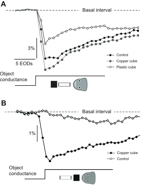
When the test object was placed at a distance of 1mm from the skin and a copper cube was placed 2mm away from its distal end (Fig.5A, inset), the amplitude of the novelty response increased by 25% compared with the control (Fig.5A, unpaired samples t-test, P<0.01, d.f.18). A plastic cube instead of the copper one caused a significant reduction (34%) of the amplitude of the novelty response compared with the control (Fig.5A, unpaired samples t-test, P<0.01, d.f.18).
When a test object was placed 23mm from the skin, the maximal change in resistance did not a evoke novelty response (Fig.5B, open symbols). When a copper cube was placed between the object and the skin (Fig.5B, inset), the maximal change in resistance of the object provoked a very clear novelty response (Fig.5B, filled symbols). This indicates that an object beyond the detectable range is detected by the interposition of the copper cube.
To pinpoint the effects of object interplay on the ability to sense the change in impedance of an object, we performed three other series of experiments in 5 fish each.
In the first series we compared extreme responses across fish. These responses were obtained with a non-conductive farthest object (2.5Mresistance, corresponding to 0.4S conductance) and a very conductive farthest object (1k, corresponding to 1000S). Statistical analysis confirmed our prediction that the responsiveness of the fish to the step in conductance of the nearest object is context dependent (Table1). Novelty responses were
always larger for farther away objects loaded by a smaller resistor (Fig.6A).
In the second series we studied the amplitude of the novelty response as a function of the step amplitude in the conductance of the nearest object (Fig.6B). As in the previous experiment, we compared the amplitude of the novelty responses evoked when the farthest object was in either the non-conductive or very conductive condition. The amplitude of the novelty response increased with the step in conductance of the nearest cylinder. In every fish, the curve obtained in the non-conductive context (Fig. 6B, 2.5M) was below that obtained in the conductive context (Fig. 6B, 1k).
3%
5 EODs
Control Copper cube Plastic cube
Object conductance
Basal interval
A
B
Control Copper cube
1%
Object conductance
[image:6.612.324.562.67.372.2]Basal interval
Table 1. Multiple comparison analysis of the effect of the conductance of the farthest object
Novelty response (% basal interval) t-test unpaired samples
High conductance Low conductance t-value d.f. Tail probability
1 8.8±0.38 8±0.45 4.07 18 <0.0005
2 12.25±1.2 10.46±0.62 3.98 13 <0.001
3 8.62±0.99 5.07±1.37 6.3 16 <0.0005
4 5.8±1.91 1.6±0.88 5.99 13 <0.0005
5 7.01±0.28 5.98±0.36 6.77 17 <0.0005
Family-wise type I error rate 1 – (1 – 0.001) ⫻(1 – 0.0005)4 <0.003
Farthest object conductance was high (1000S) or low (0.4S). Object diameter, 7mm; object length, 11mm.
Novelty response data are means ± s.d.
Fig.5. Contextual effect of a cube shown as the inter-EOD interval series.
Novelty responses were evoked by transient changes of conductance of a test, non-isotropic cylindrical object. Object resistance varied from open
(2.5M) to short (1k) circuit. (A)The test object was intercalated between
a copper or a plastic cube and the fish, as indicated in the inset. The
control novelty response in the absence of the cube is also shown. (B)The
[image:6.612.44.568.630.721.2]Using two objects of smaller diameter that have a larger effect on the image, we compared the amplitude of the novelty response provoked by a maximal step (from non-conductive to very conductive) of the nearest object as a function of the change in conductance of the farthest context object (Fig.7). We found in 5 out of 5 fish that the mean amplitude of the novelty response evoked by a maximal change in conductance of the nearest object was an increasing function of the conductance of the farthest object. Moreover, increasing the resistance of the nearest object caused a shift of the curve to lower values (Fig.7B).
Finally, we evaluated the funneling or blocking effects of an object intercalated between a test object and the skin by loading the intercalated object with different resistors. For thick objects (7mm diameter ⫻11mm length) we only obtained novelty responses for very conductive conditions of the near object (loading resistors 1 and 10k, corresponding to 1000 and 100S, respectively). Thus, to find the relationship we used narrower cylinders (4mm diameter) of different lengths (nearest object 7mm, farthest object 14mm). Under these conditions, the amplitude of the novelty response evoked by a large conductance step at the farthest object (between 0.4 and 1000S) depended on the longitudinal conductance of the interposed object in all 5 fish (Fig.8) increasing gradually as the conductance increased. In all fish, novelty responses were abolished by a loading resistance of the nearest object of above 500k
(equivalent to 5S, Fig.8, data from 2 of the studied fish), suggesting a blocking effect of the non-conductive objects.
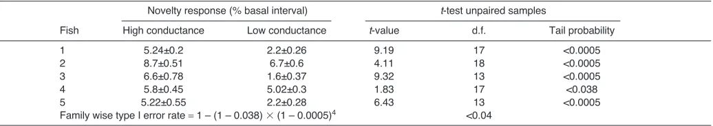
To validate these effects among fish we choose two conductance values (short circuit, 1000 and 35S) to compare mean amplitudes of the novelty responses. For every fish the mean amplitude of the novelty response at 1000S was larger than the mean amplitude of the novelty response at 35S. Statistical analysis (Table2) suggests a facilitation of the novelty response by the current funneling through the nearest object conductance.
DISCUSSION
Theoretical predictions (Rother et al., 2003; Migliaro et al., 2005; Caputi and Budelli, 2006; Pereira and Caputi, 2010) indicate that the presence of one object modifies the field in which it is placed. Thus, the field polarizing an object potentially introduced into the scene is dependent on the presence of neighboring objects. This was clearly shown by modeling in the study by Migliaro et al. when considering the presence of the fish’s body as an object (Migliaro et al., 2005) and in our previous analytical studies (Pereira and Caputi, 2010).
Here, we experimentally confirm predictions made by Rother et al. (Rother et al., 2003) that when one object is intercalated between another object and the fish’s body, there is mutual polarization and, consequently, the image is modified in a non-additive manner. If the two objects are very close (according to our results, ≤2mm apart) their mutual polarization results in changes of the image of a nearby object even though the farthest object alone is unable to yield a distinguishable image (see Figs1 and 2).
This result has two implications. First, the behavioral results confirm that object mutual polarization has significant effects on the detectability of objects. Single objects have a range of detection of about the size of the object (Pereira and Caputi, 2010). An object
8 9 10 11 12 13
Amplitude of novelty response (%)
Impedance of farthest object
Conductance of test object (μS)
10 100
5.0 7.5 10.0 12.5
1 kΩ
2.5 MΩ
A
B
1 kΩ 2.5 MΩ
Fig.6. The amplitude of the novelty response increases with the
conductance of the context object. Novelty responses were evoked by transient changes of conductance of a test, non-isotropic cylindrical object.
The resistance of this object varied from open (2.5M) to a given
resistance value. The test object was intercalated between an equal-sized
cylinder placed along the field line, which was in either the open (2.5M)
or short (1k) condition. (A)Mean amplitude of the novelty response
obtained with a maximum step in the nearest object resistance (2.5Mto
1k) when the context object was in either the open or short circuit
condition (each pair of symbols corresponds to the mean data of one fish,
sign test, P<0.032, N5). (B)Data from one fish illustrating the series of
experiments performed in each of these fish. In this plot, the amplitude of the novelty responses (mean ± s.e.m. of 10 trials for each point) is represented as a function of the conductance of the test object, either short
circuit (1k) or open circuit (2.5M).
1 10 100 1000
4 5 6 7 8 9 10
Amplitude of novelty response (%)
Conductance of farthest object (μS)
1 10 100 1000
6 8 10 12 14 16
300 Ω 50 kΩ
A
B
Fig.7. The amplitude of the novelty response increases with the
conductance of the context object. In these experiments, a small test object was intercalated between the fishʼs skin and a cylinder of equal diameter
and double length placed along the same field line (inset). (A)Novelty
responses were evoked by transient changes of conductance of the test
object from open (2.5M) to short circuit (300switch resistance).
(B)Novelty responses were evoked by transient changes of conductance of
the test object from open (2.5M)either to short circuit (300switch
resistance) or 50kswitch resistance. Each plot shows data from a
placed beyond such a distance is not detectable when it is alone but its movements or its changes in impedance can be detected if a conductive nearby object is intercalated between the first object and the fish’s body. The opposite (hiding of an object that is within the active electrolocation range) occurs if a non-conductive object is interposed between the object and the fish.
In addition, the presence of a far contextual object may also increase or decrease by small amounts the image of objects more conductive than water (see Figs3 and 4), affecting object perception. This prediction was confirmed by showing that the function relating the amplitude of the novelty response to the increase in conductance of the closest object changed in the presence of non-conductive and very conductive farthest objects. In fact, although the novelty response’s maximum change was only 10%, in all paired experiments there was a systematic difference in the amplitude range and slope of the curves when comparing the two plots (see Fig.6B). Moreover, the responses evoked by maximum conductance changes were smoothly modulated by the impedance of the farthest object. These effects of mutual polarization may be interpreted by a sensory agent in an ambiguous way. On the one hand, the farthest object increases the impedance discriminability of the closest object; on the other hand, the closest object modifies the range of electrosensory detection as a function of its impedance, shape and size. This ambiguity may be crucial for object recognition. Nevertheless, an important clue could be the presence of movements
of the fish or object. As stationary images are removed by the presence of an adaptive filter, probably at the electrosensory lobe (Caputi et al., 2003; Caputi et al., 2008), a fish may take advantage of relative movements in two ways: (1) use its own movements to maintain a closer conductive object stationary, working as a ‘telescopic device’ that artificially extends the fish’s body and consequently increases the detection range; and (2) to better discriminate the attributes of a closer object in the presence of a moving context behind the object (as for example leaves and roots of water plants) (Babineau et al., 2007; Behr et al., 2011).
CONCLUSION
Our experiments confirm the proposed short range of active electroreception and the importance of the fish’s body for active electrolocation (Migliaro et al., 2005; Pereira and Caputi, 2010; Caputi et al., 2011). Objects near the fish may sometimes act as ‘accidental auxiliary devices’ for perceiving other objects beyond the active electroreception range. The potential effects of a fish’s movements on scene polarization – thereby altering objects’ mutual polarization – is analogous to exploring an object by pressing it between the finger tip and a background object. It is virtually impossible to evaluate the consistency of a rubber eraser floating in water, but is very easy when it is placed on a table. This is because the buoyancy caused by sinking the object is much smaller and behaves differently from the reaction force of the table. We have previously reported some similarities between active electrolocation and haptic senses (Caputi et al., 2011). This is another one.
APPENDIX
Reasons for using cylindrical probes and the choice of their dimensions
As we have already reported (Pereira and Caputi, 2010), every object modulates the basal field (the field in the absence of the object) as if a set of electric sources were placed in it instead of the object. This set is called the stamp of the object. The difference between the field with the object and the basal field is called the object perturbing field (Lissmann and Machin, 1958). The electric image is a transcutaneous object perturbing field. For objects equally located relative to the fish it holds that larger stamps generate larger images.
[image:8.612.55.273.68.230.2]The stamp of an object depends on its imprimence, a parameter that depends on object size, shape and impedance. Therefore, only very simple objects are suitable for manipulation as experimental tools – among these are spheres and the cylindrical probes used in this study. These probes only conduct current in the longitudinal direction. When they are aligned to the local field their stamp has the same orientation as the field. In addition, the longitudinal conductance can be controlled at will by connecting different resistors between the bases. Objects more conductive than water facilitate the flow of current while those less conductive than water
Table 2. Multiple comparison analysis of the effect of the conductance of the nearest object
Novelty response (% basal interval) t-test unpaired samples
Fish High conductance Low conductance t-value d.f. Tail probability
1 5.24±0.2 2.2±0.26 9.19 17 <0.0005
2 8.7±0.51 6.7±0.6 4.11 18 <0.0005
3 6.6±0.78 1.6±0.37 9.32 13 <0.0005
4 5.8±0.45 5.02±0.3 1.83 17 <0.038
5 5.22±0.55 2.2±0.28 6.43 13 <0.0005
Family wise type I error rate 1 – (1 – 0.038) ⫻(1 – 0.0005)4 <0.04
Nearest object conductance was high (1000S) or low (35S). Object diameter, 4mm; object length, nearest 7mm, farthest 14mm.
Novelty response data are means ± s.d.
10 100 1000
0 1 2 3 4 5 6 7
Amplitude of novelty response (%)
Longitudinal conductance of intercalated object (μS)
Fig.8. Amplitude of the novelty response as a function of the longitudinal
conductance of an intercalated object. Inset, schematic diagram of the
experiment: two cylindrical objects of 7 and 11mm length, respectively,
were aligned with a field line. Novelty responses were evoked by transient
changes of the farthest object resistance from open (2.5M)to short
(1k) circuit. Data from two fish (open and filled symbols) are shown
[image:8.612.50.568.630.722.2]reduce the flow of current, causing an increase or a reduction of the electric image (Fig.A1A). The stamp of the object (S) is proportional to the difference between the object resistance (Ro) and
the resistance of an equal-sized cylinder of water (Rw). The
proportionality constant depends on the scene parameters ‘as seen’ from the object: the electromotive force (Es) and the internal
resistance (Rs), and also on Ro:
S[Es/ (Ro+ Rs)] ⫻(Ro– Rw) . (A1)
Shape affects Sin two ways: a doubling of diameter reduces by four times the Rothat matches Rw(compare green and blue lines in
Fig.A1B). A doubling of cylinder length increases by two times the Rovalue that matches Rwand increases also Es(compare red
and blue lines in Fig.A1B). Bearing in mind that the electrical significance of the object loading resistance (Ro) is different for every
shape, we carried out two different series of experiments; in the first we used identical objects and in the second we used thinner objects of different length to amplify the effects. Here, we only wanted to show a qualitative effect, so we did not perform a detailed study of the effect of the shape of the objects in object interplay.
ACKNOWLEDGEMENTS
We would like to thank Lic. Marcela Piffaretti for English correction of a first draft.
FUNDING
This work was partially funded by European Commission Information, Society and Media Future and Emergent Technologies (FET) [grant no. 231845] and Programa de Desarrollo de las Ciencias Básicas (PEDEClBA) (Uruguay).
REFERENCES
Aguilera, P. A. and Caputi, A. A.(2003). Electroreception in G. carapo: detection of changes in waveform of the electrosensory signals. J. Exp. Biol. 206, 989-998. Babineau, D., Longtin, A. and Lewis, J. E.(2006). Modeling the electric field of
weakly electric fish. J. Exp. Biol. 209, 3636-3651.
Babineau, D., Lewis, J. E. and Longtin, A.(2007). Spatial acuity and prey detection in weakly electric fish. PLoS Comput. Biol. 3, e38.
Behr, K., Neusel, G. and von der Emde, G.(2011). Mind the gap! Detection of gaps and locomotor strategies of electrolocating Gnathonemus petersii. Proceedings of the 104th Annual Meeting of the Deutsche Zoologische Gesellschaft (DZG). p 26. Bennett, M. V. L.(1971). Electric organs. In Fish Physiology, Vol. 5 (ed. W. S. Hoar
and D. J. Randall), pp. 347-491. London: Academic Press.
Budelli, R., Caputi, A., Gomez, L., Rother, D. and Grant, K.(2002). The electric image in Gnathonemus petersii.J. Physiol. Paris96, 421-429.
Caputi, A. A.(2011). Encyclopedia of Fish Physiology(ed. A. Farrell). Amsterdam: Elsevier.
Caputi, A. A. and Budelli, R.(2006). Peripheral electrosensory imaging by weakly electric fish. J. Comp. Physiol. A 192, 587-600.
Caputi, A., Budelli, R., Grant, K. and Bell, C. C.(1998). The electric image in weakly electric fish: physical images of resistive objects in Gnathonemus petersii. J. Exp. Biol. 201, 2115-2128.
Caputi, A. A., Aguilera, P. A. and Castelló, M. E.(2003). Probability and amplitude of novelty responses as a function of the change in contrast of the reafferent image in G. carapo. J. Exp. Biol. 206, 999-1010.
Caputi, A. A., Castelló, M. E., Aguilera, P. A., Pereira, A. C., Nogueira, J., Rodríguez-Cattaneo, A. and Lezcano, C.(2008). Active electroreception in
Gymnotus omari: imaging, object discrimination, and early processing of actively generated signals. J. Physiol. Paris 102, 256-271.
Caputi, A., Aguilera, P. and Pereira, A. C.(2011). Active electric imaging: body-object interplay and body-objectʼs ʻelectric textureʼ. PLoS ONE 6, e2279.
Castelló, M. E., Aguilera, P. A., Trujillo-Cenóz, O. and Caputi, A. A.(2000). Electroreception in Gymnotus carapo: pre-receptor processing and the distribution of electroreceptor types. J. Exp. Biol. 203, 3279-3287.
Chen, L., House, J. H., Krahe, R. and Nelson, M. E.(2005). Modeling signal and background components of electrosensory scenes. J. Comp. Physiol. A 191, 331-345.
Engelmann, J., Baceló, J., Metzen, M., Pusch, R., Bouton, B., Migliaro, A., Caputi, A., Budelli, R., Grant, K. and von der Emde, G.(2008). Electric imaging through active electrolocation: implication for the analysis of complex scenes.Biol. Cybern. 98, 519-539.
Gómez, L., Budelli, R., Grant, K. and Caputi, A. A.(2004). Pre-receptor profile of sensory images and primary afferent neuronal representation in the mormyrid electrosensory system. J. Exp. Biol. 207, 2443-2453.
Lissmann, H. W. and Machin, K. E.(1958). The mechanism of object location in
Gymnarchus niloticusand similar fish. J. Exp. Biol. 35, 451-486.
Migliaro, A., Caputi, A. A. and Budelli, R.(2005). Theoretical analysis of pre-receptor image conditioning in weakly electric fish. PLoS Comput. Biol. 1, e:16.
Nelson, M. E. (2005). Target detection, image analysis, and modeling in
electroreception. In Handbook of Auditory Research, Vol. 21 (ed. T. H. Bullock, C. D. Hopkins, A. N. Popper and R. Fay), pp. 290-317. Springer.
Nelson, M. E. and MacIver, M. A.(1999). Prey capture in the weakly electric fish
Apteronotus albifrons: sensory acquisition strategies and electrosensory consequences. J. Exp. Biol. 202, 1195-1203.
Nelson, M. E. and MacIver, M. A.(2006). Sensory acquisition in active sensing system. J. Comp. Physiol. A192, 573-586.
Pereira, A. C. and Caputi, A. A.(2010). Imaging in electrosensory systems.
Interdiscip. Sci. 2, 291-307.
Pereira, A. C., Centurión, V. and Caputi, A. A.(2005). Contextual effects of small environments on the electric images of objects and their brain evoked responses in weakly electric fish. J. Exp. Biol. 208, 961-972.
Richer de Forges, M., Crampton, W. G. R. and Albert, J. S. (2009). A new species of Gymnotus(Gymnotiformes, Gymnotidae) from Uruguay: description of a model species in neurophysiological research. Copeia2009, 538-544.
Rother, D., Migliaro, A., Canetti, R., Gómez, L., Caputi, A. and Budelli, R.(2003). Electric images of two low resistance objects in weakly electric fish. Biosystems 71, 169-177.
Sanguinetti-Scheck, J. I., Pedraja, E. F., Cilleruelo, E., Migliaro, A., Aguilera, P., Caputi, A. A. and Budelli, R.(2011). Fish geometry and electric organ discharge determine functional organization of the electrosensory epithelium. PLoS ONE6: e27470.
Sicardi, A., Caputi, A. A. and Budelli, R.(2000). Physical basis of distance discrimination in weakly electric fish. Physica A283, 86-93.
Snyder, J. B., Nelson, M. E., Burdick, J. W. and MacIver, M. A.(2007). Omnidirectional sensory and motor volumes in electric fish. PLoS Biol. 5, e301. von der Emde, G.(1990). Discrimination of objects through electrolocation in the weakly electric fish, Gnathonemus petersii. J. Comp. Physiol. A 167, 413-421. von der Emde, G. and Ronacher, B.(1994). Perception of electric properties of
objects in electrolocating weakly electric fish: two-dimensional similarity scaling reveals a City-Block metric. J. Comp. Physiol. A 175, 801-812.
Rs Ro
Es
Ro>Rw
Ro<Rw
Rs Rw
Es
S
Rs Rw
Es
S 0
Probe shape Es long cylinder
Es short cylinder
Ro
A
B
S
Fig.A1. Shape affects the ʻstampʼ (S) in
two ways. (A)Network analysis (for
explanation, see Appendix). (B)A doubling
of diameter reduces by four times the
object resistance (Ro) that matches the
resistance of an equal-sized cylinder of
water (Rw). A doubling of cylinder length
increases by two times the Rothat
matches the Rwand also increases the
electromotive force (Es). Each object has a
different equivalent Rw(water resistivity ⫻