Original Article
Prognostic value of
β1 integrin
expression in colorectal
liver metastases
Nikolaos Vassos
1,
Tilman Rau
2, Susanne Merkel
1, Fabian Feiersinger
1, Carol I Geppert
2, Michael Stürzl
3,
Werner Hohenberger
1, Roland S Croner
11Department of Surgery, University Hospital Erlangen, Germany; 2Institute of Pathology, University Hospital Erlan-gen, Germany; 3Division of Molecular and Experimental Surgery, University Hospital Erlangen, Germany
Received October 29, 2013; Accepted December 3, 2013; Epub December 15, 2013; Published January 1, 2014
Abstract: Integrins are cell surface adhesion molecules (CAM) that regulate via intercellular and cell-matrix signaling various cellular processes including wound healing, cell differentiation, division, growth, migration and metastatic dissemination. Although a correlation between carcinogenesis and changes in integrin expression, especially β1 integrin, has been reported, its role in colorectal liver metastases remains unclear. This study aimed to evaluate the
expression of β1 integrin in colorectal liver metastases and to correlate the pattern of expression with clinicopatho -logical features and to investigate the putative role of β1 integrin expression on survival of these patients. Methods:
Formalin-fixed, paraffin-embedded (FFPE) tumor samples of 81 patients who were operated because of colorectal
liver metastases without any neoadjuvant therapy were obtained and stained with hematoxylin and eosin (H & E). An immunohistochemical examination was performed using Dako, Peroxidase/DAB kit and a primary monoclonal
β1 integrin (CD29, fibronectin receptor subunit beta; ab3167, Abcam plc). β1 integrin expression was evaluated
according to the immunoreactive score of Remmele and Stegner and was related with clinicopathological features
of prognostic significance and with disease-free and overall survival as well.Statistical analysis was performed us-ing SPSS version 21.0. Results: β1 integrin was overexpressed in tumor cells in 37 (48%) patients and in stromal cell in 27 (33%) patients. The β1 expression was not statistically correlated with clinicopathological features of the primary tumors but it was statistically correlated (p=0.03) with the histological grading of liver metastases.
Kaplan-Meier survival analysis showed that there is a tendency but no statistically significant correlation in disease-free
and overall survival. Conclusion: Considering that expression of β1 integrin in colorectal liver metastases remains controversial, specially its relation with survival of patients, we showed that the β1 expression represents a reliable
prognostic factor regarding the grading of liver metastases of CRC and our findings imply that β1 integrin expres
-sion profiles may have further potential in identifying the stage of colorectal liver metastases and being a marker of
prognosis in these patients.
Keywords: Colorectal liver metastases, beta1, β1 integrin, expression, prognosis
Introduction
Colorectal cancer (CRC) is the second leading
cause of cancer incidence and
cancer-associ-ated mortality in both males and females in
Western society [1-3]. The prognosis of CRC
patients is mainly determined by the
metastat-ic spread of the tumor [1, 4]. Thus,
understand-ing the mechanisms that contribute to
metas-tasis is of fundamental importance for desi-
gning better therapeutic strategies for treating
this disease.
The liver is the most important and common
metastatic site of CRC [2, 5]. It is a unique
Amongst CAM, integrins are a versatile family
which consists of heterodimer cell surface
receptors composed of
α and β
transmem-brane subunits; each of them includes a large
extracellular, transmembrane and short
cyto-plasmic domain [13]. In mammals, 19
α
and 8
β
subunits combine with each other to form a
family of 25 cell adhesion molecules, however
splice variants have been identified for some
subunits [14, 15]. Their ligands include
compo-nents of the extracellular matrix such as fibro
-nectin, vitro-nectin, collagen, laminin and
IgSFCAMs [14, 16, 17]. Functionally integrins
contribute to intercellular adhesion and
con-tact, anchorage-dependent cell survival and
regulate via outside-in and inside-out signalling
various cellular processes including wound
healing, cell differentiation, division, growth
and migration [18, 19].
Integrins expressed by tumor cells and host
cells can directly contribute to the control and
progress of metastatic dissemination. During
tumor development, changes in integrin
expres-sion, intracellular control of integrin functions
and signals perceived from integrin ligand
bind-ing influence the ability of tumor cells to inter
-act with their environment. This enables
meta-static cells to convert from a sessile, stationary
to a migratory and invasive phenotype [20].
Therefore integrin expression can profoundly
influence formation of metastasis [21-23].
Alter-ations of integrin expression and their
recep-tors have been observed in various cancers
including colorectal cancer [23, 24]. However,
the mechanisms by which integrins participate
in the steps of the metastasis formation in vivo
are only partially understood [20].
According to their
β
subunits, integrins are
divided into four subfamilies and integrins with
β1-subunits are called β1 integrins. The β1
integrin subfamily (or Very Late Antigens, VLA)
is characterised by a
β
1 subunit associated
with at least nine a subunits (termed a1-a9,
CD49a-i) constituting the largest subfamily of
the integrins [25, 26] and can bind to collagen,
laminin and fibronectin [26]. β
1 integrin is the
mainly expressed integrin in normal and tumor
cells and controls developmental processes
including angiogenesis, tumor progression and
metastasis [27]. This integrin typically
medi-ates adhesion of epithelial cells to the
base-ment membrane and may also contribute to
cell survival of tumor cells by interacting with
others molecules. For example, it usually
acti-vates cytokine receptors or growth factors
receptors [28]. Based on these observations,
β
1 integrin has become a target of interest for
immunotherapy in several types of cancers,
including breast cancer [29-34], gastric cancer
[35], lung adenocarcinoma [36-38],
glioblasto-ma [39], prostate cancer [40], pancreatic
ade-nocarcinoma [41] and CRC [42-45].
Immunohis-tochemical studies of expression of
β
1 integrin
in CRC have shown that the expression of this
integrin has considerable heterogeneity within
the primary tumor [42-45]
and is correlated
with cellular differentiation [46], indicating that
alteration of expression of
β
1 integrin in
colorectal cancer might contribute to an
altera-tion in adhesiveness and invasiveness of
can-cer cells. But there are no studies which
inves-tigate the prognostic value of the expression of
β
1 integrin within liver metastases of CRC.
The aim of this study is to assess the
immuno-histochemical
β1 integrin expression in
colorec-tal liver metastases and its relationship with
clinicopathological features of prognostic
sig-nificance. Our additional hypothesis is that the
enhanced expression of
β
1 integrin in liver
metastases could affect the progress of
metas-tasis and may therefore be of prognostic
rele-vance influencing disease-free and overall
survival.
Material and methods
Patients
Our study included patients with colorectal liver
metastases who underwent curative hepatic
resection for the first event of the metastases
from 1995 to 2010 in the Department of
Surgery at the University Hospital Erlangen.
Patients upon whom a neoadjuvant therapy of
the primary tumor or of the liver metastases
was performed, patients with synchronous
metastases in other organs or patients who
died postoperatively were excluded. The clinical
and histopathological data of the patients were
diameter and differentiation’s grade of the liver
metastases. Tumors were classified at the
institute of pathology according to the 7
thTNM
cancer staging system of the American Joint
Committee on Cancer (AJCC) and the Union for
International Cancer Control (UICC) [47]
and
were graded as well, moderately or poorly
dif-ferentiated depending on their behavior
accord-ing to the recommendations of the World Health
Organization (WHO) [48]. Disease-free survival
and overall survival were defined as time from
the first diagnosis of liver metastasis to local
recurrence in the liver and to cancer-related
death, respectively.
The subsequent 81 patients of our study
com-prised of 45 males (56%) and 36 females (44%)
with a median age of 62 years (mean: 62, range
43-81 years) at the diagnosis of liver
metasta-ses. The clinicopathological characteristics of
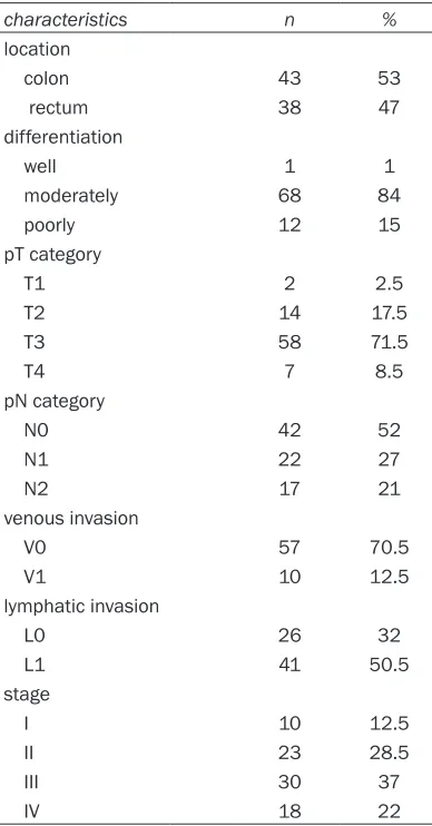
[image:3.612.91.285.93.464.2]the primary tumors are illustrated in
Table 1A
.
The liver metastases occurred synchronously
in 18 cases (22%) and metachronously in 63
cases (78%) with a median time interval of 19
months (range 4-163 months). The
characteris-tics of the hepatic metastases and the type of
the performed hepatic resection are
demon-strated in
Table 1B
. The mean follow-up for all
patients was 65 months (median follow-up: 55
months), with a range between 1 and 188
months. During the follow-up, 20 patients (25%)
received adjuvant chemotherapy. Twenty-seven
patients (33.3%) developed metachronous
recurrent hepatic metastases, 10 of them in
other organs as well (peritoneum, lymph nodes,
lung, pleura, brain). These events occurred in a
median period of 16 months (range, 4-60) after
Table 1A.
Characteristics of the primary
colorectal tumors (n=81)
characteristics n %
location
colon 43 53
rectum 38 47
differentiation
well 1 1
moderately 68 84
poorly 12 15
pT category
T1 2 2.5
T2 14 17.5
T3 58 71.5
T4 7 8.5
pN category
N0 42 52
N1 22 27
N2 17 21
venous invasion
V0 57 70.5
V1 10 12.5
lymphatic invasion
L0 26 32
L1 41 50.5
stage
I 10 12.5
II 23 28.5
III 30 37
IV 18 22
Table 1B.
Characteristics of hepatic
metasta-ses (n=81)
characteristics
n %
liver metastasis in relation to the primary tumor
synchronous 18 22
metachronous 63 78
median time internal: 19 months (range, 4-163) distribution of liver metastases
unilobular 66 81.5
bilobar 15 18.5
number of liver metastases
1 54 67
2 17 21
3 4 5
>3 6 7
type of hepatic resection
right lobectomy 23 28
extended right lobectomy 3 4
left lobectomy 9 11
trisegmentectomy 3 4
bisegmentectomy 11 14
non-anatomic bisegmentectomy 7 8
segmentectomy 15 18
non-anatomic segmentectomy 7 9
mesohepatectomy 3 4
differentiation grade
moderate 70 87
poor 9 11
unknown 2 2
hepatic tumor size mm
median 47
[image:3.612.322.519.96.502.2]the curative resection (R0) of the first event of
liver metastases.
Tumor samples and immunochemistry
Tumor blocks (samples) were taken from the
diagnostic archives of the Institute of Pathology
of the University Hospital Erlangen and the use
of materials was positively stated by the local
Ethical Committee. Tumor samples were
previ-ously fixed in 4% buffered formalin and embed
-ded in paraffin (FFPE). One section of FFPE tis
-sue of each sample was stained with
hematoxylin and eosin to confirm the presence
of the tumor by light microscopy. All stained
slides were reviewed by an experienced
pathol-ogist. Sections (4
μ
m) were obtained from the
paraffin blocks and immunohistochemical
reactions were performed with the Dako REAL
TMEnVision
TMDetection System, Peroxidase/DAB
kit (Dako Denmark A/S, Denmark). The kit was
employed in a two-step procedure. The first
step was incubation of the tissue with an
opti-mally diluted primary antibody and the second
step was incubation with Dako REAL
TMEnVision
TM/HRP reagent of the kit. This reagent
was a peroxidase-conjugated polymer, which
also carries antibodies to immunoglobulins.
The reaction was visualized by Dako REAL
TMDAB+ Chromogen. Primary monoclonal
β1
integrin
(CD29, fibronectin receptor subunit
beta) antibody [4
Β
7R] (ab3167) was obtained
from Abcam plc, Cambridge, UK (dilution 1:35).
Negative controls were prepared omitting the
primary antibody and using non-immune IgG
instead of primary antibodies showed no
staining.
Immunohistochemical assessment
Tumorous immunoreactivity was graded
depending on the percentage of cancer cells
stained: Score 0 means negative; score 1
means 1 to 10% of tumor cells positive; score 2
means 11 to 50% of tumor cells positive; score
3 means 51-80% of tumor cells positive and
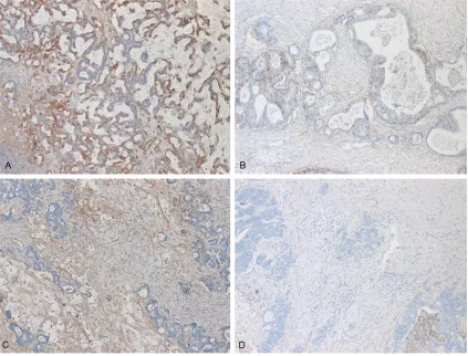
Figure 1. Immunohistochemical presentation of the expression of β1 integrin in tumor cells of colorectal liver
[image:4.612.96.519.72.394.2]score 4, >80% of tumor cells positive within the
tumors. The immunostaining intensity was
eval-uated by light microscopy and scored on the
fol-lowing scale: negative (0), weak (1), moderate
(2) and strong (3). We took into account both
the proportion of positive cells and the intensity
to give a semiquantitative estimate levels of
antigen in the tumor and in the neighboring
stroma. To identify the integrin’s expression, a
combination factor of immunoreactivity and
intensity according to the Remmele-Scoring
system was performed [49]. The theoretical
limits of this immunoreactive score range from
β1 integrin.
P
-values <0.05 were considered to
be statistically significant.
Results
Staining pattern of β1 integrin in colorectal
liver metastases
β1 integrin expression was observed throughout
the colorectal hepatic metastases. An example
of strong immunoreactivity of β1 expression in
tumor cells is shown in
Figure 1A
and, in
con-trast, a tumor sample negative for β1 expres
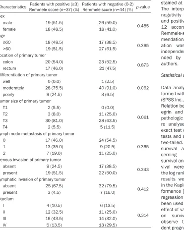
-Table 2.
Correlation of
β
1 expression in tumor cells of liver metastases
with clinicopathological findings of primary tumors
Characteristics Remmele score (n=37) (%)Patients with positive (≥3) Patients with negative (0-2) Remmele score (n=44) (%) p-value sex
male 19 (51.5) 26 (59.0) 0.485
female 18 (48.5) 18 (41.0)
age
≤60 18 (48.5) 17 (38.5)
0.365
>60 19 (51.5) 27 (61.5)
location of primary tumor
colon 20 (54.0) 23 (52.5)
0.873
rectum 17 (46.0) 21 (47.5)
differentiation of primary tumor
well 0 (0.0) 1 (2.5)
0.062 moderately 28 (75.5) 40 (91.0)
poorly 9 (24.5) 3 (6.5)
tumor size of primary tumor
T1 2 (5.5) 0 (0.0)
0.061
T2 3 (8.0) 11 (25.0)
T3 30 (81.0) 28 (63.5)
T4 2 (5.5) 5 (11.5)
lymph node metastasis of primary tumor
0 17 (46.0) 24 (54.5)
0.365
1 13 (35.0) 9 (20.5)
2 7 (19.0) 11 (25.0)
venous invasion of primary tumor
absent 9 (24.5) 17 (38.5)
0.343
present 19 (51.5) 22 (50.0)
lymphatic invasion of primary tumor
absent 25 (67.5) 32 (79.5)
0.412
present 3 (4.5) 7 (16.0)
stadium
I 4 (10.5) 6 (13.5)
0.314
II 12 (32.5) 11 (25.0)
III 16 (43.5) 14 (32.0)
IV 5 (13.5) 13 (29.5)
0 (0% of cells stained)
to 12 (>80% of the cells
stained at 3+ intensity).
The interpretation was
negativity from 0 to 2
and positivity from 3 to
12 according to the
Remmele-score
recom-mendations [50].
Evalu-ation was performed
independently and
bli-nded by two of the
authors.
Statistical analysis
[image:5.612.96.467.95.559.2]sion in
Figure 1B. Within metastases β1 integ
-rin was expressed by tumor stroma as well but
in less amounts compared to the tumor cells.
Samples positive and negative for
β1 expression
in stromal cells are presented in
Figure 1C
,
1D
,
correspondingly. Specifically, expression of the
β
1 integrin was increased in tumors in 37 (48%)
patients, 24 of whom had a negative
expres-sion in stroma, whereas the
β
1 integrin was
increasingly expressed in 27 patients (33%), 14
of whom had a negative expression in tumor.
metastases and weak in moderate
differenti-ated metastases, as showed in
Figure 1
, where
the expression of
β1 integrin in poorly
differentiated metastases (
Figure 1A
) is really
stronger than this one in moderated
differenti-ated metastases (
Figure 1C
). These
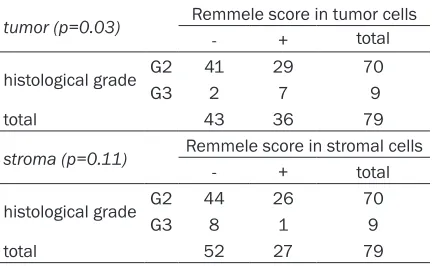
differenc-es were statistically significant (p=0.03). In
contrast, the expression of
β
1 integrin in tumor
stromal cells shows no correlation between
his-topathological grading and β1 integrin expres
-sion (p=1.12) (Table 4).
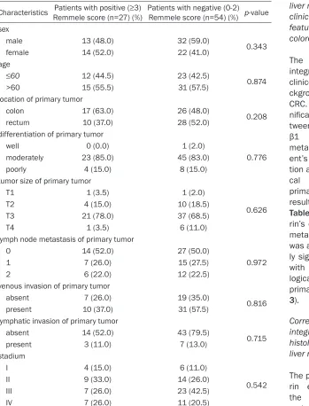
Table 3.
Correlation of
β
1 expression in stromal cells of liver metastases
with clinicopathological findings of primary tumors
Characteristics Remmele score (n=27) (%)Patients with positive (≥3) Patients with negative (0-2) Remmele score (n=54) (%) p-value sex
male 13 (48.0) 32 (59.0) 0.343
female 14 (52.0) 22 (41.0)
age
≤60 12 (44.5) 23 (42.5)
0.874
>60 15 (55.5) 31 (57.5)
location of primary tumor
colon 17 (63.0) 26 (48.0)
0.208
rectum 10 (37.0) 28 (52.0)
differentiation of primary tumor
well 0 (0.0) 1 (2.0)
0.776 moderately 23 (85.0) 45 (83.0)
poorly 4 (15.0) 8 (15.0)
tumor size of primary tumor
T1 1 (3.5) 1 (2.0)
0.626
T2 4 (15.0) 10 (18.5)
T3 21 (78.0) 37 (68.5)
T4 1 (3.5) 6 (11.0)
lymph node metastasis of primary tumor
0 14 (52.0) 27 (50.0)
0.972
1 7 (26.0) 15 (27.5)
2 6 (22.0) 12 (22.5)
venous invasion of primary tumor
absent 7 (26.0) 19 (35.0)
0.816
present 10 (37.0) 31 (57.5)
lymphatic invasion of primary tumor
absent 14 (52.0) 43 (79.5)
0.715
present 3 (11.0) 7 (13.0)
stadium
I 4 (15.0) 6 (11.0)
0.542
II 9 (33.0) 14 (26.0)
III 7 (26.0) 23 (42.5)
IV 7 (26.0) 11 (20.5)
Correlation of β1
integrin expression in
liver metastases with
clinicopathological
features of the primary
colorectal cancer
The expression of
β
1
integrin was related to
clinicopathological
ba-ckground of the primary
CRC. There was no
sig-nificant relation
be-tween the expression of
[image:6.612.95.444.94.552.2]β
1 integrin in liver
metastases with
pati-ent’s sex or age,
loca-tion and
histopathologi-cal features of the
primary tumors. These
results are shown in
Table 2
. The β1 integ
-rin’s expression in liver
metastases stroma
was also not
statistical-ly significant correlated
with the
clinicopatho-logical features of the
primary tumors (Table
3).
Correlation of β1
integrin expression with
histological grading of
liver metastases
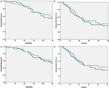
Prognostic value of integrin β1 expression
We examined whether there was an
associa-tion between β1 expression and patient surviv
-al. We analyzed the data by considering only
disease-related death as an event, censoring
deaths unrelated to disease and the patients
who were alive when they were last seen. To
determine differences in the disease-free and
overall survival among patients who differed in
β1 expression, the log-rank test was performed
and the corresponding Kaplan-Meier plot is
shown in Figure 2. The log-rank test indicated
there was a tendency but no significant differ
-ence in disease-free and overall survival
between patients with score 0-2 and patients
with score 3-12 in tumor cells (p=0.198 and
p=0.340 respectively). Similarly, comparing the
survival of the 81 patients according to the
integrin
β
1 expression in stroma, the
disease-free and overall survival rate for patients who
had positive immunoreactive score of β1 integ
-rin in stromal cells was not significantly better
compared to individuals who had negative
immunolabeling (p=0.890 and p=0.740 respe-
ctively).
We also examined if there was a prognostic
rel-evance of integrin
β
1 expression in patients
with positive
β
1 expression in tumor cells and
negative expression in stromal cells in
compari-son with the patients with negative β1 expres
-sion in tumor cells and positive expres-sion in
stromal cells. A statistical significance was not
confirmed neither for the disease-free survival
(p=0.890) nor for the overall survival (p=0.511).
Multivariate regression analysis using the cox
proportional hazards model was performed to
observe the independent prognostic value of
β1 integrin expression in tumor cells with
several prognostic factors such sex and age of
the patient, clinicopathological parameters of
primary tumor (location, differentiation, tumor
size, nodal status, venous and lymphatic
inva-sion), differentiation of liver metastases and
time interval between primary tumor and liver
metastases, but no correlation was found
(Supplementary Table 1).
Discussion
The development of liver metastasis is an
omi-nous event in the natural history and
progres-sion of CRC. The fact that not all disseminated
CRC cells develop into macrometastases
indi-cates that subpopulations of malignant cells
evolve a genetic advantage to adhere, migrate,
and invade through the ECM, and survive in the
new liver environment [53]. This advantage is
associated with the up-regulation of various
CAM, and in particular integrins [53, 54], which
are the prime example of bidirectional receptor
signalling.
Analysing cell adhesion, current research
attempts to clarify the numerous cell
interac-tions which are evident within the liver
sinu-soids and thus control the outcome of the
whole metastatic process [55, 56]. During the
last years a number of reports and multiple
research studies have investigated different
integrins (
α
vβ
3,
α
vβ
5,
α
vβ
1,
α
vβ
6,
α
vβ
8,
α
2) in CRC
and its metastases, showing that blockade of
them in cell lines of primary or metastatic
can-cer caused a significant reduction of tumour
cell proliferation and prevention of increase of
colorectal hepatic metastases [57-62]. There
are also immunohistochemical studies in
colorectal cancer samples showing that the
expression of integrins is remarkably altered in
CRC [42-45, 63, 64], since patients with the
highest levels of e.g.
α
vβ
6or
α
vβ
3protein
expres-sion on immunostaining had the poorest
sur-vival [63, 65, 66] and moreover the
expres-sion‘s level of these integrins in colorectal
cancer samples were correlated with the
devel-opment of liver metastases [65].
[image:7.612.89.305.106.239.2]Concerning the CRC, it is known that blockade
of
β
1 integrin-mediated cell adhesion reduced
adhesive properties of CRC cells within the
hepatic microcirculation [67]. In vitro studies
on human colorectal cancer cell lines revealed
Table 4. Correlation of β1-integrin immunolabel
-ing with the differentiation grade of liver
metas-tases
tumor (p=0.03) Remmele score in tumor cells
- + total
histological grade G2 41 29 70
G3 2 7 9
total 43 36 79
stroma (p=0.11) Remmele score in stromal cells
- + total
histological grade G2 44 26 70
G3 8 1 9
that
β1
integrin regulates differentiation toward
cancer progression and invasion [67-69].
Furthermore immunohistochemical studies in
colon or colorectal cancer specimens have
con-sistently revealed altered β1 integrin expres
-sion in the development and progres-sion of this
disease [42, 44] which was also correlated with
lymph node metastasis or depth of invasion
[27]. A few reports have shown correlations
between integrin expression and tumor
progno-sis or clinical stage too [70-78].
But the prognostic significance of integrins›
deposition around liver metastases remains
unclear since there are only a few studies in
metastatic cancer samples consisting of a very
small number of patients which examine the
expression of
β
1 integrin in hepatic metastatic
cancer samples [43, 79-81]; however without
any investigation of the clinicopathological
parameters and without any correlation with
tumor prognosis. This is the first study which
examines the expression of
β
1 integrin in liver
metastases samples in a large group of
patients and correlates it with different
parameters.
For the evaluation of the β1 integrin’s expres
-sion we choose the score proposed by Remmele
and Stegner [49]
based on the level of signal
and percentage of tumor cells expressing the
[image:8.612.93.518.71.414.2]signal. It is the first study which evaluates inte
-grins’ expression in CRC with this score. We
found increased cell surface expression of the
integrin
β
1 protein in tumor and stromal cells in
hepatic metastases compared to normal liver
tissue. Although a number of studies have
implicated integrin expression in the
metastat-ic process, including liver metastases, the find
-ings presented here are the first to directly
show a role for integrin
β
1 in selective liver
metastasis since our results suggest that
β
1 in
human colorectal cancer mediates the
poten-tial of liver colonization and liver metastasis,
because of its high expression in metastatic
tissues of liver and supports the idea that
β
1
integrin in CRC correlates with disease
progres-sion and poorer clinical outcome.
We found that the stroma, which is
character-ized with a dense vascular network and a loose
connective tissue, was also intensely positive
for
β
1 but in low levels in comparison with
tumor. This difference in
β
1-expression in tumor
and stromal cells of the metastatic liver could
be due to the fact that the
β
1 integrin receptor,
fibronectin, is found in the surrounding tissue
around the tumor but
β
1 integrin are anchored
to the membrane of tumor cells, leading to a
stronger expression [82, 83]. Gulubova and
Vlaykova [81] showed that the intensity of
immune reaction of
α
5β
1and
α
9β
1integrins in
liver metastases tended to be lower than that
in primary tumor. Similar findings were observed
by Dueck too [84]. This could be due to the fact
that stroma-forming cells in the liver are far
fewer in number than those in the connective
tissue stroma of the primary tumor [77], but till
now there is no explanation about the
differ-ence of
β
1 integrin’s expression in tumor and in
stromal cells within liver metastases.
Furthermore, it has already been showed that
the formation of a fibrotic capsule around the
hepatic lesions in CRC liver metastases is
asso-ciated with better prognosis, since they showed
that
α
5β
1and
α
9β
1integrins were significantly
increased in cases without a fibrotic capsule
and linked with shorter survival [80].
An additional aim of our study was to
investi-gate the relationship between the levels of
immunoreactivity (expression) of
β
1-integrin
with the clinicopathological characteristics of
the primary tumors, but there was no
correla-tion between them. Interesting finding in this
respect was the observed association between
the staining levels of
β
1 integrin and the grade
of differentiation in liver metastases, since the
alteration of
β
1 integrin expression in hepatic
tumor cells was statistically significant corre
-lated with the grading of liver metastases. This
indicates a significant role of β
1 integrin in the
differentiation of the liver metastases in it and
implies that the poorer is the differentiation
grade of the metastatic liver, the higher is the
expression of
β
1 integrin in it.
We found that β1 integrin expression in colorec
-tal liver metastases does not represent any
reli-able prognostic factor neither for other
histo-pathological features except grading nor for
patients’ survival. This can be explained by the
fact that patients who were treated with a
neo-adjuvant (radio)-chemotherapy either for the
primary or for the metastatic disease or
suf-fered from synchronous metastases in other
organs were excluded from our group; meaning
that we had a high selective group of patients.
An interesting finding was also the different
expression of
β
1 integrin in tumor and stromal
cells; this difference is of biological implication
and needs further investigation.
It bears no question that integrin biology is
interwoven with each and every step of
tumori-genesis, but critically affects cell cycle,
apopto-sis and aggressiveness. Treatments targeting
all these processes have to take into account
the role of integrins in CRC. It is more likely that
combinatorial therapeutics which considers
redundant pathways may be more efficacious
that single-agent treatments. Therefore further
research in the integrin field may ultimately
suggest more rational treatment combinations
[23].
Nowadays, therapies directed at influencing
integrin cell expression and function are being
explored for inhibition of tumor growth,
metas-tasis and angiogenesis. Such therapeutic
strat-egies include anti-integrin monoclonal
antibod-ies, peptidic inhibitors (cyclic and linear),
calcium-binding protein antagonists, proline
analogs, apoptosis promotors, and antisense
oligonucleotides. Moreover, platelet
aggrega-tion induced by tumor cells, which facilitates
metastatic spread, can be inhibited by the
dis-integrins, a family of viper venom-like peptides.
Therefore, adhesion molecules from the
integ-rin family and components of angiogenesis
might be useful as tumor progression markers
for diagnostic and prognostic purposes.
cur-rently, anti-integrin antibodies [85], disintegrins
and synthetic peptides [86, 87] have been
reported to be effective antimetastatic agents
and inhibited invasion and metastasis in in
vitro and in vivo models. Surely, integrin
inhibi-tion alone and with other targeted therapeutic
approaches should be further investigated in
clinical trials in patients with CRC in order to
identify new molecular markers and signalling
pathways that are characteristic of aggressive
colorectal carcinomas and, in turn, provide
novel therapeutic candidates.
Conclusion
Although there was no statistically significant
prognostic association between
β
1 integrin
expression with the different
clinicopathologi-cal parameters, the relationship in CRC liver
metastases needs to be better understood,
and further studies are needed to clarify the
molecular basis involved in this process.
However, the finding that the alteration of
expression of β1 integrin was statistically col
-lated with the differentiation grade of liver
metastases indicate that development of
integ-rin cell expression profiles for individual tumors
may have further potential in identifying a cell
surface signature for a specific tumor type and/
or stage. Thus, recent advances in elucidating
the structure, function, ECM binding, and
sig-nalling pathways of the
β
1 integrin can be led to
new and exciting modalities for colorectal
met-astatic cancer therapeutics and diagnoses.
Disclosure of conflict of interest
None.
Address correspondence to: Dr. Nikolaos Vassos, Department of Surgery, University Hospital Erlangen, Krankenhausstrasse 12, D-91054 Erlangen, Germany. Tel: 85-33296; Fax: +49-9131-85-37078; E-mail: [email protected]
References
[1] Weitz J, Koch M, Debus J, Hohler T, Galle PR, Buchler MW. Colorectal cancer. Lancet 2005; 365: 153-165.
[2] McMillan DC, McArdle CS. Epidemiology of colorectal liver metastases. Surg Oncol 2007; 16: 3-5.
[3] Rothbarth J, van de Velde CJ. Treatment of liver metastases of colorectal cancer. Ann Oncol 2005; 16 Suppl 2: 144-149.
[4] McLoughlin JM, Jensen EH, Malafa M. Resec-tion of colorectal liver metastases: current per-spectives. Cancer Control 2006; 13: 32-41. [5] Bird NC, Mangnall D, Majeed AW. Biology of
colorectal liver metastases: a review. J Surg Oncol 2006; 94: 68-80.
[6] Hahn E, Wick G, Pencev D, Timpl R. Distribu-tion of basement membrane proteins in
nor-mal and fibrotic human liver: collagen type IV, laminin, and fibronectin. Gut 1980; 21: 63-71.
[7] Martinez-Hernandez A, Amenta PS. The hepat-ic extracellular matrix. I. Components and dis-tribution in normal liver. Virchows Arch A Pathol Anat Histopathol 1993; 423: 1-11.
[8] Fidler IJ. Critical factors in the biology of hu-man cancer metastasis: twenty-eighth G.H.A. Clowes memorial award lecture. Cancer Res 1990; 50: 6130-6138.
[9] Fidler IJ. The pathogenesis of cancer metasta-sis: the ‘seed’ and ‘soil’ hypothesis revisited. Nat Rev Cancer 2003; 3: 453-458.
[10] Nicolson GL. Cancer metastasis: tumor cell and host properties important in colonization
of specific secondary sites. Biochim Biophys
Acta 1988; 948: 175-224.
[11] Weiss L. Biomechanical interactions of cancer cells with the microvasculature during hema-togenous metastasis. Cancer Metastasis Rev 1992; 11: 227-235.
[12] Haier J, Nasralla M, Nicolson GL. Different ad-hesion properties of highly and poorly meta-static HT-29 colon carcinoma cells with extra-cellular matrix components: role of integrin expression and cytoskeletal components. Br J Cancer 1999; 80: 1867-1874.
[13] Hynes RO. Integrins: versatility, modulation and signaling in cell adhesion. Cell 1992; 69: 11-25.
[14] Hynes RO. Integrins: bidirectional, allosteric signaling machines. Cell 2002; 110: 673-687. [15] Gilcrease MZ. Integrin signaling in epithelial
cells. Cancer Lett 2007; 247: 1-25.
[16] van der Flier A, Sonnenberg A. Function and interactions of integrins. Cell Tissue Res 2001; 305: 285-298.
[17] Danen EH. Integrins: regulators of tissue func-tion and cancer progression. Curr Pharm Des 2005; 11: 881-891.
[18] Chung J, Kim TH. Integrin-dependent transla-tional control: Implication in cancer progres-sion. Microsc Res Tech 2008; 71: 380-386. [19] Hynes RO, Zhao Q. The evolution of cell
adhe-sion. J Cell Biol 2000; 150: 89-96.
[20] Felding-Habermann B. Integrin adhesion re-ceptors in tumor metastasis. Clin Exp Metasta-sis 2003; 20: 203-213.
metastat-ic gastrointestinal carcinomas. Histochem J 2002; 34: 67-77.
[22] Felding-Habermann B, O’Toole TE, Smith JW, Fransvea E, Ruggeri ZM, Ginsberg MH, Hughes PE, Pampori N, Shattil SJ, Saven A, Mueller BM. Integrin activation controls metastasis in human breast cancer. Proc Natl Acad Sci U S A 2001; 98: 1853-1858.
[23] Moschos SJ, Drogowski LM, Reppert SL, Kirk-wood JM. Integrins and cancer. Oncology (Wil-liston Park) 2007; 21: 13-20.
[24] Le Tourneau C, Faivre S, Raymond E. The role of integrins in colorectal cancer. Oncology (Wil-liston Park) 2007; 21: 21-24.
[25] Takada Y, Ye X, Simon S. The integrins. Ge-nome Biol 2007; 8: 215.
[26] Arnaout MA, Goodman SL, Xiong JP. Structure and mechanics of integrin-based cell adhe-sion. Curr Opin Cell Biol 2007; 19: 495-507. [27] Fujita S, Watanabe M, Kubota T, Teramoto T,
Kitajima M. Alteration of expression in integrin beta1-subunit correlates with invasion and metastasis in colorectal cancer. Cancer Lett 1995; 91: 145-149.
[28] Desgrosellier JS, Cheresh DA. Integrins in can-cer: biological implications and therapeutic op-portunities. Nat Rev Cancer 2010; 10: 9-22. [29] dos Santos PB, Zanetti JS, Ribeiro-Silva A,
Bel-trao EL. Beta 1 integrin predicts survival in breast cancer: a clinicopathological and immu-nohistochemical study. Diagn Pathol 2012; 7: 104.
[30] Yao ES, Zhang H, Chen YY, Lee B, Chew K,
Moore D, Park C. Increased β1 integrin is as -sociated with decreased survival in invasive breast cancer. Cancer Res 2007; 67: 659-664. [31] Gonzalez MA, Pinder SE, Wencyk PM, Bell JA,
Elston CW, Nicholson RI, Robertson JF, Blamey RW, Ellis IO. An immunohistochemical exami-nation of the expression of E-cadherin, alpha- and beta/gamma-catenins, and alpha2- and beta1- integrins in invasive breast cancer. J Pathol 1999; 187: 523-529.
[32] Lanzafame S, Emmanuele C, Torrisi A. Correla-tion of alpha 2 beta 1 integrin expression with histological type and hormonal receptor status in breast carcinomas. Pathol Res Pract 1996; 192: 1031-1038.
[33] Berry MG, Gui GP, Wells CA, Carpenter R. Inte-grin expression and survival in human breast cancer. Eur J Surg Oncol 2004; 30: 484-489. [34] Petricevic B, Vrbanec D, Jakic-Razumovic J,
Brcic I, Rabic D, Badovinac T, Ozimec E, Bali V. Expression of Toll-like receptor 4 and beta 1 integrin in breast cancer. Med Oncol 2012; 29: 486-494.
[35] Shen Z, Ye Y, Kauttu T. Seppänen H, Vainion-pää S, Wang S, Mustonen H, Puolakkainen P. Novel focal adhesion protein kindling-2
pro-motes the invasion of gastric cancer cells through phosphorylation of integrin β1 and β3. J Surg Oncol 2013; 108: 106-112.
[36] Zhang PF, Zeng GQ, Yi RZ, Liu JP, Wan XX, Qu JQ, Li JH, Li C, Tang CE, Hu R, Ye X, Chen Y,
Chen ZC, Xiao ZQ. Identification of integrin β1 as a prognostic biomarker for human lung ad-enocarcinoma using 2D-LC-MS/MS combined with iTRAQ technology. Oncol Rep 2013; 30: 341-349.
[37] Guo L, Zhang F, Cai Y, Liu T. Expression profiling
of integrins in lung cancer cells. Pathol Res Pract 2009; 205: 847-853.
[38] Dingemans AM, van den Boogaart V, Vosse BA,
van Suylen RJ, Griffioen AW, Thijssen VL. Integ
-rin expression profiling identifies integ-rin al -pha5 and beta1 as prognostic factors in early stage non-small cell lung cancer. Mol Cancer 2010; 9: 1-9.
[39] Carbonell WS, DeLay M, Jahangiri A, Park CC,
Anhi MK. β1 integrin targeting potentiates anti -angiogenic therapy and inhibits the growth of-bevacizumab-resistant glioblastoma. Cancer Res 2013; 73: 3145-3154.
[40] Pontes-Junior J, Reis ST, Bernardes FS, Oliveira LC, de Barros EA, Dall’Oqlio MF, Timosczuk LM, Ribeiro-Filho LA, Srouqi M, Leite KR.
Correla-tion between β1 integrin expression and prog -nosis in clinically localized prostate cancer. Int Braz J Urol 2013; 39: 335-343.
[41] Bottger TC, Maschek H, Lobo M, Gottwohl RG, Brenner W, Junginger T. Prognostic value of im-munohistochemical expression of beta-1 inte-grin in pancreatic carcinoma. Oncology 1999; 56: 308-313.
[42] Pignatelli M. Smith ME, Bodmer WF. Low ex-pression of collagen receptors in moderate and poorly differentiated colorectal adenocar-cinomas. Br J Cancer 1990; 61: 636-638. [43] Koretz K, Schlag P, Boumsell L, Möller P.
Ex-pression of alpha 2, alpha 6 and VLA-beta 1 chains in normal mucosa and adeno-mas of the colon, and in colon carcinoadeno-mas and their liver metastasis. Am J Pathol 1991; 138: 741-750.
[44] Stallmach A, von Lampe B, Matthes H, Born-höft G, Riecken EO. Diminished expression of integrin adhesion molecules on human colonic epithelial cells during the benign to malign tu-mour transformation. Gut 1992; 33: 342-346. [45] Nigam AK, Savage FJ, Boulos PB, Stamp GW,
Liu D, Pignatelli M. Loss of cell and cell-matrix adhesion molecules in colorectal can-cer. Br J Cancer 1993; 68: 507-514.
[46] Ricart AD, Tolcher AW, Liu G, Holen K, Schwartz G, Albertini M, Weiss G, Yazji S, Ng C, Wilding G. Volociximab, a chimeric monoclonal
cor-relative study. Clin Cancer Res 2008; 14: 7924-7929.
[47] Edge SB, Byrd DR, Compton CC, Fritz AG, Greene FL, Trotti A, editors. AJCC cancer stag-ing manual. 7th ed. New York, NY: Sprstag-inger, 2010.
[48] Jass JR, Sobin LH, Watanabe H. The World
Health Organization’s histologic classification
of gastrointestinal tumors. A commentary on the second edition. Cancer 1990; 66; 2162-2167.
[49] Remmele W and Stegner HE.
Recommenda-tion for uniform definiRecommenda-tion of an immunoreac -tive score (IRS) for immunohistochemical es-trogen receptor detection (ER-ICA) in breast cancer tissue. Pathologe 1987; 8: 138-140. [50] Kaplan EL, Meier P. Nonparametric estimation
from incomplete observations. J Am Stat Assoc 1958; 53: 457-481.
[51] Peto R, Pike MC, Armitage P, Breslow NE, Cox DR, Howard SW, Mantel N, McPherson K, Peto J, Smith PG. Design and analysis of random-ized clinical trials requiring prolonged observa-tion of each patient. II. Analyses and examples. Br J Cancer 1977; 35: 1-39.
[52] Cox DR. Regression models and life tables. J R Stat Soc B 1972; 34: 187-220.
[53] Cairns RA, Khokha R, Hill RP. Molecular mech-anisms of tumor invasion and metastasis: an integrated view. Curr Mol Med 2003; 3: 659-671.
[54] Ngan CY, Yamamoto H, Seshimo I, Ezumi K, Terayama M, Hemmi H, Takemasa I, Ikeda M, Sekimoto M, Monden M. A multivariate analy-sis of adhesion molecules expression in as-sessment of colorectal cancer. J Surg Oncol 2007; 95: 652-662.
[55] Varner JA, Cheresh DA. Integrins and cancer. Curr Opin Cell Biol 1996; 8: 724-730.
[56] Guo W, Pylayeva Y, Pepe A, Yoshioka T, Muller WJ, Inghirami G, Giancotti FG. Beta 4 integrin
amplifies ErbB2 signaling to promote mam -mary tumorigenesis. Cell 2006; 126: 489-502.
[57] Conti JA, Kendall TJ, Bateman A, Armstrong TA, Papa-Adams A, Xu Q, Packham G, Primrose JN, Benyon RC, Iredale JP. The desmoplastic reac-tion surrounding hepatic colorectal adenocar-cinoma metastases aids tumor growth and surgical via alphav integrin ligation. Clin Can-cer Res 2008; 14: 6405-6413.
[58] van der Bij GJ, Oosterling SJ, Bögels M, Bhoelan F, Fluitsma DM, Beelen RH, Meijer S, van Eg-mond M. Blocking alpha2 integrins on rat CC531s colon carcinoma cells prevents opera-tion-induced augmentation of liver metastases outgrowth. Hepatology 2008; 47: 532-543. [59] Enns A, Korb T, Schlüter K, Gassmann P,
Spie-gel HU, Senninger N, Mitjans F, Haier J.
Alphav-beta5-integrins mediate early steps of metas-tasis formation. Eur J Cancer 2005; 41: 1065-1072.
[60] Kikkawa H, Kaihou M, Horaguchi N, Uchida T, Imafuku H, Takiquchi A, Yamazaki Y, Koike C, Kuruto R, Kakiuchi T, Tsukada H, Takada Y, Matsuura N, Oku N. Role of integrin alpha(v) beta3 in the early phase of liver metastasis: PET and IVM analyses. Clin Exp Metastasis 2002; 19: 717-725.
[61] Wai PY, Mi Z, Guo H, Sarraf-Yazdi S, Gao C, Wei J, Marroquin CE, Clary B, Kuo PC. Osteopontin silencing by small interfering RNA suppresses in vitro and in vivo CT26 murine colon adeno-carcinoma metastasis. Carcinogenesis 2005; 26: 741-751.
[62] Yoshimura K, Meckel KF, Laird LS, Chia CY, Park JJ, Olino KL, Tsunedomi R, Harada T, Iizu-ka N, Hazama S, Kato Y, Keller JW, Thompson JM, Chang F, Romer LH, Jain A, Iacobuzio-Do-nahue C, Oka M, Pardoll DM, Schulick RD. Inte-grin alpha2 mediates selective metastasis to the liver. Cancer Res 2009; 69: 7320-7328. [63] Bates RC, Bellovin DI, Brown C, Maynard E, Wu
B, Kawakatsu H, Sheppard D, Oettgen P, Mer-curio AM. Trascriptional activation of integrin beta6 during the epithelial-mesenchymal
tran-sition defines a novel prognostic indicator of
aggressive colon carcinoma. J Clin Invest 2005; 115: 339-347.
[64] McCarty JH. Alphav integrins lead the way for colorectal metastases. Clin Cancer Res 2008; 14: 6351-6353.
[65] Vonlaufen A, Wiedle G, Borisch B, Birrer S, Lud-er P, Imhof BA. Integrin alpha(v)beta(3) expres-sion in colon carcinoma correlates with surviv-al. Mod Pathol 2001; 14: 1126-1132. [66] Yang GY, Xu KS, Pan ZQ, Zhang ZY, Mi YT, Wang
JS, Chen R, Niu J. Integrin alpha v beta 6 medi-ates the potential for colon cancer cells to colo-nize in and metastasize to the liver. Cancer Sci 2008; 99: 879-887.
[67] Enns A, Gassmann P, Schlüter K, Korb T, Spie-gel HU, Senninger N, Haier J. Integrins can di-rectly mediate metastatic tumor cell adhesion within the liver sinusoids. J Gastrointest Surg 2004; 8: 1049-1059.
[68] Kirkland SC, Ying H. Alpha2β1 integrin regu -lated lineage commitment in multipotent hu-man colorectal cancer cells. J Biol Chem 2008; 283: 27612-27619.
[69] Shi X, Ma YQ, Tu Y, Chen K, Wu S, Fukuda K, Qin J, Plow EF, Wu C. The MIG-2/integrin inte-raction strengthnes cell-matrix adhension and modulates cell motility. J Biol Chem 2007; 282: 20455-20466.
[71] Lindmark G, Gerdin B, Pahlman L, Glimelius B, Gehlsen K, Rubin K. Interconnection of integ-rins alpha 2 and alpha 3 and structure of the basal membrane in colorectal cancer: relation to survival. Eur J Surg Oncol 1993; 19: 50-60. [72] Agrez MV, Bates RC. Colorectal cancer and the
integrin family of cell adhesion receptors: Cur-rent status and future directions. Eur J Cancer 1994; 14: 2166-2170.
[73] Streit M, Schmidr R, Hilgenfeld RU, Thiel E, Kreuse ED. Adhesion receptors in malignant transformation and dissemination of gastroin-testinal tumors. Recent Results Cancer Res 1996; 142: 19-50.
[74] Haier J, Nasralla M, Nicolson GL. Cell surface molecules and their prognostic values in as-sessing colorectal carcinomas. Ann Surg 2000; 231: 11-24.
[75] Haier J, Nasralla M, Nicolson GL. Different ad-hesion properties of highly and poorly meta-static HT-29 colon carcinoma cells with extra-cellular matrix components: Role of integrin expression and cytoskeletal components. Br J Cancer 1999; 80: 1867-1874.
[76] Haier J, Nasralla MY, Nicolson GL. Beta1-integ-rin-mediated dynamic adhesion of colon carci-noma cells to extracellular matrix under
lami-nar flow. Clin Exp Metastasis 1999; 17:
377-387.
[77] Hanamura N, Yoshida T, Matsumoto E,
Kawara-da Y, Sakakura T. Expression of fibronectin and tenascin-C mRNA by myofibroblasts, vascular
cells and epithelial cells in human colon ade-nomas and carciade-nomas. Int J Cancer 1997; 73: 10-15.
[78] Gulubova MV, Vlaykova T. Tenascin immunore-activity in the large bowel and the liver in pa-tients with colorectal cancer. Histochem J 2001; 33: 111-120.
[79] Koretz K, Fietz T, Laque M, Bruderlein S, Henne
C, Moller P. Fibronectin and fibronection-recep -tors of the integrin type in normal colon muco-sa, adenomas and carcinoma. Int J Oncol 1994; 5: 1315-1323.
[80] Gulubova MV, Vlaykova TI. Significance of te
-nascin-C, fibronectin, laminin, collagen IV, al -pha5beta1 and alpha9β1 integrins and fibrotic
capsule formation around liver metastases originating from cancers of the digestive tract. Neoplasma 2006; 53: 372-383.
[81] Gulubova M, Vlaykova T.
Immunohistochemi-cal assessment of fibronectin and tenascin
and their integrin receptors alpha5beta1 and alpha9beta1 in gastric and colorectal cancers with lymph node and liver metastases. Acta Histochem 2006; 108: 25-35.
[82] De Wever O, Marrel M. Role of tissue stroma in cancer cell invasion. J Pathol 2003; 200: 429-447.
[83] Albrecht M, Renneberg H, Wennemuth G, Möschler O, Janssen M, Aumüller G, Konrad L. Fibronectin in human prostatic cells in vivo and in vitro: expression, distribution and
path-ological significance. Histochem Cell Biol
1999; 112: 51-61.
[84] Dueck M, Riedl S, Hinz U, Tandara A, Möller P, Herfarth C, Faissner A. Detection of tenascin-C isoforms in colorectal mucosa, ulcerative coli-tis, carcinomas and liver metastases. Int J Can-cer 1999; 82: 477-483.
[85] Fujita S, Suzuki H, Kinoshita M, Hirohashi S. Inhibition of cell attachment, invasion and me-tastasis of human carcinoma cells by anti-inte-grin beta 1 subunit antibody. Jpn J Cancer Res 1992; 83: 1317-1326.
[86] Saiki I, Murata J, Matsuno K, Ogawa R, Nishi N, Tokura S, Azuma I. Anti-metastatic and anti-in-vasive effects of polymeric Arg-Gly-Asp (RGD) peptide, poly(RGD), and its analogues. Jpn J Cancer Res 1990; 81: 660-667.
[87] Humphries MJ, Olden K, Yamada K. A synthetic
peptide from fibronectin inhibits experimental
Supplementary Table 1.
Disease-free and overall survival rate of patients with colorectal liver
metas-tases according to clinicopathologic characteristics of primary and metastatic tumor and Remmele’s
immunoreactive score of
β
1-integrin
Characteristics Disease-free survival rate (%) Overall survival rate (%)
Immunoreactive score p-value Immunoreactive score p-value positive score
(3-12) negative score (0-2) positive score (3-12) negative score (0-2)
All 41.7 (8.2) 36.4 (7.5) 0.605 52.3 (9.0) 53.8 (7.6) 0.501
sex
male 44.4 (11.7) 30.8 (9.4) 0.269 54.4 (12.1) 53.3 (9.9) 0.626 female 38.9 (11.5) 43.7 (11.9) 0.674 50.9 (13.3) 55.0 (11.9) 0.657 age
≤60 38.9 (11.5) 23.5 (10.3) 0.404 59.6 (13.3) 58.8 (11.9) 0.461
>60 44.4 (11.7) 45.0 (10.0) 0.979 46.1 (11.8) 50.6 (9.8) 0.898 location of primary tumor
colon 42.1 (11.3) 43.1 (11.1) 0.943 46.1 (11.9) 65.2 (9.9) 0.955 rectum 41.2 (11.9) 28.6 (9.9) 0.398 59.4 (13.4) 42.9 (10.8) 0.367 differentiation of primary tumor
well
moderately 39.3 (9.2) 35.1 (7.8) 0.707 49.8 (10.3) 54.6 (7.9) 0.646 poorly 50.0 (17.7) 33.3 (27.2) 0.281 62.2 (17.8) 33.3 (27.2) 0.357 unknown
tumor size of primary tumor
T1 50.0 (35.4) 50.0 (35.4)
T2 0 27.3 (13.4) 0.950 66.7 (27.2) 36.4 (14.5) 0.481
T3 48.3 (9.3) 47.4 (9.7) 0.970 50.8 (10.2) 67.1 (9.0) 0.724
T4 0 0 0.765 50.0 (35.4) 20.0 (17.9) 0.419
lymph node metastasis of primary tumor
0 43.8 (12.4) 51.4 (10.6) 0.737 37.8 (12.5) 61.7 (10.1) 0.367 1 53.8 (13.8) 22.2 (13.9) 0.204 76.9 (11.7) 53.3 (17.3) 0.080 2 14.3 (13.2) 20.0 (12.6) 0.834 45.7 (22.4) 40.0 (14.5) 0.810 venous invasion of primary tumor
absent 41.7 (10.1) 41.3 (8.9) 0.777 54.4 (11.2) 51.9 (9.0) 0.476 present 33.3 (27.2) 28.6 (17.1) 0.822 50.0 (35.4) 71.4 (17.1) 0.675 lymphatic invasion of primary tumor
absent 37.5 (17.1) 52.9 (12.1) 0.727 17.8 (15.6) 68.6 (11.7) 0.081 present 42.1 (11.3) 26.5 (9.9) 0.335 72.4 (10.5) 45.5 (10.6) 0.033 stage
I 25.0 (21.7) 16.7 (15.2) 0.347 50.0 (25.0) 33.3 (19.2) 0.840 II 45.5 (15.0) 70.0 (14.5) 0.307 25.0 (14.4) 62.3 (15.0) 0.220 III 31.3 (11.6) 21.4 (11.0) 0.945 61.7 (14.6) 50.0 (13.4) 0.413 IV 80.0 (17.9) 36.9 (13.8) 0.112 80.0 (17.9) 59.3 (14.3) 0.198 differentiation of liver metastases
well
moderately 41.4 (9.1) 39.6 (7.8) 0.835 53.1 (10.4) 55.4 (7.9) 0.539 poorly 50.0 (20.4) 0 0.267 57.1 (18.7) 50.0 (35.4) 0.249 unknown
time interval between primary tumor and liver metastasis