Original Article
Co-expression of delta-catenin and RhoA is significantly
associated with a malignant lung cancer phenotype
Di Zhang1, Jun-Yi Zhang2, Shun-Dong Dai1, Shu-Li Liu1, Yang Liu1, Na Tang1, En-Hua Wang1
1Department of Pathology, The First Affiliated Hospital and College of Basic Medical Sciences of China Medical
University, Shenyang, China; 2Department of Pathology, The Medical School and The Affiliated Hospital of Chifeng
College, Chifeng, China
Received April 5, 2014; Accepted June 23, 2014; Epub June 15, 2014; Published July 1, 2014
Abstract: Delta-catenin, a member of the p120-catenin subfamily, and the Rho GTPase RhoA both have roles in the regulation of the cytoskeleton. In this study, we found that delta-catenin positive expression and RhoA over-expres-sion is consistently found in non-small cell lung cancer, but not in normal lung tissue, and that their co-expresover-expres-sion was significantly associated with histological type, differentiation, pTNM stage, lymphatic metastasis and a poor prognosis. We also demonstrate that delta-catenin can directly interact with RhoA and regulate its activity, which in turn mediates tumor invasion and metastasis.
Keywords: RhoA, delta-catenin, non-small cell lung cancer, prognosis, GTPase, co-expression
Introduction
Delta-catenin is an important member of p120-catenin (p120ctn) subfamily, which also in- cludes p120ctn, ARVCF (armadillo repeat pro-tein deleted in velo-cardio-facial syndrome), p0071, and plakophilins1/2/3 [1]. It is mainly expressed in the central nervous system and promotes synaptic growth. Delta-catenin con-tains ten Armadillo repeats, and is similar to p120 in terms of both structure and function [2, 3]. It can directly bind to the juxtamembrane
domain (JMD) of E-cadherin, forming an
E-cadherin/catenin complex that modulates intercellular adhesion [4], and it can also inter-act with presenilin-1 that is associated with Alzheimer’s disease [5].
In addition, delta-catenin has been shown to modulate the cytoskeleton by regulating Rho GTPase (including RhoA) activity [6, 7]. RhoA is a core member of the Rho GTPase family and a Rho GTP-binding protein of the Ras super-fami-ly, and alternates between activated (GTP-bound) and inactivated (GDP-(GTP-bound) states. RhoA as a molecular switch dynamically regu-lates actin cytoskeleton assembly [8], and functions in a number of key cellular activities, including morphological changes, chemotaxis
and motility [9, 10]. Therefore, we hypothesized that there may be a relationship between delta-catenin and RhoA in lung cancer.
In this study, we examined the expression of delta-catenin and RhoA in 128 cases of non-small cell lung cancer (NSCLC), and discussed the relationship between their expression and clinicopathological factors. We also found that delta-catenin in lung cancer cell lines regulated the expression and activity of RhoA, thereby
influencing the invasion and metastasis of can -cer cells.
Materials and methods
Materials
Primary pulmonary squamous cell carcinoma and adenocarcinoma samples were obtained from 128 patients (surgical resection
speci-mens at the First Hospital of China Medical
University between 1998 and 2005). There were 68 male and 60 female patients, with a mean age of 58 years. Based on the WHO
histo-logical classification of lung cancer, 65 cases
dif-Delta-catenin and RhoA in lung cancer
ferentiated, and 37 cases were poorly
differen-tiated. Tumors were staged using the TNM stag -ing standard revised version produced by the International Union Against Cancer (UICC) in 2002, on the basis of which there were 22 cases of stage I, 43 cases of stage II, 57 cases of stage III and 6 cases of stage IV disease. Seventy of these cases had complete follow-up records.
In addition, 30 samples of fresh lung cancer and adjacent normal lung tissue were obtained for the extraction of protein and RNA.
Immunohistochemistry
The expression of delta-catenin and RhoA was evaluated using streptavidin peroxidase(SP) immunohistochemistry in all 128 NSCLC sam-ples. All specimens were cut into 6-µm-thick sections. After de-waxing and hydration, anti-gen retrieval was performed by heating under pressurein citrate buffer (pH 6.0), endogenous peroxidase was blocked with 3% H2O2, and then
non-specific binding sites were blocked using
non-immune serum. The sections were incu-bated with monoclonal antibodies against del-ta-catenin (l:50, Santa, USA) and RhoA (1:100, Santa, USA) at 4°C overnight. The secondary antibody and DAB were then successively added.
We observed the expression of delta-catenin in tumor tissue, and calculated the percentage of
positively stained cells in a field of 400 tumor
cells. Staining intensity was scored as 0 for negative, 1 for weak, 2 for moderate, and 3 for strong, and the proportion of positive cells was scored as 0 for absent, 1 for 1~25%, 2 for
26~50%, 3 for 51~75%, and 4 for ≥76%. The final score was the product of the two ratings, and negative expression was defined as a score <2, and positive expression as a score ≥2 [11].
Based on a previous study of RhoA expression in ovary tumors by Horiuchi, the intensity of
Cell culture, plasmids, and transfection
The human lung cell lines H460 and LK2 were
cultured in RPMI 1640 or DMEM culture medi -um containing 10% fetal bovine ser-um, 2.3 g/L NaHCO3, and 100 U/ml green streptomycin, at 37°C in 5% CO2. Every two days the cells were passaged using 0.25% trypsin digestion.
Lipofectamine 2000 (Invitrogen, USA) was used to transfect full-length human
delta-catenin cDNA plasmids
(pCMV5-FLAG/delta-catenin) into LK2 cells that only expressed endogenous delta-catenin at a low level. The empty vector was used as a negative control. The designed delta-catenin siRNA (5’-CUACGU- UGACUUCUACUCAUU-3’, 5’-UGAGUAGAAGUCA- ACGUAGUU-3’) was transfected into H460 cells that expressed a high level of delta-catenin,
while non-specific siRNA was used as a nega -tive control.
Western blot
Lung cancer tissue and cells were lysed in lysis buffer, sonicated on ice, and then centrifuged at high speed (16000 rpm, 4°C, 30 minutes). The supernatant was collected. The lysates were separated by SDS-PAGE, and then trans-ferred to a PVDF membrane. The membrane was incubated with anti-delta-catenin antibody (1:200) and the anti-RhoA antibody (l:300) at 4°C overnight, and then incubated with the sec-ondary antibody at 37°C for 2 hours. The pro-teins were visualized using ECL and detected using BioImaging Systems. GAPDH expression was used as an internal control.
RT-PCR
Total RNA was extracted from lung tissue and cells using Trizol reagent (Invitrogen). RT-PCR
was carried out using the RNA PCR (AMV)
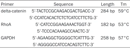
[image:2.612.91.345.84.180.2]Ver.3.0 kit (TaKaRa Bio Inc., Dalian, Liaoning, Table 1. Primer sequences and reaction conditions
Primer Sequence Length Tm
delta-catenin 5’-TACTCCGCAAGACGACTGACC-3’ 284 bp 59°C 5’-CCATCACACTCTCTCATCCTTCTG-3’
RhoA 5’-CATCCGGAAGAAACTGGT-3’ 182 bp 53°C
5’-TCCCACAAAGCCAACTC-3’
GAPDH 5’-AGAAGGCTGGGGCTCATTTG-3’ 258 bp 57°C 5’-AGGGGCCATCCACAGTCTTC-3’
RhoA cytoplasmic staining was scored as strong (+++), moderate (++), faint (+), or negative (-). Scores of – or +
were defined as normal expression, and higher scores were defined as
over-expression [12].
The simultaneous occurrences of del-ta-catenin positive expression and
RhoA over-expression were defined as
China). Primer sequences and reaction condi-tions are shown in Table 1. RT-PCR products were separated on a 1.5% agarose gel, and then grey values were determined using Biolmaging Systems. GAPDH mRNA was use-das an internal control.
RhoA activity assay
RhoA activity was determined using the RhoA G-LISA™ activation assay kit (BK124, Cyto- skeleton, Denver, USA). GTP-bound RhoA activ-ity was measured using a spectrometer (BD Transduction Laboratories) at the wave length of 490 nm.
Matrigel invasion assay (Transwell assay)
Matrigel basement membrane gel (100 μl,
diluted 1:4) (BD Biosciences) was added to the
upper chamber that was incubated at 37°C in 5% CO2 for 2 hours. Then 100 μl of cell suspen -sion was added to the upper chamber (3×10⁵
cells/ml). The lower chamber was filled with RPMI1640 medium containing 10% fetal
bovine serum. Transwell microporous
mem-brane (8 μm pore size, Corning, USA) was
placed between the upper and lower chamber. After transfection (24 hours), tumor cells were seeded into the upper chamber and incubated at 37°C in 5% CO2 for 24 hours. The cells on the
lower surface of the membrane were fixed with
100% methanol for 30 minutes and stained with hematoxylin, and then counted under a
microscope in five fields (×400).
Immunoco-localization
The lung cancer-derived cell lines H460 and LK2 were digested with 0.25% trypsin, and
fixed with 4% paraformaldehyde for 30 min -utes. The cells were treated with 0.5% Triton X-100 for 30 minutes at room temperature, and then incubated with anti-delta-catenin antibody or anti-RhoA antibody at 4°C overnight. The
cells were then incubated with the fluorescently
[image:3.612.90.522.73.292.2]labeled secondary antibody at 37°C for lhour, stained with 5 pg/ml DAPI for 2 minutes, and then examined using laser scanning confocal microscopy.
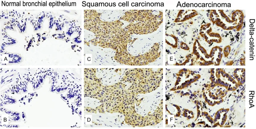
Figure 1. Delta-catenin and RhoA expression in normal bronchial epithelium, lung squamous cell carcinoma and adenocarcinoma. Delta-catenin (A) and RhoA (B) was weakly expressed in the cytoplasm of normal bronchial epi-thelial cells. Positive delta-catenin expression was detected in the cytoplasm of squamous cell carcinoma (C), and RhoA was strongly expressed in these cells (D). Delta-catenin was expressed (E) and RhoA (F) was over-expressed in adenocarcinoma.
Table 2. Correlation between delta-catenin and RhoA in lung cancer
delta-catenin P-value negative positive
RhoA
Normal expression 34 11 <0.001
[image:3.612.91.295.397.465.2]Delta-catenin and RhoA in lung cancer
Co-immunoprecipitation
The H460 and LK2 cells were digested with 0.25% trypsin, collected by centrifugation (12,000 rpm), lysed in lysis buffer for 1 hour, and then centrifuged at 14,000×g for 30
min-taining immune complexes were washed five
times with PBS buffer. Normal rabbit IgG was used as a negative control.
Statistical analysis
All data were analyzed with SPSS for Windows 18.0 (SPSS Inc., Chicago, IL, USA). Immuno- histochemical results were analyzed using Pearson’s Chi-Square test. The results of the western blot and RT-PCR were analyzed by
vari-ance analysis and the t-test. The Kaplan Meier
method was used to compare survival bet- ween different patient groups, and differences in survival were tested using the Log-Rank test. A value of P<0.05 was considered statistically
significant.
Results
Delta-catenin and RhoA co-expression is asso-ciated with a poor prognosis in NSCLC patients
Among 128 lung cancer cases, delta-catenin was expressed in 64.06% (82/128) of cases; and RhoA was over-expressed in 64.84%
(83/128) of cases. There was a significant
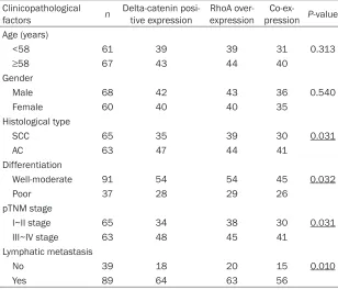
[image:4.612.90.398.97.361.2]association between RhoA over-expression and delta-catenin positive expression (P<0.001) Table 3. Relationship between co-expression and clinicopathological
factors
Clinicopathological
factors n Delta-catenin posi-tive expression RhoA over-expression pressionCo-ex- P-value Age (years)
<58 61 39 39 31 0.313
≥58 67 43 44 40
Gender
Male 68 42 43 36 0.540
Female 60 40 40 35
Histological type
SCC 65 35 39 30 0.031
AC 63 47 44 41
Differentiation
Well-moderate 91 54 54 45 0.032
Poor 37 28 29 26
pTNM stage
I~II stage 65 34 38 30 0.031
III~IV stage 63 48 45 41
Lymphatic metastasis
No 39 18 20 15 0.010
Yes 89 64 63 56
utes at 4°C. The super-natant was extracted, and the protein concen-tration was determined. The extracts (containing 1 mg of total protein)
were incubated with 1 μl
of normal rabbit serum
and 30 μl of protein G
Plus/protein A agarose beads at 4°C for 1 hour, and then centrifuged at 12,000×g for 5 minutes to remove the beads. Anti-delta-catenin (or RhoA) antibody together
with 30 μl protein G
Plus/protein A agarose beads were added to the supernatant, and this was incubated at 4°C overnight. The mixtures were then centrifuged at 12,000×g for 3 minutes to remove the superna-tant, and the beads
[image:4.612.90.287.167.562.2](Figure 1, Table 2). We also analyzed the rela-tionship between co-expression (concurrent delta-catenin positive expression and RhoA over-expression) and clinicopathological fac-tors (Table 3). This revealed that the co-expres-sion rate in lung adenocarcinoma (65.08%,
41/63) was significantly higher than in squa -mous cell carcinoma (46.15%, 30/65) (P<0.05). Furthermore, the co-expression rate in stage III-IV disease (65.08%, 41/63) was higher than in stage I-II disease (46.15%, 30/65) (P<0.05), and the co-expression rate in well to moderate-ly differentiated tumors (70.27%, 26/37) was higher than in poorly differentiated tumors (49.45%, 45/91) (P<0.05). The co-expression rate in tumors with lymph node (LN) metastasis (62.92%, 56/89) was higher than in those with-out LN metastasis (38.46%, 15/39) (P<0.05).
There were no significant differences in
co-expression with respect to patient age and sex (P>0.05).
In cases where delta-catenin and RhoA were co-expressed in tumors, the average survival
time and 5-year survival rate were significantly
lower than when tumors did not co-express delta-catenin and RhoA (P<0.05 for both). The
co-expression of these proteins was thus
sig-nificantly associated with a poor prognosis
(Figure 2).
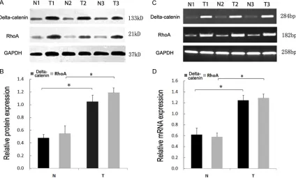
Delta-catenin and RhoA expression was el-evated in lung cancer at both the protein and RNA level
Western blot analysis revealed that the protein expression of both RhoA and delta-catenin in
adjacent normal lung tissue was significantly
lower than in lung cancer (Figure 3A & 3B). RT-PCR analysis also revealed that delta-cateninand RhoARNA levels were significantly
higher in lung cancer compared to normal lung tissue (Figure 3C & 3D).
A direct interaction between delta-catenin and
RhoA
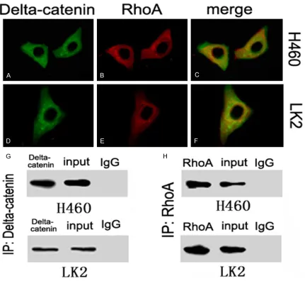
The co-localization of delta-catenin and RhoA in the cytoplasm of H460 and LK2 cells was
confirmed using laser scanning confocal
[image:5.612.94.518.75.330.2]microscopy (Figure 4A-F). Furthermore, the co-immunoprecipitation analysis demonstrated that delta-catenin and RhoA could directly interact with each other in both cell types (Figure 4G & 4H).
Delta-catenin and RhoA in lung cancer
Figure 4. Interaction of delta-catenin and RhoA in lung cancer cells. The sub-cellular location of delta-catenin (A) and RhoA (B) in H460 cells, and their co-localization (C). The sub-cellular location of delta-catenin (D) and RhoA (E) in LK2 cells, and their co-localization (F). Co-immunoprecipitation of delta-catenin and RhoA in H460 and LK2 cells (G, H).
[image:6.612.95.519.533.680.2]Delta-catenin enhances RhoA expression but
suppresses RhoA activity
Western blot and RT-PCR analysis revealed that the protein and mRNA expression of RhoA was reduced after delta-catenin knock-down (Figure 5A & 5B), and enhanced after the forced expression of delta-catenin (Figure 5C & 5D). We also measured RhoA activity after the forced expression or silencing of delta-catenin. G-LISA analysis showed that RhoA activity was markedly decreased after up-regulating delta-catenin in LK2 cells (P<0.05) (Figure 5E), whilst it was strongly increased after down-regulating delta-catenin in H460 cells (Figure 5F).
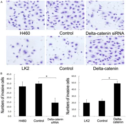
Delta-catenin promotes lung cancer cell
inva-sion
The invasive ability of lung cancer cells after the forced expression or silencing of
delta-catenin was evaluated using the Matrigel inva -sion assay (Figure 6). Forty-eight hours after the forced expression of delta-catenin, the average number of invasive cells (48.65) was
significantly higher than in the control group
[image:7.612.92.521.70.492.2](20.54), whilst 48 hours after the silencing of delta-catenin, the average number of invasive cells (14.96) was lower than in the control group (35.04). Hence, delta-catenin is required for cancer cell invasion.
Delta-catenin and RhoA in lung cancer
Discussion
The expression of delta-catenin and RhoA has been studied in a number of malignant tumors, including those of prostate, ovary, and lung cancer, and glioma [11, 13-20]. However, an association between delta-catenin and RhoA expression has not been established in lung cancer, and it was also uncertain whether there was a relationship between their co-expression and the prognosis of NSCLC patients.
In this study, we found elevated RNA and pro-tein delta-catenin expression in lung cancer cells compared to normal lung tissue. RhoA was also over-expressed in lung squamous cell carcinoma and adenocarcinoma, often concur-rently with delta-catenin expression. Their co-expression was associated with a higher degree of malignancy (high stage, adenocarcinoma, poor differentiation and lymph node metasta-sis), and was closely associated with a poor prognosis for NSCLC patients.
RhoA has been shown to be expressed at a low level in normal tissue, and is mainly cytoplas-mic. It is activated by extracellular stimulation, where upon it is transported to the plasma membrane, and once there it can stabilize intercellular junctions. Delta-catenin was more highly expressed in the cytoplasm of tumor cells, maintaining RhoA in an inactivate state but inducing its expression. This in turn inter-rupts adhesion signals, leading to the degrada-tion of proteins that inhibit migradegrada-tion, including
stress fibers and focal adhesions [12, 21, 22].
Therefore, the immunopositive stainingin the cytoplasm of lung cancer cells might represent an accumulation of GDP-bound RhoA. RhoA activity was suppressed by delta-catenin, and the inactivated RhoA would enhance tumor invasion and metastasis.
We found a significant association between
delta-catenin positive expression and RhoA over-expression, and that these proteins inter-act directly. Both proteins affect cell-cell adhe-sion, and thus delta-catenin was considered to be an important direct regulator of RhoA activi-ty. It has also been reported that RhoA activity is regulated by proteins such as GEF, GAP and GDI [23], and that delta-catenin could indirectly regulate RhoA activity through interacting with these factors [24]. However, the exact nature of these regulatory interactions remains unclear, and further study is therefore needed.
Acknowledgements
We sincerely thank Dr. Shun-ichi Nakamura at Kobe University, Japan, for kindly providing
pCMV5-FLAG/delta-catenin plasmid. This work
was supported by the National Natural Science Foundation of China (no.81101779 to Di Zhang,
MD; no.30870977 and 81071905 to En-Hua Wang, MD; no.81201844 to Jun-Yi Zhang, MD;
no.81372338 to Shu-Li Liu).
Disclosure of conflict of interest
None.
Address correspondence to: Dr. En-Hua Wang, Department of Pathology, The First Affiliated Hospital and College of Basic Medical Sciences of China Medical University, Shenyang, 110001, China. Tel: (+ 86 24) 23261638; Fax: (+ 86 24) 23261638; E-mail: wangeh@hotmail.com
References
[1] Hatzfeld M. The p120 family of cell adhesion molecules. Eur J Cell Biol 2005; 84: 205-14. [2] Kosik KS, Donahue CP, Israely I, Liu X, Ochiishi
T. delta-catenin at the synaptic-adherens junc-tion. Trends Cell Biol 2005; 15: 172-178. [3] Kawamura Y, Fan QW, Hayashi H, Michikawa
M, Yanagisawa K, Komano H. Expression of the mRNA for two isoforms of neural pla-kophilin-related arm-repeat protein/delta-catenin in rodent neurons and glial cells. Neu-rosci Lett 1999; 277: 185-8.
[4] Lu Q, Paredes M, Medina M, Zhou J, Cavallo R, Peifer M, Orecchio L, Kosik KS. delta-catenin, an adhesive junction associated protein which promotes cell scattering. J Cell Biol 1999; 144: 519-532.
[5] Koutras C, Lessard CB, Lévesque G. A nuclear function for the presenilin 1 neuronal partner NPRAP/δ-catenin. J Alzheimers Dis 2011; 27: 307-16.
[6] Kim K, Sirota A, Chen Yh YH, Jones SB, Dudek R, Lanford GW, Thakore C, Lu Q. Dendrite-like process formation and cytoskeletal remodel-ing regulated by delta-catenin expression. Exp Cell Res 2002; 275: 171-84.
[7] Abu-Elneel K, Ochiishi T, Medina M, Remedi M, Gastaldi L, Caceres A, Kosik KS. A delta-catenin signaling pathway leading to dendritic protrusions. J Biol Chem 2008; 283: 32781-91.
control of neuronal morphology. Cell Biol 1997; 137: 1603-13.
[9] Azab AK, Azab F, Blotta S, Pitsillides CM, Thompson B, Runnels JM, Roccaro AM, Ngo HT, Melhem MR, Sacco A, Jia X, Anderson KC, Lin CP, Rollins BJ, Ghobrial IM. RhoA and Rac1 GTPases play major and differential roles in stromal cell-derived factor-1-induced cell ad-hesion and chemotaxis in multiple myeloma. Blood 2009; 114: 619-29.
[10] Struckhoff AP, Rana MK, Kher SS, Burow ME, Hagan JL, Del Valle L, Worthylake RA. PDZ-Rho-GEF (PRG) is essential for CXCR4-driven breast tumor cell motility through spatial regulation of RhoA. J Cell Sci 2013; 126: 4514-26.
[11] Zhang JY, Wang Y, Zhang D, Yang ZQ, Dong XJ, Jiang GY, Zhang PX, Dai SD, Dong QZ, Han Y, Zhang S, Cui QZ, Wang EH. Delta-Catenin pro-motes malignant phenotype of non-small cell lung cancer by non-Competitive binding to E-cadherin with p120ctn in cytoplasm. J Pathol 2010; 222: 76-88.
[12] Horiuchi A, Imai T, Wang C, Ohira S, Feng Y, Ni-kaido T, Konishi I. Up-regulation of small GT-Pases, RhoA and RhoC, is associated with tu-mor progression in ovarian carcinoma. Lab Invest 2003; 83: 861-70.
[13] Burger MJ, Tebay MA, Keith PA, Samaratunga HM, Clements J, Lavin MF, Gardiner RA. Ex -pression analysis of delta-catenin and pros-tate-specific membrane antigen: their poten -tial as diagnostic markers for prostate cancer. Int J Cancer 2002; 100: 228-37.
[14] Lu Q, Dobbs LJ, Gregory CW, Lanford GW, Rev-elo MP, Shappell S, Chen YH. Increased ex -pression of delta-catenin/neural plakophilin-related armadillo protein is associated with the down-regulation and redistribution of E-cad-herin and p120ctn in human prostate cancer. Hum Pathol 2005; 36: 1037-48.
[15] Lu Q, Zhang J, Allison R, Gay H, Yang WX, Bhow-mick NA, Frelix G, Shappell S, Chen YH. Identi-fication of extracellular delta-catenin accumu -lation for prostate cancer detection. Prostate 2009; 69: 411-8.
[16] Fang Y, Li Z, Wang X, Zhang S. Expression and biological role of δ-catenin in human ovarian cancer. J Cancer Res Clin Oncol 2012; 138: 1769-76.
[17] Li XY, Liu SL, Cha N, Zhao YJ, Wang SC, Li WN, Wang EH, Wu GP. Transcription expression and clinical significance of dishevelled-3 mRNA and δ-catenin mRNA in pleural effusions from patients with lung cancer. Clin Dev Immunol 2012; 2012: 904-946.
[18] Zhang JY, Zhang D, Wang EH. Overexpression of small GTPases directly correlates with ex-pression of δ-catenin and their coexex-pression predicts a poor clinical outcome in non-small cell lung cancer. Mol Carcinog 2013; 52: 338-47.
[19] Dai SD, Wang Y, Zhang JY, Zhang D, Zhang PX, Jiang GY, Han Y, Zhang S, Cui QZ, Wang EH. Upregulation of delta-catenin is associated With poor prognosis and enhances transcrip-tional activity through Kaiso in non-small-cell lung cancer. Cancer Sci 2011; 102: 95-103. [20] Wang M, Dong Q, Zhang D, Wang Y. Expression
of delta-catenin is associated with progression of human astrocytoma. BMC Cancer 2011; 11: 514.
[21] Fritz G, Brachetti C, Bahlmann F, Schmidt M, Kaina B. Rho GTPases in human breast tu-mours: expression and mutation analyses and correlation with clinical parameters. Br J Can-cer 2002; 87: 635-44.
[22] Anastasiadis PZ, Reynolds AB. Regulation of Rho GTPases by p120-catenin. Curr Opin Cell Biol 2001; 13: 604-10.
[23] Falkenberg CV, Loew LM. Computational analy -sis of Rho GTPase cycling. PLoS Comput Biol 2013; 9: e1002831.