Evaluation of equine cortical bone transplantation
in a canine fracture model
S.H. Heo
1, C.S. Na
2, N.S. Kim
11College of Veterinary Medicine, Chonbuk National University, Jeonju, Republic of Korea 2College of Agriculture, Chonbuk National University, Jeonju, Republic of Korea
ABSTRACT: Freeze-dried bovine bone transplantation is commonly used for orthopaedic surgery. Equine bone,
which is available in great quantity, can be obtained as easily as bovine bone, and so represents a potential source of bone for transplantation. In the present study freeze-dried equine cortical bones were transplanted into experimentally-induced fibular defects in canines to evaluate xenogenic implantation of equine bone. Cortical bones that had been freed of antigens and defatted with chloroform and methanol were freeze-dried at –80 °C for preservation of bone morphogenetic protein, sterilized with ethylene oxide gas and stored at room temperature. The experimental osteotomy was performed in a 15 mm-long bilateral region of each proximal metaphyseal fibula. The area of defect in eight beagle dogs (n = 16) received a transplanted freeze-dried equine cortical implant. The control group consisting of two beagles dogs (n = 4) received an autograft of a similar implant. The experiment region was radiographically monitored for bone union and host serum osteocalcin level was determined to assess osteoblast activity every two weeks for 24 weeks. In 14 of the 16 experimental cases, the graft was not associ-ated with new bone formation. Resorption after new bone formation and remodelling with new bone formation each occurred in a single case. The results support the potential of using freeze-dried equine cortical bones as a xenogenic bone graft material in canines.
Keywords: equine bone; bone graft; xenogenic bone; bone healing; canine model
In the reconstruction of skeletal defects, it is of-ten necessary to transplant cancellous or cortical bone to restore skeletal integrity and enhance bone healing. The materials used in bone grafting can be divided into autografts, allografts, xenografts, and synthetic materials (Finkemeier, 2002; Parikh, 2002; Cho and Lee, 2006).
In clinical medicine, autologous bone grafting is the best choice for enhancing bone repair and reconstructive bone defects (Burchardt, 1987; Finkemeier, 2002; Parikh, 2002). However, the use of autografts is hampered by the limited supply of bone grafts, the necessity of an additional incision site, donor site morbidity, nerve damage, and injury of the donor site (Younger and Chapman, 1989; Finkemeier, 2002).
Allografts are frequently used in orthopaedic sur-gery for the treatment of bone nonunion,
enhanc-ing the repair of spine fracture, and reconstruction of bone defects (Sinibaldi, 1989; Berrey et al., 1990; Brady et al., 1999; Haddad and Duncan, 2003). The advantages of allografts include the absence or min-imization of donor site morbidity and the unlimited choice of graft shape and size (Finkemeier, 2002; Cho and Lee, 2006). In human medicine, allografts are commonly used, although their expanded use is limited by the supply of bone and the potential for disease transmission (Buck et al., 1989; Hofmann et al., 1995). These limitations have spurred interest in substitute materials.
Recent therapeutic technologies concerning bone substitutes and alternatives to biocompatible scaf-folds include growth factor, calcium phosphate, hy-droxyapatite, tricalcium phosphate, type I collagen, bioactive glasses, and synthetic polymers. Such ma-terials have been used to fill bone defects in
mental animal studies and also clinically (Shand and Heggie, 2000; Finkemeier, 2002; Cho and Lee, 2006; Hing et al., 2006; Komaki et al., 2006). The synthetic materials are not osteoinductive and do not induce formation of new bone.
Xenograft bone represents an unlimited supply of available material. Bovine bone transplantation is very commonly used for orthopaedic treatment in periodontal, maxillofacial, and neurosurger-ies. Xenograft bone or collagen has been experi-mentally explored as a bone substitute (Marchesi, 2000; El-Sabban et al., 2007; Stievano et al., 2008). However, the use of bovine bone transplantation is currently limited in human medicine because of the potential for introduction of infection (Boneva et al., 2001; Wenz et al., 2001). In seeking an alter-native, equine bone is an attractive potential op-tion. It is available in large quantities and can be obtained as readily as bovine bone. However, the use of equine bone graft in canines has not been studied. This study was undertaken to investigate the use of freeze-dried equine cortical bones as a xenogenic implant in experimentally-induced fibu-lar defects in beagle dogs.
MATERIAL AND METHODS
Animals and experimental design
Ten Beagle dogs weighing 9–12 kg (average 10.1 ± 1.1 kg) and 2–3-years-of-age (average 2.1 ± 1.7 years) were used. Physical and radiographic ex-aminations were performed to ensure the absence of orthopaedic diseases. A canine fibula segmental
defect model (Burchardt et al., 1977; Burchardt, 1987) was used to determine the efficacy of the union implants and to compare healing follow-ing bone graftfollow-ing. The basic experimental model consisted of a 15 mm-long, bilateral region proxi-mal metaphyseal fibula segmental defect, which was created in each fibula using an oscillating saw under continuous saline irrigation. Eight dogs re-ceived the transplanted freeze-dried equine corti-cal implant. The remaining two dogs constituted the control, and received an autograft of the trans-planted freeze-dried cortical implant. Therefore, the experimental group consisted of 16 cases and the control group consisted of four cases.
Preparation of freeze-dried cortical bone materials
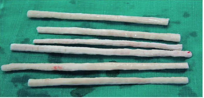
[image:2.595.128.476.555.722.2]Cortical bone was obtained from a six-year-old, castrated, male racehorse. The ends of the long bones were cut away with a bone band saw. The midshafts were mechanically scraped clean of soft tissues including cartilage, periosteum, and bone marrow, and washed in cold Ringer’s lactate solu-tion. The bone segment (3–5 thick, 20–30 mm-long) was cut to fibula size. The bones were dried and washed with deionized water for 1 h at 4 °C, with periodic stirring. The bones were defatted in a 1 : 1 ratio of chloroform and methanol at 24 °C for seven days, and kept in flowing air for 1 h at room temperature to remove the residual chloroform and methanol by evaporation. The defatted bones were washed by stirring in deionized water for 2 h at 4 °C, and were then frozen at –80 °C. The cortical bone
plate were placed in the drying chamber of a model OPR-FDC-8032 freeze-drier (Operon Engineering, Seoul, Korea) pre-cooled to –80 °C , and a vacuum of 10–2 was applied. Complete freeze-drying took about six days, after which the moisture content of the bone was about 5%. The freeze-dried bone was double-wrapped in sterilized packs (one side paper, one side plastic film) and sterilized by exposure to ethylene oxide gas. Sterilized samples were stored at room temperature until use (Figure 1).
Surgical procedure
Prior to surgery, each patient received 25 mg/kg cephalexin intramuscularly (Methilexin®; Union Korea Pharm, Seoul, Korea) for prophylaxis. The experimental animals were premedicated with 0.02 mg/kg subcutaneous atropine sulfate (Atropine Sulfate Daewon®; Dae Won Pharm, Seoul, Korea), anaesthesia was induced using 5 mg/kg intravenous propofol (Anepol IN®; Ha Na Pharm, Seoul, Korea), and was maintained with 2–3.5% isoflurane and oxygen. The animals were placed in a lateral re-cumbent position and all surgical procedures were conducted under sterile conditions. A lateral lon-gitudinal incision was made over the fibula and the underlying muscles were retracted, exposing the femur shaft. A 15 mm-long segmental defect was created proximal to the metaphyseal of the fibula using a model BL-F3 electric handpiece saw (Osada Electric, Tokyo, Japan) under continuous saline ir-rigation (Figure 2). In the experimental group of
eight dogs, each fibular segmental defect (n = 16) received a freeze-dried equine cortical implant. In the two control animals, each segmental defect (n = 4) was inverted and received an autograft of freeze-dried cortical implant. The muscle attach-ment was repaired and the skin was closed in layers. Postoperatively a radiography was obtained for all operated ulna to ensure adequate implant place-ment and alignplace-ment. The animals were permitted to freely weight bear immediately following surgery and were allowed to eat.
Haematology and clinical chemistry analyses
Analyses were obtained on the day of surgery, and every two-weeks for 24 weeks. Clinical and labo-ratory assessment in all dogs included a physical examination, a complete blood count performed using a Vet Abc automated animal blood counter (ABX Diagnostic, Montpellier, France), a blood smear examination, and basic serum chemistry pro-file using a SpotchemTM SP-4410 automated chem-istry analyzer (Daiichi Kagaku, Tokyo, Japan).
Osteocalcin analysis
[image:3.595.74.518.532.723.2]Jugular venous blood samples of each dog were col-lected every two weeks. Samples were individually collected in a 10 ml serum tube. All samples were centrifuged at 3000 rpm at 4 °C for 10 min within 1 h
after sampling. Serum was stored in 3 ml aliquots in a deep freezer at –81 °C until assayed. A Gla-type osteo-calcin enzyme-linked immunoassay (EIA) kit (Takara Shuzou, Kyoto, Japan) was used for determination of Gla type osteocalcin concentration according to the manufacturer’s protocol. In brief, 100 ml of each test sample was dispensed into wells of a microtitre plate precoated with bovine Gla-osteocalcin standard solu-tion. Following incubation at room temperature for 2 h and washing, detection antibody (mouse
[image:4.595.128.465.82.406.2]mono-clonal anti-Gla-osteocalcin) conjugated to horserad-ish peroxidase was applied to each well and incubated for 1 h at room temperature. After washing, 100 ml of substrate solution containing 3,3',5,5'-tetrameth-ylbenzine was added to each well. The plates were incubated for 15 min at room temperature, followed by addition of 100 ml of stop solution. After stopping the reaction with 100 µl of 1N H2SO4, the test ab-sorbance at 450 nm was measured with a microplate reader (Labsystem Japan, Tokyo, Japan).
Figure 3. Serial radiographs of hindlimbs with transplanted equine freeze-dried cortical bone in the fibular of dogs. No remarkable changes at the implanted area were observed in 14 cases (A). Transplants with different absorption patterns were found in two cases (B and C)
[image:4.595.108.455.620.726.2]Plain radiography
The fate of the graft was examined by X-ray every two weeks for 24 weeks. Examined parameters in-cluded new bone formation, union of the gap de-fect, and absorption of bone material.
Histological evaluation
After 24 weeks, each dog was euthanatized by in-travenous administration of sodium pentobarbital (Entobar Inj®; Han Lim Pharm, Seoul, Korea). Each
fibula was carefully dissected from the surrounding soft tissue to preserve the bone. Each sample was fixed with 10% formalin, demineralized with 10% nitric acid for 3–5 days, and processed routinely for light microscopy. In brief, the demineralized samples were treated with ascending graded alco-hol solutions (70% to absolute alcoalco-hol). After treat-ment with xylene, the samples were embedded in paraffin and sectioned. The 5 µm-thick sections were stained with Masson trichrome and observed under a light microscope. Sections were examined for evidence of inflammation and foreign body re-action.
0 2 4 6 8 10 12
0 2 4 6 8 10 12 14
Weeks
(n
g/
m
l)
Control
Experimental
* * *
*
* *
*
[image:5.595.64.326.82.256.2]* *
Figure 5. Changes in osteocalcin values in serum after transplantion of freeze-dried cortical equine bone into dogs
*significant difference in comparison with pre-operative values at weeks 0; (P < 0.05)
[image:5.595.69.521.440.703.2]Statistical analysis
Results are presented as means ± SD. Statistical analysis of data was performed using Student’s t-test. Values were considered statistically signifi-cant when a P < 0.05 or < 0.01 was obtained.
RESULTS
Surgical follow-up
All animals recovered fully with no infection or wound inflammation evident. All clinical signs in the study period were within normal ranges.
Haematology and clinical chemistry analyses
No significant changes were observed. Post-surgical mean values for white blood cell count and liver enzymes were higher in the two control dogs than in the eight experimental dogs at two weeks, but were not statistically significant (P > 0.05)
Plain radiography
In the xenograft group, no remarkable changes at the site of bone transplant were observed in 14 of the 16 cases without any evidence of heal-ing (Figure 3A). In the remainheal-ing two cases, the transplant was gradually resorbed (Figures 3B and C). In the autograft group, no evidence of union was found in any of the four cases and the transplants tended to be absorbed more slowly (Figure 4).
Osteocalcin analysis
During the bone graft period, the osteocalcin serum concentration tended to be lower than the pre-operative level in all groups. In the two control dogs, serum osteocalcin values decreased significantly between weeks 2–14 (P < 0.05). In the experimental group, serum osteocalcin values decreased significantly between weeks 4–10 (P < 0.05). However, no significantly differences were evident between the groups (Figure 5).
Histological evaluation
No evidence of inflammation and foreign body reaction were observed in either group. In the xe-nograft group, a longitudinal section in 14 cases demonstrated no evidence of bone bridging, filling of the middle gap with collagenous fibrous or bone union (Figure 6), while in two cases, the longitu-dinal section demonstrated an irregular margin of bone defect due to resorption and a lack of heal-ing of the bone defect (Figure 7). In the autograft group, the longitudinal section demonstrated no evidence of bone bridging, resorption of cortical bone, or tissue (Figure 8).
DISCUSSION
The absorption of transplanted bone can take place early following transplantation, can occur later in time, or may be a long lasting absorption without new bone formation (Burchardt et al., 1977; Burchardt, 1987; Choi et al., 1996). In this study, 14 out of 16 equine bone grafts showed no absoption and were without remarkable changes. Implant resorption was found in two cases only.
A rabbit study conducted over three decades ago demonstrated that decalcified cortical bone can induce bone formation intramuscularly in an ectopic site (Urist, 1965). Xenograft experiments conducted in a variety of animal species and graft sites, and with different bank methods have re-ported enhanced bone formation and bone fracture repair (Anderson et al., 1965; Fujinaga and Koike, 1976; Jensen et al., 2006; Nienhuijs et al., 2006). Mare cortical bone xenografts of low antigenicity have been successfully produced by a cycle of boil-ing and freezboil-ing (Fujinaga and Koike, 1976). Gupta et al. (1982) compared defatted and decalcified xenogenic bone implants in large cortical defects repair and reported a high success rate. Zhao et al. (1998) reported a successful result using bo-vine bone graft for the reconstruction of anterior base defects in humans. The present results echo those found using bone transplantation allografts. Although less union and formation of callus bridges was observed, the union of distal host bone did oc-cur. No appreciable graft antigenicity was observed either clinically or histologically.
osteocacin-Figure 7. In the xenograft group, the section shows non-union of bones. Both bone margins are resorbed without evidence of reactive bone formation and the gap between the margins are filled with dense collagenous fibrous con-nective tissue. There is no evidence of inflammatory reactions (trichrome stain, inlet original magnification ×100)
Figure 8. In the autograft group, the gap between the margins of the bone defect is filled with dense collagenous fibrous connective tissue. The margin of the bone defect shows irregular ends due to resorption without evidence of bone healing. No inflammatory reaction is noted (trichrome stain, inlet original magnification ×100)
deficient mice has been reported (Ducy et al., 1996), indicating a suppressive role of osteocacin in bone formation. In contrast, other studies conducted
[image:7.595.67.520.459.709.2]Xenografts can be processed to minimize an-tigenicity. Successful processes include freezing, decalcification, freeze-drying, and deproteiniza-tion, with freeze-drying and deep-freezing being most common. Preservation by either freezing or freeze-drying allows extended storage but reduces graft immunogenicity and may alter its mechanical strength. Bone graft preservation aims to maintain the osteoindutive and osteoconductive capacity of material, as well as to reduce its immunogenicity (Friedlaender and Mankin, 1981; Pelker et al., 1984; Burchardt, 1987). The present graft preparations and storage regimens were consistent with these aims.
Lipid extraction by chloroform methanol increas-es the incorporation of frozen bone grafts (Thoren et al., 1995), perhaps due to a decreased immuno-logic response. The effect may be also caused by the eradication of major histocompatibility antigens because of cell membrane dissolution (Friedlaender and Mankin, 1981). In this study, inflammation and foreign body response were not observed in the fibula surgical site. However, in the experimental group, repair was characterized by unincorporated, non-union of host bone. The implanted equine cor-tical bone grafts were not resorbed and no com-plications ensued.
In conclusion, freeze-dried equine cortical bone has potential as a bone graft material in dogs to alleviate serious bone defects.
REfERENCES
Anderson KJ, Fry LR, Clawson DK, Sakurai O (1965): Ex-perimental comparison of autogenous, homogenous, and heterogenous bone grafts: a planimetric measure-ment study. Annals of Surgery 161, 263–271.
Berrey BH Jr, Lord CF, Gebhardt MC, Mankin HJ (1990): Fractures of allografts. Frequency, treatment, and end-results. Journal of Bone and Joint Surgery (American) 72, 825–833.
Boneva RS, Folks TM, Chapman LE (2001): Infectious disease issues in xenotransplantation. Clinical Micro-biology Reviews 14, 1–14.
Brady OH, Garbuz DS, Masri BA, Duncan CP (1999): The treatment of periprosthetic fractures of the femur using cortical onlay allograft struts. Orthopedic Clin-ics of North America 30, 249–257.
Buck BE, Malinin TI, Brown MD (1989): Bone trans-plantation and human immunodeficiency virus. An estimate of risk of acquired immunodeficiency
syn-drome (AIDS). Clinical Orthopaedics and Related Research 240, 129–136.
Burchardt H (1987): Biology of bone transplantation. Orthopedic Clinics of North America 18, 187–196. Burchardt H, Glowczewskie FP, Enneking WF (1977):
Allogeneic segmental fibular transplants in azathio-prine-immunosuppressed dogs. Journal of Bone and Joint Surgery (American) 59, 881–894.
Cho TJ, Lee KS (2006): Bone graft substitute. Journal of the Korean Fracture Society 19, 109–116.
Choi IH, Kim HG, Kim NS, Sasaki N (1996): Effective-ness of freeze-dried bone grafts on the non-union fracture of dogs. Korean Journal of Veterinary Re-search 36, 495–511.
Ducy P, Desbois C, Boyce B, Pinero G, Story B, Dunstan C, Smith E, Bonadio J, Goldstein S, Gundberg C, Bra-dley A, Karsenty G (1996): Increased bone formation in osteocalcin-deficient mice. Nature 382, 448–452. El-Sabban ME, El-Khoury H, Hamdan-Khalil R,
Sindet-Pedersen S, Bazarbachi A (2007): Xenogenic bone matrix extracts induce osteoblastic differentiation of human bone marrow-derived mesenchymal stem cells. Regenerative Medicine 2, 383–390.
Finkemeier CG (2002): Bone-grafting and bone-graft substitutes. Journal of Bone and Joint Surgery (Amer-ican) 84, 454–464.
Friedlaender GE, Mankin HJ (1981): Bone banking: cur-rent methods and suggested guidelines. Instructional Course Lectures 30, 36–55.
Fujinaga T, Koike T (1976): An examination of graft al-teration and recipient response to processed mare cortical bone xenografting. Japanese Journal of Vet-erinary Research 24, 1–12.
Gupta D, Khanna S, Tuli SM (1982): Bridging large bone defects with a xenograft composited with autologous bone marrow. An experimental study. International Orthopaedics 6, 79–85.
Haddad FS, Duncan CP (2003): Cortical onlay allograft struts in the treatment of periprosthetic femoral frac-tures. Instructional Course Lectures 52, 291–300. Hing KA, Revell PA, Smith N, Buckland T (2006): Effect
of silicon level on rate, quality and progression of bone healing within silicate-substituted porous hydroxya-patite scaffolds. Biomaterials 27, 5014–5026.
Hofmann GO, Kirschner MH, Wangemann T, Falk C, Mempel W, Hammer C (1995): Infections and immu-nological hazards of allogeneic bone transplantation. Archives of Orthopaedic and Trauma Surgery 114, 159–166.
phosphate. A histologic and histomorphometric study in the mandibles of minipigs. Clinical Oral Implants Research 17, 237–243.
Kim NS, Kim SM, Kang CW, Choi OK, Choi IH (2002): The changes of osteocacin and procollagen carboxy-terminal propetide on healing in canine fracture model. Journal Veterinary Clinics 19, 405–410. Komaki H, Tanaka T, Chazono M, Kikuchi T (2006):
Repair of segmental bone defects in rabbit tibiae using a complex of beta-tricalcium phosphate, type I col-lagen, and fibroblast growth factor-2. Biomaterials 27, 5118–5126.
Marchesi DG (2000): Spinal fusions: bone and bone sub-stitutes. European Spine Journal 9, 372–378.
Lammens J, Liu Z, Aerssens J, Dequeker J, Fabry G (1998) Distraction bone healing versus osteotomy healing: a comparative biochemical analysis. Journal of Bone and Mineral Research 13, 279–286.
Nienhuijs ME, Walboomers XF, Merkx MA, Stoelinga PJ, Jansen JA (2006): Bone-like tissue formation using an equine COLLOSS E-filled titanium scaffolding ma-terial. Biomaterials 27, 3109–3114.
Parikh SN (2002): Bone graft substitutes: past, present, future. Journal of Postgraduate Medicine 48, 142–148. Pelker RR, Friedlaender GE, Markham TC, Panjabi MM,
Moen CJ (1984): Effects of freezing and freeze-drying on the biomechanical properties of rat bone. Journal of Orthopaedic Research 1, 405–411.
Shand JM, Heggie AA (2000): Use of a resorbable fixation system in orthognathic surgery. British Journal of Oral and Maxillofacial Surgery 38, 335–337.
Sinibaldi KR (1989): Evaluation of full cortical allografts in 25 dogs. Journal of the American Veterinary Med-ical Association 194, 1570–1577.
Stievano D, Di Stefano A, Ludovichetti M, Pagnutti S, Gazzola F, Boato C, Stellini E (2008): Maxillary sinus lift through heterologous bone grafts and simultane-ous acid-etched implants placement. Five year fol-low-up. Minerva Chirurgica Journal 63, 79–91. Thoren K, Aspenberg P, Thorngren KG (1995): Lipid
extracted bank bone. Bone conductive and mechanical properties. Clinical Orthopaedics and Related Re-search 311, 232–246.
Urist MR (1965): Bone: formation by autoinduction. Sci-ence 150, 893–899.
Wenz B, Oesch B, Horst M (2001): Analysis of the risk of transmitting bovine spongiform encephalopathy through bone grafts derived from bovine bone. Bio-materials 22, 1599–1606.
Younger EM, Chapman MW (1989): Morbidity at bone graft donor sites. Journal of Orthopaedic Trauma 3, 192–195.
Zhao Y, Symington JM, Listrom RD (1998): Experimen-tal study of cattle bone grafting in combination with implant insertion. Chinese Journal of Stomatology 33, 76–78.
Received: 2010–10–25 Accepted after corrections: 2011–03–13
Corresponding Author:
Professor Dr. Nam Soo Kim, Chonbuk National University, College of Veterinary Medicine, Department of Veterinary Surgery, Jeonju 561-756, Republic of Korea