N
-(2,4,6-Trimethylphenyl)formamide
Marile´ Landman, Belinda van der Westhuizen, Daniela I. Bezuidenhout and David C. Liles*
Department of Chemistry, University of Pretoria, Private Bag X20, Hatfield 0028, South Africa
Correspondence e-mail: dave.liles@up.ac.za
Received 6 December 2010; accepted 8 December 2010
Key indicators: single-crystal X-ray study;T= 293 K; mean(C–C) = 0.003 A˚;
Rfactor = 0.048;wRfactor = 0.138; data-to-parameter ratio = 11.0.
The title compound, C10H13NO, was obtained as the unexpected, almost exclusive, product in the attempted synthesis of a manganese(I)–N-heterocyclic carbene (NHC) complex. The dihedral angle between the planes of the formamide moiety and the aryl ring is 68.06 (10). In the
crystal, molecules are linked by N—H O hydrogen bonds, forming infinite chains along thecaxis.
Related literature
For background to formamide formation from NHCs, see: Denket al.(2001). The rotation of the formamide entity out of the plane of the aryl ring and the hydrogen-bonding motif displayed by this structure are similar to those observed for the related compound N-(2,6-dimethyl)-formamide, see: Hansonet al.(2004); Omondiet al.(2005).
Experimental
Crystal data
C10H13NO
Mr= 163.21 Monoclinic,P21=c
a= 8.0659 (7) A˚
b= 15.9004 (13) A˚ c= 8.4290 (7) A˚
= 119.361 (1)
V= 942.17 (14) A˚3
MoKradiation
= 0.07 mm1
0.440.380.28 mm
Data collection
Bruker (Siemens) P4 diffractometer fitted with a SMART 1K CCD detector
Absorption correction: multi-scan (SADABS; Bruker, 2001) Tmin= 0.946,Tmax= 0.979
4988 measured reflections 1778 independent reflections 1607 reflections withI> 2(I) Rint= 0.026
Refinement
R[F2> 2(F2)] = 0.048
wR(F2) = 0.138
S= 1.09 1778 reflections
161 parameters
All H-atom parameters refined max= 0.21 e A˚
3
min=0.21 e A˚ 3
Table 1
Hydrogen-bond geometry (A˚ ,).
D—H A D—H H A D A D—H A
N1—H1 O1i
0.83 (2) 2.05 (2) 2.8775 (18) 171.4 (19)
Symmetry code: (i)x;yþ1 2;z
1 2.
Data collection:SMART(Bruker, 2001); cell refinement:SAINT
(Bruker, 2001); data reduction:SAINT; program(s) used to solve structure: SHELXTL (Sheldrick, 2008); program(s) used to refine structure:SHELXTL andSHELXL97(Sheldrick, 2008); molecular graphics:POV-RAY(Cason, 2004) andMercury(Brunoet al., 2002); software used to prepare material for publication:SHELXL97and
PLATON(Spek, 2009).
Funding received for this work from the University of Pretoria, and the National Research Foundation is acknowl-edged.
Supplementary data and figures for this paper are available from the IUCr electronic archives (Reference: BT5433).
References
Bruker (2001).SMART,SAINTandSADABS. Bruker AXS Inc., Madison, Wisconsin, USA.
Bruno, I. J., Cole, J. C., Edgington, P. R., Kessler, M., Macrae, C. F., McCabe, P., Pearson, J. & Taylor, R. (2002).Acta Cryst.B58, 389–397.
Cason, C. J. (2004).POV-RAY for Windows. Persistence of Vision, Raytracer Pty. Ltd, Victoria, Australia. URL: http://www.povray.org.
Denk, M. K., Rodenzo, J. M., Gupta, S. & Lough, A. (2001).J. Organomet. Chem.617–618, 242–253.
Hanson, J. R., Hitchcock, P. B. & Rodriguez-Medina, I. C. (2004).J. Chem. Res. pp. 664–666.
Omondi, B., Fernandes, M. A., Layh, M., Levendis, D. C., Look, J. L. & Mkwizu, T. S. P. (2005).CrystEngComm,7, 690–700.
Sheldrick, G. M. (2008).Acta Cryst.A64, 112–122. Spek, A. L. (2009).Acta Cryst.D65, 148–155.
Structure Reports
Online
supporting information
Acta Cryst. (2011). E67, o120 [https://doi.org/10.1107/S1600536810051469]
N
-(2,4,6-Trimethylphenyl)formamide
Maril
é
Landman, Belinda van der Westhuizen, Daniela I. Bezuidenhout and David C. Liles
S1. Comment
N-(2,4,6-Trimethyl-phenyl)-formamide (N-mesityl-formamide) (1) was formed as an unexpected product in the attempted synthesis of a manganese(I)—N-heterocyclic carbene (NHC) complex. Instead of the target complex, the mesityl
formamide was obtained almost exclusively. The ylidene molecule, formed by deprotonation of
1,3-bis(2,4,6-trimethyl-phenyl)-imidazolium chloride (IMesHCl) by a strong base, is prone to undergo side reactions. Thus the strong base, and
the subsequent addition of Mn(CO)5Br, resulted in the formation of N,N′-bis-mesityl-N-vinyl-formamidine and after
hydrolysis of this molecule the NC—N bond dissociated to form 1 and a mesityl-vinyl-amine fragment which was not isolated. Denk et al. (2001) have reported the hydrolysis of NHCs, with formamide formation via ring opening, resulting
in an acyclic product.
The molecular structure of the title compound (1) (Fig. 1) is similar to that of the related compound, N-(2,6-dimethyl-phenyl)-formamide, the structure of which has been reported at 173 K (Hanson, et al., 2004) and 293 K (Omondi, et al.,
2005). Owing to the influence of the bulky methyl substituents in the 2 and 6 positions, the formamide moiety is rotated
out of the plane of the aryl ring: in 1, the angle between the planes of the formamide moiety (C1, N1, C10, O1) and the aryl ring is 68.06 (10)°. This compares with 64.75 (12)° (173 K) and 66.45 (12)° (293 K) found for the 2,6-dimethyl
analogue.
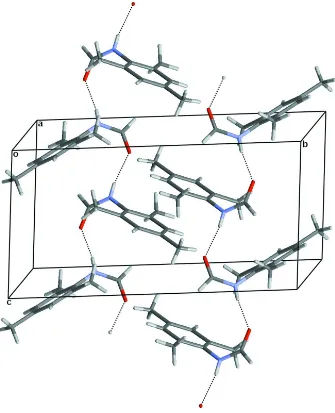
In the formamide moieties of both structures the O atom is trans to N—H thus allowing the molecules to be linked to
form infinite chains by N—H···O hydrogen bonds. However the spatial arrangements within the chains differ. In the
2,6-dimethyl analogue (space group P212121), the axis of each chain is parallel to the a unit cell axis and neighbouring
molecules within a chain are related by the a-axial unit cell translation. Thus the aryl ring of each molecule is parallel to
those of its neighbours within the chain and they are stacked one above the other but with a step-wise offset. In contrast,
in 1, the axis of each chain is parallel to the c unit cell axis and neighbouring molecules within a chain are related by a c-glide plane. Thus neighbouring molecules in a chain are arranged on opposite sides of the chain axis and the aryl rings are
not mutually parallel (Fig. 2).
S2. Experimental
Mn(CO)5Br (3 mmol, 0.74 g) and Me3NO (2.8 mmol, 0.21 g) were stirred in thf resulting in a red solution. IMesHCl (3
mmol, 1.02 g) was deprotonated in thf by the addition of base (3 mmol) and the ylidene was added to the solution and
stirred overnight. The thf solvent was removed and the products were separated on an aluminium oxide 90 (alox) column.
Elution with dichloromethane (dcm) and thf yielded starting material and a yellow fraction respectively. The yellow
fraction was crystallized from a saturated chloroform solution to give an unexpected organic product,
N-mesityl-formamide (1, C10H13NO). 1H NMR (δ, p.p.m.), C6D6: 2.24 (br, 9H), 3.85 (br, 1H), 6.65 (br, 2H), 8.32 (br, 1H); 13C NMR
The coordinates and individual Uiso parameters for all H atoms were freely refined.
Figure 1
Figure 2
Packing diagram of 1 viewed approximately down the a-axis and showing O—H···O hydrogen bonding interactions which link molecules to form infinite chains.
N-(2,4,6-Trimethylphenyl)formamide
Crystal data
C10H13NO
Mr = 163.21 Monoclinic, P21/c
Hall symbol: -P 2ybc a = 8.0659 (7) Å b = 15.9004 (13) Å c = 8.4290 (7) Å β = 119.361 (1)° V = 942.17 (14) Å3
Z = 4
F(000) = 352 Dx = 1.151 Mg m−3
Mo Kα radiation, λ = 0.71073 Å Cell parameters from 3714 reflections θ = 2.8–26.4°
µ = 0.07 mm−1
Bruker P4 diffractometer
Radiation source: fine-focus sealed tube Graphite monochromator
Detector resolution: 8.3 pixels mm-1
φ and ω scans
Absorption correction: multi-scan (SADABS; Bruker, 2001) Tmin = 0.946, Tmax = 0.979
4988 measured reflections 1778 independent reflections 1607 reflections with I > 2σ(I) Rint = 0.026
θmax = 26.4°, θmin = 2.6°
h = −9→10 k = −14→18 l = −10→5
Refinement
Refinement on F2
Least-squares matrix: full R[F2 > 2σ(F2)] = 0.048
wR(F2) = 0.138
S = 1.09 1778 reflections 161 parameters 0 restraints 0 constraints
Primary atom site location: structure-invariant direct methods
Secondary atom site location: difference Fourier map
Hydrogen site location: difference Fourier map All H-atom parameters refined
w = 1/[σ2(F
o2) + (0.0738P)2 + 0.186P]
where P = (Fo2 + 2Fc2)/3
(Δ/σ)max = 0.004
Δρmax = 0.21 e Å−3
Δρmin = −0.21 e Å−3
Special details
Geometry. All e.s.d.'s (except the e.s.d. in the dihedral angle between two l.s. planes) are estimated using the full covariance matrix. The cell e.s.d.'s are taken into account individually in the estimation of e.s.d.'s in distances, angles and torsion angles; correlations between e.s.d.'s in cell parameters are only used when they are defined by crystal symmetry. An approximate (isotropic) treatment of cell e.s.d.'s is used for estimating e.s.d.'s involving l.s. planes.
Refinement. Refinement of F2 against ALL reflections. The weighted R-factor wR and goodness of fit S are based on F2,
conventional R-factors R are based on F, with F set to zero for negative F2. The threshold expression of F2 > 2σ(F2) is
used only for calculating R-factors(gt) etc. and is not relevant to the choice of reflections for refinement. R-factors based on F2 are statistically about twice as large as those based on F, and R- factors based on ALL data will be even larger.
Fractional atomic coordinates and isotropic or equivalent isotropic displacement parameters (Å2)
x y z Uiso*/Ueq
H8C 0.611 (4) −0.0151 (19) 0.415 (5) 0.116 (9)* C9 1.0959 (2) 0.08575 (13) 0.1626 (3) 0.0571 (4) H9A 1.083 (3) 0.0893 (13) 0.040 (3) 0.073 (6)* H9B 1.161 (4) 0.0329 (16) 0.214 (3) 0.088 (7)* H9C 1.179 (3) 0.1299 (15) 0.234 (3) 0.078 (6)* N1 0.85323 (19) 0.22763 (8) 0.01026 (18) 0.0489 (4) H1 0.868 (3) 0.2161 (12) −0.078 (3) 0.060 (5)* C10 0.9063 (3) 0.30398 (11) 0.0798 (2) 0.0579 (5) H10 0.947 (2) 0.3419 (11) 0.007 (2) 0.056 (5)* O1 0.9086 (2) 0.33069 (8) 0.21640 (18) 0.0792 (5)
Atomic displacement parameters (Å2)
U11 U22 U33 U12 U13 U23
C1 0.0488 (8) 0.0413 (7) 0.0360 (7) −0.0034 (6) 0.0240 (6) −0.0044 (5) C2 0.0491 (8) 0.0497 (8) 0.0436 (8) 0.0045 (6) 0.0247 (6) −0.0013 (6) C3 0.0446 (8) 0.0618 (10) 0.0526 (8) −0.0046 (7) 0.0287 (7) −0.0060 (7) C4 0.0568 (9) 0.0480 (8) 0.0452 (8) −0.0113 (7) 0.0277 (7) −0.0062 (6) C5 0.0549 (9) 0.0397 (8) 0.0463 (8) 0.0015 (6) 0.0256 (7) −0.0010 (6) C6 0.0443 (7) 0.0450 (8) 0.0387 (7) −0.0013 (6) 0.0218 (6) −0.0057 (5) C7 0.0683 (12) 0.0706 (13) 0.0771 (13) 0.0254 (10) 0.0400 (10) 0.0154 (11) C8 0.0884 (15) 0.0662 (13) 0.0719 (12) −0.0216 (11) 0.0480 (12) 0.0007 (10) C9 0.0499 (9) 0.0671 (11) 0.0600 (10) 0.0033 (8) 0.0313 (8) −0.0011 (8) N1 0.0682 (8) 0.0471 (7) 0.0426 (7) −0.0035 (6) 0.0359 (6) −0.0011 (5) C10 0.0854 (12) 0.0489 (9) 0.0507 (8) −0.0124 (8) 0.0422 (9) 0.0005 (7) O1 0.1406 (13) 0.0553 (8) 0.0656 (8) −0.0296 (8) 0.0691 (9) −0.0155 (6)
Geometric parameters (Å, º)
C1—C2 1.394 (2) C7—H7B 0.95 (3) C1—C6 1.396 (2) C7—H7C 0.95 (3) C1—N1 1.4303 (18) C8—H8A 1.00 (4) C2—C3 1.385 (2) C8—H8B 0.95 (4) C2—C7 1.508 (2) C8—H8C 0.92 (3) C3—C4 1.382 (2) C9—H9A 0.99 (2) C3—H3 0.94 (2) C9—H9B 0.97 (3) C4—C5 1.387 (2) C9—H9C 0.95 (2) C4—C8 1.506 (2) N1—C10 1.325 (2) C5—C6 1.390 (2) N1—H1 0.83 (2) C5—H5 0.96 (2) C10—O1 1.219 (2) C6—C9 1.503 (2) C10—H10 1.023 (18) C7—H7A 0.94 (4)
C4—C3—C2 122.68 (14) C4—C8—H8C 108.2 (19) C4—C3—H3 120.4 (13) H8A—C8—H8C 101 (3) C2—C3—H3 117.0 (13) H8B—C8—H8C 111 (3) C3—C4—C5 117.91 (14) C6—C9—H9A 113.2 (12) C3—C4—C8 120.91 (16) C6—C9—H9B 112.6 (14) C5—C4—C8 121.18 (16) H9A—C9—H9B 105.7 (19) C4—C5—C6 121.78 (14) C6—C9—H9C 110.0 (13) C4—C5—H5 121.0 (11) H9A—C9—H9C 107.6 (18) C6—C5—H5 117.1 (11) H9B—C9—H9C 107.3 (19) C5—C6—C1 118.48 (13) C10—N1—C1 124.38 (12) C5—C6—C9 120.70 (14) C10—N1—H1 116.2 (13) C1—C6—C9 120.83 (14) C1—N1—H1 119.1 (13) C2—C7—H7A 113 (2) O1—C10—N1 126.09 (15) C2—C7—H7B 110.9 (19) O1—C10—H10 120.0 (10) H7A—C7—H7B 111 (3) N1—C10—H10 113.9 (10)
C6—C1—C2—C3 1.1 (2) C4—C5—C6—C1 −0.7 (2) N1—C1—C2—C3 178.93 (13) C4—C5—C6—C9 179.23 (14) C6—C1—C2—C7 −177.86 (16) C2—C1—C6—C5 −0.4 (2) N1—C1—C2—C7 0.0 (2) N1—C1—C6—C5 −178.22 (12) C1—C2—C3—C4 −0.9 (2) C2—C1—C6—C9 179.75 (14) C7—C2—C3—C4 178.09 (16) N1—C1—C6—C9 1.9 (2) C2—C3—C4—C5 −0.1 (2) C2—C1—N1—C10 70.1 (2) C2—C3—C4—C8 −179.94 (16) C6—C1—N1—C10 −112.01 (18) C3—C4—C5—C6 0.9 (2) C1—N1—C10—O1 −1.5 (3) C8—C4—C5—C6 −179.26 (15)
Hydrogen-bond geometry (Å, º)
D—H···A D—H H···A D···A D—H···A
N1—H1···O1i 0.83 (2) 2.05 (2) 2.8775 (18) 171.4 (19)