organic papers
Acta Cryst.(2007). E63, o247–o249 doi:10.1107/S1600536806052652 Fernandeset al. C
8H12N+C10H11O2
o247
Acta Crystallographica Section EStructure Reports
Online
ISSN 1600-5368
Powder study of (
R
)-1-phenylethylammonium
(
R
)-2-phenylbutyrate form 2
Philippe Fernandes,aAlastair Florence,a* Kenneth Shankland,b Panagiotis G. Karamertzanis,c Ashley T. Hulmecand Parathay Anandamanoharand
aSolid-State Research Group, Department of
Pharmaceutical Sciences, University of Strathclyde, 27 Taylor Street, Glasgow G4 0NR, Scotland,bISIS Facility, Rutherford Appleton Laboratory, Chilton, Didcot, Oxon OX11 0QX, England,cChristopher Ingold Laboratory, Department of Chemistry, University College London, 20 Gordon Street, London WC1H 0AJ, England, anddDepartment of Chemical
Engineering, University College London, Torrington Place, London WC1E 7JE, England
Correspondence e-mail: alastair.florence@strath.ac.uk
Key indicators
Powder X-ray study
T= 295 K
Mean(C–C) = 0.002 A˚
Rfactor = 0.025
wRfactor = 0.029
For details of how these key indicators were automatically derived from the article, see http://journals.iucr.org/e.
Received 1 November 2006 Accepted 5 December 2006
#2007 International Union of Crystallography All rights reserved
The crystal structure of a new polymorph of the title compound, C8H12N
+
C10H11O2
, was solved by simulated annealing from laboratory X-ray powder diffraction data, collected at 295 K. Subsequent Rietveld refinement using data collected to 1.54 A˚ resolution yielded an Rwp of 0.029. The
compound crystallized with one (R)-1-phenylethylammonium cation and one (R)-2-phenylbutyrate anion in the asymmetric unit.
Comment
The structure of the title compound, (I), was first reported by Brianso (1978), hereafter referred to as form 1. Crystallization from ethanol yielded a second polymorph, which is reported here.
The crystal structure of the new form (form 2) was solved by simulated annealing using laboratory capillary X-ray powder diffraction data. The compound crystallizes in the ortho-rhombic space group P212121 with one (R
)-1-phenylethyl-ammonium cation and one (R)-2-phenylbutyrate anion in the asymmetric unit (Fig. 1).
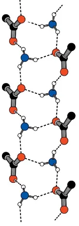
The ion pairs in this new polymorph pack to form a hydrogen-bonded ladder parallel to theaaxis (Fig. 2). Each ladder consists ofR3
4(10) (Etter, 1990) hydrogen-bonded rings
comprising four alternating ammonium and carboxylate groups linked by N—H O C contacts (Table 1). O1 forms a bifurcated hydrogen bond to H1NBand H1NC, while O2 forms just one hydrogen bond to H1NA. All strong hydrogen-bond donors and acceptors are satisfied.
Experimental
(R)-2-phenylbutyric acid (Lancaster, 97% purity) and (R )-1-phenyl-ethylammine (Alfa Aesar, 99+% purity) were used without further purification. The product was crystallized as a fine powder byeva-poration of an ethanol solution with a starting ratio of 2:1 acid:base. The sample was loaded into a 0.7 mm borosilicate glass capillary and rotated throughout the data collection to minimize preferred orientation effects. Data were collected using a variable count time (VCT) scheme in which the step time is increased with 2(Shankland
et al., 1997; Hill & Madsen, 2002).
Crystal data
C8H12N+C10H11O2
Mr= 285.37
Orthorhombic,P212121
a= 6.0620 (1) A˚
b= 16.7794 (3) A˚
c= 16.8881 (4) A˚
V= 1717.80 (6) A˚3 Z= 4
Dx= 1.104 Mg m
3
CuK1radiation
Wavelength of incident radiation: 1.54056 A˚
= 0.56 mm1
T= 295 K
Specimen shape: cylinder 120.70.7 mm Specimen prepared at 380 K Particle morphology: needle, white
Data collection
Bruker AXS D8 Advance diffractometer
Specimen mounting: 0.7 mm borosilicate capillary
Specimen mounted in transmission mode
Scan method: step Absorption correction: none 2min= 6.0, 2max= 60.0
Increment in 2= 0.017
Refinement
Rp= 0.025 Rwp= 0.029 Rexp= 0.015 RB= 0.022
S= 2.03
Profile function: Fundamental parameters with axial divergence correction.
161 parameters
Only H-atom coordinates refined
w= 1/(Yobs)2
(/)max= 0.005
Preferred orientation correction: A spherical harmonics-based preferred orientation correction (Ja¨rvinen, 1993) was applied with
TOPAS(Coelho, 2003) during the Rietveld refinement
Table 1
Hydrogen-bond geometry (A˚ ,).
D—H A D—H H A D A D—H A
N1—H1NA O2 0.964 (5) 1.863 (6) 2.732 (3) 148.5 (5) N1—H1NB O1i 0.976 (6) 1.907 (7) 2.744 (3) 142.2 (5) N1—H1NC O1ii
0.956 (5) 1.932 (7) 2.797 (5) 149.4 (6) C10—H10 O2iii
0.953 (5) 2.492 (6) 3.426 (3) 166.8 (4)
Symmetry codes: (i)xþ1;y;z; (ii)xþ1 2;yþ
3
2;z; (iii)x1;y;z.
organic papers
o248
Fernandeset al. C [image:2.610.311.566.69.259.2]8H12N+C10H11O2 Acta Cryst.(2007). E63, o247–o249
Figure 1
[image:2.610.63.275.74.269.2]The asymmetric unit of (I), with the atom-numbering scheme. Displace-ment spheres are shown at the 50% probability level (Bruker, 2000). The dashed line indicates a hydrogen bond.
Figure 2
The hydrogen-bonded ladder motif observed in form 2. Atoms not directly involved in hydrogen-bond contacts have been omitted for clarity.
Figure 3
Final observed (points), calculated (line) and difference [(yobsycalc)/
[image:2.610.126.207.331.561.2]The diffraction pattern indexed to a monoclinic cell [M(19) = 30.7,
F(19) = 63.0;DICVOL91; Boultif & Loue¨r, 1991] and the space group
P212121was assigned from volume considerations and a statistical
consideration of the systematic absences (Markvardsenet al., 2001). The data set was background subtracted and truncated to 59.52for
Pawley fitting (Pawley, 1981;2Pawley= 6.10) and the structure solved
using the simulated annealing (SA) global optimization procedure, described previously (Davidet al., 1998), that is now implemented in theDASHcomputer program (Davidet al., 2001). The SA structure solution used 311 reflections and involved the optimization of two fragments totaling 14 degrees of freedom (six positional and orien-tational for each fragment present in the asymmetric unit plus a torsion angle for each fragment). All degrees of freedom were assigned random values at the start of the simulated annealing. The best SA solution had a favourable2
SA/ 2
Pawleyratio of 3.41 and a
chemically reasonable packing arrangement, with no significant misfit to the diffraction data.
The solved structure was then refined against the data in the range 6–59.72using a restrained Rietveld (1969) method as implemented
in TOPAS (Coelho, 2003), with Rwp falling to 0.029 during the
refinement. All atomic positions (including H atoms) for the structure of (I) were refined, subject to a series of restraints on bond lengths, bond angles and planarity. The refined C—H distances were 0.949 (5)–0.973 (5) A˚ .Uisovalues for H atoms were constrained to
equal 0.076 A˚2.
The restraints were set such that bonds and angles did not deviate more than 0.01 A˚ and 0.8, respectively, from their initial values
during the refinement. Atoms C16, C15, C14, C13, C18, C17, H16, H15, H14, H18 and H17 (phenylethylammonium) and atoms C5, C6, C7, C8, C9, C10, H6, H7, H8, H9 and H10 (phenylbutyrate) were restrained to lie in respective planar groups. A spherical harmonics (fourth order) correction of intensities for preferred orientation was applied in the final refinement (Ja¨rvinen, 1993). The refined final spherical harmonics coefficients were consistent with mild preferred orientation effects in the sample. The observed and calculated diffraction patterns for the refined crystal structure are shown in Fig. 3.
Data collection: DIFFRAC plus XRD Commander (Kienle & Jacob, 2003); cell refinement:TOPAS(Coelho, 2003); data reduction:
DASH (David et al., 2001); program(s) used to solve structure:
DASH; program(s) used to refine structure: TOPAS; molecular graphics: Mercury (Macrae et al., 2006) and SHELXTL (Bruker, 2000); software used to prepare material for publication: enCIFer
(Version 1.1; Allenet al., 2004).
We thank the Basic Technology programme of the UK Research Councils for funding under the project Control and
Prediction of the Organic Solid State (http://
www.cposs.org.uk).
References
Allen, F. H., Johnson, O., Shields, G. P., Smith, B. R. & Towler, M. (2004).J. Appl. Cryst.37, 335–338.
Boultif, A. & Loue¨r, D. (1991).J. Appl. Cryst.24, 987–993. Brianso, M.-C. (1978).Acta Cryst.B34, 679–680.
Bruker (2000). SHELXTL. Version 6.10. Bruker AXS Inc., Madison, Wisconsin, USA.
Coelho, A. A. (2003).TOPAS User Manual.Version 3.1. Bruker AXS GmbH, Karlsruhe, Germany.
David, W. I. F., Shankland, K., Cole, J., Maginn, S., Motherwell, W. D. S. & Taylor, R. (2001).DASH. Version 3.0 User Manual. Cambridge Crystal-lographic Data Centre, Cambridge, England.
David, W. I. F., Shankland, K. & Shankland, N. (1998).Chem. Commun.
pp. 931–932.
Etter, M. C. (1990).Acc. Chem. Res.23, 120–126.
Hill, R. J. & Madsen, I. C. (2002).Structure Determination from Powder Diffraction Data, edited by W. I. F. David, K. Shankland, L. B. McCusker & Ch. Baerlocher, pp. 114–116. Oxford: Oxford University Press.
Ja¨rvinen, M. (1993).J. Appl. Cryst.26, 525–531.
Kienle, M. & Jacob, M. (2003).DIFFRAC plus XRD Commander.Version 2.3. Bruker AXS GmbH, Karlsruhe, Germany.
Macrae, C. F., Edgington, P. R., McCabe, P., Pidcock, E., Shields, G. P., Taylor, R., Towler, M. & van de Streek, J. (2006).J. Appl. Cryst.39, 453–457. Markvardsen, A. J., David, W. I. F., Johnson, J. C. & Shankland, K. (2001).Acta
Cryst.A57, 47–54.
Pawley, G. S. (1981).J. Appl. Cryst.14, 357–361. Rietveld, H. M. (1969).J. Appl. Cryst.2, 65–71.
Shankland, K., David, W. I. F. & Sivia, D. S. (1997).J. Mater. Chem.7, 569–572.
organic papers
Acta Cryst.(2007). E63, o247–o249 Fernandeset al. C
supporting information
sup-1
Acta Cryst. (2007). E63, o247–o249
supporting information
Acta Cryst. (2007). E63, o247–o249 [https://doi.org/10.1107/S1600536806052652]
Powder study of (
R
)-1-phenylethylammonium (
R
)-2-phenylbutyrate form 2
Philippe Fernandes, Alastair Florence, Kenneth Shankland, Panagiotis G. Karamertzanis, Ashley
T. Hulme and Parathay Anandamanoharan
(R)-1-phenylethylammonium (R)-2-phenylbutyrate
Crystal data
C8H12N+·C10H11O2−
Mr = 285.37
Orthorhombic, P212121
Hall symbol: P 2ac 2ab
a = 6.0620 (1) Å
b = 16.7794 (3) Å
c = 16.8881 (4) Å
V = 1717.80 (6) Å3
Z = 4
F(000) = 616.0
Dx = 1.104 Mg m−3
Cu Kα1 radiation, λ = 1.54056 Å
µ = 0.56 mm−1
T = 295 K
Particle morphology: needles white
cylinder, 12 × 0.7 mm
Specimen preparation: Prepared at 380 K
Data collection
Bruker AXS D8 Advance diffractometer
Radiation source: sealed X-ray tube, Bruker-AXS D8
Primary focussing, Ge 111 monochromator
Specimen mounting: 0.7 mm borosilicate capillary
Data collection mode: transmission Scan method: step
2θmin = 6.0°, 2θmax = 60.0°, 2θstep = 0.017°
Refinement
Least-squares matrix: selected elements only
Rp = 0.025
Rwp = 0.029
Rexp = 0.015
RBragg = 0.022
3176 data points
Profile function: Fundamental parameters with axial divergence correction.
161 parameters 101 restraints 1 constraint
Only H-atom coordinates refined
Weighting scheme based on measured s.u.'s 1/σ(Yobs)2
(Δ/σ)max = 0.005
Background function: Chebyshev polynomial Preferred orientation correction: A spherical
harmonics-based preferred orientation correction (Järvinen, 1993) was applied with TOPAS (Coelho, 2003) during the Rietveld refinement.
Special details
supporting information
sup-2
Acta Cryst. (2007). E63, o247–o249
Fractional atomic coordinates and isotropic or equivalent isotropic displacement parameters (Å2)
x y z Uiso*/Ueq
supporting information
sup-3
Acta Cryst. (2007). E63, o247–o249
Geometric parameters (Å, º)
O1—C1 1.282 (3) C6—H6 0.949 (5)
O2—C1 1.227 (3) C7—H7 0.955 (4)
N1—C12 1.4831 (14) C8—H8 0.950 (6)
N1—H1NC 0.956 (5) C9—H9 0.958 (5)
N1—H1NA 0.964 (5) C10—H10 0.953 (5)
N1—H1NB 0.976 (6) C11—C12 1.5125 (14)
C1—C2 1.5069 (17) C12—C13 1.5123 (18)
C2—C3 1.5252 (17) C13—C18 1.3870 (17)
C2—C5 1.504 (2) C13—C14 1.4076 (18)
C3—C4 1.5326 (14) C14—C15 1.3818 (15)
C5—C6 1.390 (2) C15—C16 1.3927 (17)
C5—C10 1.3885 (19) C16—C17 1.3767 (17)
C6—C7 1.390 (3) C17—C18 1.3848 (16)
C7—C8 1.394 (3) C11—H11A 0.971 (5)
C8—C9 1.388 (3) C11—H11B 0.969 (5)
C9—C10 1.392 (3) C11—H11C 0.956 (5)
C2—H2 0.954 (5) C12—H12 0.952 (5)
C3—H3B 0.955 (6) C14—H14 0.960 (6)
C3—H3A 0.952 (5) C15—H15 0.970 (7)
C4—H4A 0.965 (5) C16—H16 0.952 (5)
C4—H4B 0.973 (5) C17—H17 0.967 (5)
C4—H4C 0.970 (5) C18—H18 0.970 (5)
H1NA—N1—H1NB 106.7 (5) C8—C7—H7 118.1 (3)
H1NA—N1—H1NC 108.5 (5) C9—C8—H8 121.8 (4)
H1NB—N1—H1NC 108.5 (5) C7—C8—H8 120.0 (4)
C12—N1—H1NC 108.6 (3) C10—C9—H9 117.6 (3)
C12—N1—H1NA 109.8 (3) C8—C9—H9 121.0 (3)
C12—N1—H1NB 114.7 (3) C5—C10—H10 119.9 (3)
O2—C1—C2 119.36 (18) C9—C10—H10 119.7 (3)
O1—C1—O2 123.6 (2) C11—C12—C13 112.89 (10)
O1—C1—C2 116.92 (14) N1—C12—C13 110.06 (11)
C1—C2—C3 110.29 (10) N1—C12—C11 109.80 (9)
C3—C2—C5 111.74 (10) C12—C13—C14 120.66 (10) C1—C2—C5 111.02 (12) C12—C13—C18 119.72 (11) C2—C3—C4 111.22 (10) C14—C13—C18 119.53 (12) C6—C5—C10 118.57 (14) C13—C14—C15 119.61 (11) C2—C5—C6 119.88 (12) C14—C15—C16 119.93 (11) C2—C5—C10 121.51 (12) C15—C16—C17 120.48 (10) C5—C6—C7 120.82 (12) C16—C17—C18 119.92 (10) C6—C7—C8 120.68 (14) C13—C18—C17 120.38 (12)
C7—C8—C9 118.2 (2) C12—C11—H11A 108.6 (3)
supporting information
sup-4
Acta Cryst. (2007). E63, o247–o249
C3—C2—H2 105.8 (3) H11B—C11—H11C 109.0 (4)
C2—C3—H3A 106.1 (3) N1—C12—H12 106.7 (3)
C2—C3—H3B 106.9 (3) C11—C12—H12 109.6 (3)
C4—C3—H3A 110.7 (3) C13—C12—H12 107.6 (3)
C4—C3—H3B 111.4 (3) C13—C14—H14 119.3 (3)
H3A—C3—H3B 110.4 (4) C15—C14—H14 121.1 (3) H4A—C4—H4B 112.0 (4) C14—C15—H15 116.8 (4) H4A—C4—H4C 104.6 (4) C16—C15—H15 122.8 (4)
C3—C4—H4C 109.6 (3) C15—C16—H16 120.1 (4)
C3—C4—H4A 108.2 (3) C17—C16—H16 119.2 (4)
C3—C4—H4B 110.0 (3) C16—C17—H17 119.7 (3)
H4B—C4—H4C 112.3 (4) C18—C17—H17 120.3 (3)
C7—C6—H6 117.8 (3) C13—C18—H18 118.2 (3)
C5—C6—H6 121.4 (3) C17—C18—H18 120.9 (3)
C6—C7—H7 121.0 (3)
O1—C1—C2—C5 89.7 (3) C7—C8—C9—C10 1.2 (3) O2—C1—C2—C3 38.2 (3) C8—C9—C10—C5 1.5 (3) O2—C1—C2—C5 −86.2 (3) N1—C12—C13—C14 64.12 (18) C1—C2—C3—C4 61.18 (12) N1—C12—C13—C18 −119.26 (15) C5—C2—C3—C4 −174.82 (9) C11—C12—C13—C14 −58.95 (19) C1—C2—C5—C6 78.06 (16) C11—C12—C13—C18 117.68 (14) C1—C2—C5—C10 −99.50 (15) C12—C13—C14—C15 179.65 (15) C3—C2—C5—C6 −45.52 (17) C18—C13—C14—C15 3.0 (3) C3—C2—C5—C10 136.93 (14) C12—C13—C18—C17 −178.80 (14) C2—C5—C6—C7 −178.62 (15) C14—C13—C18—C17 −2.1 (3) C10—C5—C6—C7 −1.0 (2) C13—C14—C15—C16 −4.0 (3) C2—C5—C10—C9 175.95 (16) C14—C15—C16—C17 4.0 (3) C6—C5—C10—C9 −1.6 (2) C15—C16—C17—C18 −3.1 (3) C5—C6—C7—C8 3.8 (3) C16—C17—C18—C13 2.2 (3) C6—C7—C8—C9 −3.8 (3)
Hydrogen-bond geometry (Å, º)
D—H···A D—H H···A D···A D—H···A
N1—H1NA···O2 0.964 (5) 1.863 (6) 2.732 (3) 148.5 (5) N1—H1NB···O1i 0.976 (6) 1.907 (7) 2.744 (3) 142.2 (5)
N1—H1NC···O1ii 0.956 (5) 1.932 (7) 2.797 (5) 149.4 (6)
C10—H10···O2iii 0.953 (5) 2.492 (6) 3.426 (3) 166.8 (4)