1
J. J. Dempsey*, I. Wilson, P. T. N. Spencer-Phillips and D. Arnold 2
Centre for Research in Biosciences, University of the West of England, Bristol, BS16 1QY 3
* Corresponding author tel.: +353 877673447.Email: [email protected] 4
Running head: Suppression of M. nivale by phosphite 5
Abstract 6
The ascomycete fungus Microdochium nivale is a major pathogen of many species of the 7
gramineae. Control measures rely heavily on chemical fungicides, making alternative means 8
of disease reduction desirable. Phosphite (PO33-,) has proven efficacy in reducing susceptibility
9
of different species of gramineae to oomycetes, and has adverse effects on the in vitro growth 10
of numerous other pathogens. The effect of phosphorous acid (H3PO3), phosphoric acid
11
(H3PO4), dihydrogen potassium phosphite (KH2PO3), dihydrogen potassium phosphate
12
(KH2PO4), and potassium hydroxide (KOH) on the in vitro mycelial growth and development
13
of M. nivale was determined. Radial growth on amended Potato Dextrose Agar (PDA) was 14
used to calculate mean daily growth and percent inhibition. PO33- had a significant inhibitory
15
effect on mycelial growth with EC50 values ranging between 35.9 and 40.99 μg/ml-1, whilst
16
PO43- and KOH had no significant inhibitory effect. Microscopic examination of mycelia
17
showed morphological deformities in hyphae growing on PO33- amended PDA, whilst hyphal
18
growth was normal on PO43- and KOH amended PDA. Conidial germination of M. nivale was
19
significantly reduced following immersion in solutions of 50, 100 and 250 μg/ml of PO33-,
20
PO43- and KOH at same concentrations induced no inhibitory affect. These results show that
21
PO33- is a significant inhibitor of the growth of M. nivale and may have the potential to be used
22
as a chemical control agent in the field. 23
Keywords: Microdochium nivale, turfgrass, in vitro, phosphite, disease suppression 24
2
Microdochium nivale (teleomorph Monographella nivalis (Schafnitt)) is an ascomycete 26
pathogen and causal agent for many disease complexes in numerous graminaceous species 27
(Smiley et al., 1992; Tronsmo et al., 2001). Microdochium nivale produces conidia in large 28
numbers which are readily dispersed by wind and rain splash and, along with soil borne 29
mycelium, are the main source of inoculum (Tronsmo et al., 2001). In turfgrasses, M. nivale is 30
regarded as the most damaging pathogen of temperate climates, infecting and causing disease 31
in most cool season species, causing pink snow mould and microdochium patch (Vargas, 32
2005). Chemical protectants represent the foremost tool used to control this pathogen (Smiley 33
et al., 1992; Yang et al., 2011) and while the efficacy and safety of these plant protection 34
products is not disputed, development of alternative means of reducing susceptibility is 35
desirable. Phosphite is an attractive alternative to established turfgrass plant protectants for a 36
number of reasons, to date there has been no issues regarding resistance, it is highly mobile 37
within the plant, its ability to induce plant defence responses and its reported enhancement of 38
turfgrass quality. While phosphite is registered as a fungicide in some legislations, in many it 39
is regarded as a biostimulant. However it is the alternative mode of action in suppressing 40
numerous plant pathogens that is of interest here. 41
Phosphite (PO33-) is a reduced form of phosphorus (P) derived from the alkali metal salts of
42
phosphorous acid (H3PO3) (Guest and Grant, 1991). The pH of phosphorous acid is modified
43
to prevent phytotoxicity, commonly by combining with potassium hydroxide (KOH), forming 44
potassium dihydrogen phosphite (KH2PO3) or dipotassium hydrogen phosphite (K2HPO3).
45
Phosphite is chemically similar to phosphate (PO43-), but the different tetrahedral molecular
46
structure of phosphite ensures that enzymes, which react with phosphate to catalyse 47
metabolic processes, do not bind to phosphite in the same manner ensuring that phosphite 48
does not supply a metabolically usable form of P (Mcdonald et al., 2001). Phosphite, 49
3
1984). The mode of suppression remains a subject of debate (Abbasi and Lazarovits, 2006) 51
with research showing it as acting both directly on the pathogen and indirectly by stimulating 52
host defences (Guest and Grant, 1991). 53
The use of in vitro studies is an established method to assess a compound’s ability either to 54
reduce or inhibit the growth of, or to kill plant pathogenic organisms (Mann, 2002; Glynn et
55
al., 2008; Hofgaard et al., 2010). When compiling a disease protection programme an 56
important factor is determining whether a compound is fungicidal or fungistatic. It is possible 57
that at sufficient concentrations, fungistatic compounds will prevent fungal growth and 58
sporulation fully but, upon removal, the effects are reversed and growth will re-commence. 59
This would have a significant bearing on the application rate and interval. 60
Most studies on phosphite mediated inhibition of plant pathogens have been on its effects on 61
oomycetes. Suppression of Pythium by phosphite under field conditions was reported by 62
Sanders (1983), but when no in vitro inhibition was demonstrated it was concluded that 63
control resulted from enhanced host defences. However, Fenn and Coffey (1984, 1987) 64
demonstrated that phosphite inhibited four Pythium spp.and Phytophthora cinnamomi in
65
vitro. Phytophthora cinnamomi exhibited sensitivity to phosphite with EC50 values (Effective
66
Concentration which reduces growth by 50% of control growth) ranging from 4 to 148 μg ml
-67
1 (Wilkinson et al., 2001). In a later study Pythium spp. were inhibited with EC
50 values
68
between 38.7 and 220.8 μg/ml-1 (Cook et al., 2009). This direct mode of inhibition seems to
69
involve disruption of the pathogen’s metabolism. For example, a study with three 70
Phytophthora species showed that phosphite interfered with phosphate metabolism in 71
pathogen cells by causing an accumulation of polyphosphate and pyrophosphate, diverting 72
ATP from other metabolic pathways, resulting in reduced growth (Niere et al., 1994). Other 73
4
pathways, disrupting phosphorus metabolism in P. palmivora by competing with phosphate 75
as an allosteric regulator on several enzymes (Stehmann and Grant, 2000). 76
Less has been published on the in vitro effects of phosphite on fungal pathogens. Reuveni et
77
al. (2003) showed inhibition of Alternaria alternata mycelial growth and conidial 78
germination, while Burpee (2005) reported suppression of in vitro growth of Colletotrichum
79
cereale (Colletotrichum graminicola). Mills et al. (2004) demonstrated that H2PO3 not only
80
reduced mycelial growth but caused complete inhibition of sporulation of A. alternata, 81
Botrytis cinerea and Fusarium solani. Growth of F. culmorum and F. graminearum was 82
reduced on KH2PO3 amended PDA (Hofgaard et al., 2010). The same study included the
83
effects of phosphite on Microdochium majus, and found thatmycelial growth was reduced by 84
more than 90% at the lowest KH2PO3 concentration used (10 μg ml−1), with full inhibition at
85
concentrations of 100 μg ml−1 (Hofgaard et al., 2010)(Hofgaard et al., 2010)(Hofgaard et al., 86
2010). However, there has been no published data on the in vitro effect phosphite may have 87
on M. nivale.
88
Data from turfgrass field trials conducted to evaluate M. nivale suppression by KH2PO3,
89
determined that phosphite significantly (p < 0.05) suppressed disease symptom expression 90
(Dempsey et al., 2012). The success of these trials led to this current research to discover 91
possible modes of suppression. The aims of this research, therefore, were to determine the 92
effect phosphite may have on the in vitro mycelial growth and conidial germination of M.
93
nivale, and to determine if phosphite has fungistatic or fungicidal properties. 94
Materials and methods 95
Microdochium nivale mycelial and conidial inoculum 96
Four isolates of M. nivale were assessed. Two isolates were obtained from infected Poa annua
97
golf greens on Irish golf courses, the remainder from the Sports Turf Research Institute, 98
5
Carlow, using molecular biology techniques as described by Glynn et al. (2005). Conidiation 100
was induced by incubating mycelia in darkness for 48 hours and then exposing to UV light 101
(Jewell and Hsiang, 2013). Conidia were then collected by flooding the plate with sterile 102
distilled water (SDW) and scraping with a sterile rod, immediately before use in experiments. 103
PDA amendments, H3PO3, H3PO4, KH2PO3, KH2PO4 and KOH 104
Phosphorous acid (H3PO3) and phosphoric acid (H3PO4), were obtained from 1 M reagent
105
grade solutions (supplied by Lennox Laboratory Supplies, Dublin). Dihydrogen potassium 106
phosphite (KH2PO3)and dihydrogen potassium phosphate (KH2PO4) amendments were
107
prepared by titrating 1 M solution phosphorus and phosphoric acids with 6 M reagent-grade 108
potassium hydroxide (KOH) to pH 6.5. KOH amendments were prepared from 6 M 109
potassium hydroxide, and all amendments were serial diluted to required concentrations. 110
Unamended PDA, containing no additional chemicals, were used as controls. All 111
experimental compounds were filter sterilised and added to autoclaved Potato Dextrose Agar 112
(PDA, 19 g/l, Himedia Potato Dextrose Agar, Sparks Laboratory Supplies, Dublin), after 113
cooling to 50o C to ensure no oxidation of phosphite to phosphate (Komorek and Shearer, 114
1997). 115
Measurement of mycelial growth on solid media 116
Experiments were a randomised complete design with six replications. Measurement of 117
mycelial growth of M. nivale isolates, incubated on PDA amended with 0 (unamended 118
control), 10, 50, 100 and 250 μg/ml of H3PO3, H2PO4, KH2PO3, KH2PO4 and KOH were used
119
to calculate mean daily growth (MDG), percent relative growth (PRG), percent inhibition and 120
colony diameters. Agar plugs, 5 mm in diameter, were cut from margins of actively-growing 121
colonies of M. nivale, and transferred to the centre of plates of amended PDA then incubated 122
in darkness in a growth chamber maintained at 18° +/- 20 C. Mycelial growth rate was 123
6
initial inoculum to the extreme outer margin area of fungal mycelial development and growth 125
rates (mm day−1) calculated. Radial growth measurements were taken 1, 2, 3, 4, 5, 6, 7, 8, 9, 126
and 10 days post inoculation (dpi). Mean values of each of the six replicates were used to 127
calculate MDG and PRG on amended compared to unamended control PDA. PRG was 128
calculated as (radial growth on amended PDA/radial growth on unamended control PDA) × 129
100, and was used to calculate percent inhibition (calculated as 100-PRG = percent 130
inhibition). The effective concentrations that reduced mycelial growth by 50% (EC50) and
131
90% (EC90) were determined by probit transforming the PRG and regressing against the
132
Log10 of amendment concentrations. This experiment was repeated three times with similar
133
results obtained each time. 134
Determination of fungistatic properties of phosphite 135
Experiments were a randomised complete design with six replications. Mycelial plugs, 136
prepared as before, were placed into 10 mL SDW containing 0 (control), 10, 50, 100 and 250 137
μg/ml of H3PO3, H2PO4, KH2PO3, KH2PO4 and KOH (n=6), and incubated in darkness in a
138
growth chamber maintained at 18° +/- 20 C for 10 days. The plugs were retrieved, rinsed twice 139
in SDW and transferred onto fresh unamended PDA and grown in darkness at 18° +/- 20 C 140
(n=6) for 10 dpi. Growth responses were measured and the presence or absence of growth 141
determined if the concentrations were fungicidal or fungistatic. Colony diameters, as 142
determined above on solid media, were also used to assess the fungistacity of phosphite over 143
10 dpi. This experiment was repeated twice with similar results each time. 144
Microscopic analysis of the effect of phosphite on hyphal morphology 145
Microdochium nivale hyphal morphology was examined by bright field and fluorescence 146
microscopy using a Bresser epifluorescence microscope. Mycelia, sampled from the outer 147
margins of actively growing colonies, growing on PDA amended with 0 (unamended control), 148
10, 50, 100 and 250 μg/ml of H3PO3, H2PO4, KH2PO3, KH2PO4 and KOH were examined. The
7
fluorescent dye, Calcofluor White, was used to visualise hyphae as in Dubas et al. (2010). 150
Images were captured using a Canon D1100 camera and processed by Adobe Photoshop 151
version 5.0 LE (Adobe Systems, Inc., San Jose, CA). 152
Effects of phosphite on conidial germination 153
Experiments were a randomised complete design with six replications. Microdochium nivale
154
conidial suspensions were filtered through sterile cheesecloth, to remove mycelium, and 50 μl 155
aliquots were transferred to 1.5 ml tubes and mixed with 1 ml solutions of 0 (control), 10, 50, 156
100 and 250 μg/ml concentrations of H3PO3, H2PO4, KH2PO3, KH2PO4 and KOH. Aliquots
157
(50 μl) of the mixtures were pipetted onto depressions in cavity microscope slides and 158
immediately placed on moist tissue paper in 9 cm Petri dishes and sealed (n=6). Following 159
incubation in darkness in a growth chamber maintained at 18° +/- 20 C for 48 h, the samples 160
were agitated using an orbital shaker for 1 h then 20 μl pipetted onto fresh slides. The number 161
of germinating conidia was counted and percent germination calculated (conidia 162
germinated/total conidia x 100). Conidia were considered to be germinated when the germ 163
tube extended to at least twice the length of the conidium (Mills et al., 2004). This experiment 164
was repeated twice with similar results each time. 165
Data analysis 166
Data were analysed using the statistical programme SPSS Statistics 21. Anova assessed for 167
significant differences among the four isolates of M. nivale used. Data were assessed prior to 168
analyses to ensure they met the requirements for the relevant statistical methods used. Residual 169
analyses were performed to test for the assumptions of the two-way Anova, outliers assessed 170
by inspection of boxplots, normality assessed using Shapiro-Wilk's normality test and 171
homogeneity of variances was assessed by Levene's test. Two-way Anova, assessed significant 172
effects and interactions on MDG, percent inhibition, the fungicidal or fungistatic properties of 173
8
significant effects or interactions, one-way Anova, followed by Tukey HSD post hoc tests, at 175
a significance level of p = 0.05, were used to determine and separate statistical differences. For 176
calculation of EC50 and EC90 values, probit analysis was used to transform percent inhibition
177
from sigmoid to linear data and then regress against the Log10 of amendment concentrations. 178
One-way Anova was then assessed for significant differences among compounds. Where 179
required, data were suitably transformed prior to analyses and back-transformed for 180
presentation of charts. 181
Results 182
Effects of phosphite on in vitro mycelial growth of M. nivale on solid media 183
Measurement of mycelial growth of M. nivale isolates grown on amended PDA were carried 184
out from 1 to 10 dpi. Anova determined no significant (p > 0.05) differences in responses 185
among the four isolates used and therefore the data were pooled to produce mean daily growth 186
rates (MDG). Percent relative growth (PRG) rates of M. nivale grown on amended PDA were 187
used to determine the percent inhibition. The analyses determined a significant (p < 0.05) 188
difference in growth inhibition among compounds and rates of concentrations used, (Fig.1). 189
Both H3PO3 and KH2PO3 caused significant inhibition of mycelial growth compared to all other
190
compounds. EC50 and EC90 values, calculated at 5 dpi, were 40.99 and 80.90 μg/ml for the
191
H3PO3 and 35.95 and 77.68 μg/mlfor the KH2PO3, respectively. In contrast, there was no
192
significant (p > 0.05) growth inhibition with H3PO4, KH2PO4 and KOH amendments.
193
Statistical analysis determined the KH2PO3 PRG growthvalueswere significantly (p < 0.05)
194
lower than the H3PO3. Mycelial growth of M. nivale was suppressed by PO33- presence when
195
compared to plates amended with H3PO4, KH2PO4 and KOH (Fig. 2).
196
Fungistatic properties of phosphite 197
Colony diameters of the M. nivale isolates, which had been immersed in a range of compound 198
9
diameters with concentrations of 0 (control) and 10 μg/mlhad no significant (p > 0.05) effect. 200
While there were significant (p < 0.05) differences in growth determined following immersion 201
in the 50, 100, 250 and 500 μg/ml concentrations, with some suppression of growth, there was 202
no complete inhibition. Further evidence of the fungistatic rather than fungicidal properties of 203
phosphite was determined by measurement of colony diameters growing on H3PO3 and
204
KH2PO3 amended PDA at 10 dpi. Evidence that phosphite reduces rather than fully inhibits
205
growth can be seen in Fig 4, which show that colonies continued to grow to the end of the 10 206
dpi experimental period. 207
Effects of phosphite on hyphal morphology 208
Microdochium nivale hyphae, viewed using brightfield microscopy at 100x magnification in 209
unamended control PDA (Fig. 5 A) showed normal morphology, as evidenced by the smooth 210
hyphal outlines. Hyphae grown on H3PO4 (Fig. 5 B) and KOH (Fig. 5 C) amended PDA,
211
appeared similar to those on unamended controls. M. nivale hyphae grown on H3PO3 at
212
concentrations of 75 and 100 μg/ml amended PDA, displayed an altered hyphal morphology 213
(Figs 5 D and 5 E). In the presence of phosphite, M. nivale hyphae appeared swollen, short-214
branched and stunted, compared to hyphae grown on PO43- and KOH amended plates.
215
216
Effects of phosphite on conidial germination 217
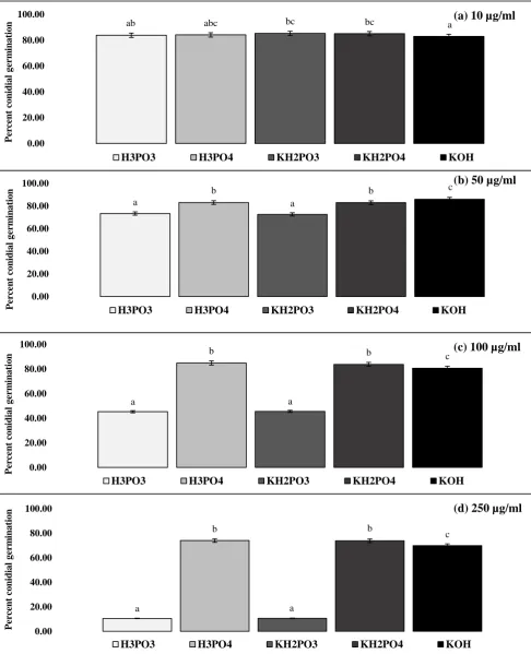
Microdochium nivale conidia in amended solutions were incubated in darkness and conidial 218
germination assessed. Conidia in all the 0 μg/ ml-1 unamended controls did not achieve 100%
219
germination, with the highest rate of 85.6% determined in one of the sets of 6 replicates. 220
Whilst there were only minor differences in germination rates in the 10 μg/ ml-1
221
concentrations of all compounds, at the 50, 100 and 250 μg/ml concentrations, germination 222
rates in the H3PO3 and KH2PO3 amended plates were significantly (p < 0.05) less than with
223
10
Discussion 225
The majority of research with phosphite for controlling plant pathogens has been with 226
oomycetes (Coffey and Bower, 1984; Smillie et al., 1989; Cook et al., 2005; Garbelotto et al., 227
2008). In contrast, relatively few studies have focused on phosphite suppressing the in vitro
228
growth of ascomycetes (Reuveni et al., 2003; Burpee, 2005). Numerous assessments of M.
229
nivale mycelial growth on amended PDA were conducted, and bright field and fluorescence 230
microscopy was used to assess effects on individual hyphae and conidial structures. These 231
studies have shown that phosphite reduces mycelial growth, interferes with morphological 232
development and reduces spore germination. Whilst the effects of phosphite on M. majus were 233
investigated by Hofgaard et al. (2010), the present study is the first to provide equivalent data 234
for M. nivale, the more significant pathogen of turf grasses. Significant growth suppression 235
of M. nivale was shown in the presence of phosphite with no statistical (p > 0.05) difference 236
between the four M. nivale isolates, despite being sourced from different geographical 237
locations. Replication of these studies using a wider pathogen population would be of value 238
as it would verify the findings here that all isolates are affected to similar levels. 239
Phosphite significantly suppressed in vitro mycelial growth of M. nivale. This inhibitory 240
effect was also reflected in the disruption of hyphal morphology and the reduction in percent 241
conidial germination. This sensitivity of M. nivale to phosphite was further evident from EC50
242
and EC90 values of 40.99 and 80.90 μg/ml for the H3PO3 and 35.95 and 77.68 μg/ml for the
243
KH2PO3, respectively, at 5 dpi.
244
While both H3PO3 and KH2PO3 inhibited growth, the EC values highlight significant (p < 0.05)
245
differences between these compounds. The differences in EC values could be attributed to 246
combinations of compounds used, where there were significant (p < 0.05) differences between 247
the inhibitory effects of both compounds at all concentrations used, with the exception of the 248
11
mobile polyphosphate into fungal cells, maintaining that it helps retain the charge balance and 250
pH of the fungal cell and is the counter ion to the transport of polyphosphates into the vacuole. 251
Darakis et al. (1997) concluded the presence of K facilitated phosphite uptake into 252
Phytophthora capsici hyphae. If mycelial growth suppression is used as an indicator of 253
increased phosphite assimilation, then this enhanced assimilation of phosphite in the presence 254
of K may have occurred, as statistically KH2PO3 produced significantly (p < 0.05) greatly
255
inhibition compared to H3PO3. Compared to phosphite amendments, concentrations of H3PO4,
256
KH2PO4 and KOH induced no similar significant inhibitory effects. The inhibitory effects of
257
phosphate, at concentrations of 50 μg/mland above, while significantly (p < 0.05) less than 258
that of phosphite, were not unexpected. Reuveni et al. (1996) studying the infection of 259
cucumber (Cucumis sativus L.) by the ascomycete pathogen Sphaerotheca fuliginea
260
(Schlecht.:Fr.), demonstrated that disease symptoms were suppressed by a foliar spray 261
treatment of KH2PO4. Howard (2001) confirmed that phosphate had fungicidal properties
262
against a number of fungal species in vitro. 263
The effect of KOH on mycelial growth inhibition is an area of particular interest. Levels of K, 264
currently recommended for management of cool-season amenity turfgrasses, appeared to 265
increase susceptibility to M. nivale, when compared to lower K inputs (Soldat and Koch, 2016). 266
As phosphite is most commonly pH adjusted with KOH, the results here (Fig. 1) showed that 267
KOH concentrations of 100 and 250 μg/mlsignificantly inhibited mycelial growth compared 268
to similar concentrations of H3PO4 and KH2PO4. This inhibitory effect possibly due to the
269
increased pH of KOH amendments. 270
To date, there have been no published data specifically on the growth suppression of M. nivale, 271
by phosphite in vitro. The results here, however, reflect the findings of Cook et al. (2009), 272
who carried out a series of in vitro studies using KH2PO3 and KH2PO4 amended growth
12
medium, inoculated with the oomycete pathogen Pythium aphanidermatum. Whilst KH2PO3
274
inhibited growth of mycelia, KH2PO4 had no effect on growth, comparable to the results found
275
here with M. nivale suppression. The closest related research to the present study was by 276
Hofgaard et al. (2010), who examined the in vitro mycelial growth of M. majus on PDA 277
amended with a range of concentrations of a foliar fertiliser containing 731 g/l of a 50% 278
KH2PO3 solution. At 10 μg/ml, mycelial growth was reduced by more than 90% and at
279
concentrations above 50 μg/ml, growth was inhibited fully. Their results appear to show 280
phosphite as having significantly lower EC50 values than those reported here, either perhaps
281
because M. majus is more susceptible to phosphite than M. nivale, or possibly due to 282
differences in experimental methods. 283
The mode of action by which phosphite inhibits mycelial growth has been the subject of a 284
number of studies. Most conclude that inhibition involves disruption of phosphorus 285
metabolism and inhibition of enzymes involved in the glycolytic and phosphogluconate 286
pathways (Grant et al., 1990; Niere et al., 1994; Stehmann, 2000; Mcdonald et al., 2001). 287
Barchietto et al. (1992) demonstrated that phosphite interacts with phosphate for the catalytic 288
site of phosphorylating enzymes, and concluded that in Phytophthora spp. the activity of 289
phosphite produced a physiological state similar to that produced as a result of P limitation. 290
The disruption to hyphal morphology in M. nivale may be due to P deficiency in the presence 291
of phosphite. This malformation of hyphae induced by phosphite/phosphate antagonism was 292
also seen by Wong (2006), who studied the effect of phosphite on the hyphal morphology of 293
Phytophthora spp. In the presence of phosphite, hyphae were stunted and swollen, again in a 294
manner similar to those of M. nivale. This P deficiency view is supported by the findings of 295
Niere et al. (1994), who concluded that phosphite inhibition in Phytophthora spp. was due to 296
13
pyrophosphate and polyphosphate. They concluded that increased accumulation of phosphite 298
interfered with phosphate metabolism and diverted ATP from other pathways of metabolism, 299
resulting in decreased mycelial growth rates. Furthermore, they suggest that accumulation of 300
pyrophosphate and polyphosphate also alters the ion balance concentrations of potassium, 301
magnesium, calcium and iron, influencing the activity of enzymes catalysing essential steps 302
in metabolism. 303
An important aspect of this study was to determine if phosphite acted as a fungicide and killed 304
the pathogen or was fungistatic, reducing or slowing hyphal growth. Evidence of the 305
fungistatic properties of phosphite were clearly demonstrated when, after being immersed in 306
a range of phosphite concentrations for 10 days, M. nivale recommenced growth after transfer 307
to un-amended PDA, without displaying any major malformation and in a manner similar to 308
the samples immersed in phosphate and KOH. Complimenting these data, and supporting the 309
fungistatic rather than fungicidal properties of phosphite, are that when plated on phosphite 310
amended PDA, M. nivale growth, while significantly reduced, was not fully suppressed, but 311
continued to grow at a reduced rate over 10 dpi. 312
The ability of oomycetes and fungi to tolerate the presence of phosphite and maintain a 313
suppressed growth rate can be explained by Dunstan et al. (1990), who found that P. palmivora
314
was able to remove phosphite from its mycelium. Similarly, Smillie et al. (1989) found that 315
phosphite accumulated in P. palmivora during the first 5 days of growth, but showed a 316
subsequent decrease in cellular phosphite. Results of a metabolite profile study of 317
Phytophthora spp. by Grant et al. (1990) led them to conclude that phosphite accumulation in 318
mycelium was transient, as within 9 days phosphite had completely disappeared from the 319
mycelium. This supports the findings in this present study, were we found full suppression of 320
growth 5 dpi in PDA amended with phosphite at 250 μg/ml. However, from 6 to 10 dpi growth 321
14
This area merits further research as to the means by which this occurs. It may be that as 323
phosphite is assimilated by the fungus phosphite to phosphate ratio in the media is altered and 324
as Smillie et al. (1989) concluded phosphate significantly influences the take up of phosphite 325
This determination of phosphite as a fungistat rather than a fungicide has significant relevance 326
to disease control programmes and to the marketing of phosphite products. Depending on the 327
active ingredient and its biochemical mode of action, a fungicide can be applied either as a 328
preventative measure or as a curative to control disease infection. With a fungistatic compound, 329
which slows the growth rather than kills the pathogen, the control programme usually requires 330
treatment as a preventative measure, therefore requiring continuous sequential applications. 331
The sequential application programme would ensure the phosphite was always present in
332
planta, in order to continually suppress pathogen growth. 333
Conidial production is vital in the spread of inoculum, therefore any reduction would have a 334
significant impact on disease spread and incidence. The results here show that the inclusion 335
of phosphite in the propagating solution led to a significant reduction in conidial germination. 336
This inhibition of spore germination by phosphite has been well documented in oomycetes, 337
but less so in ascomycetes (Reuveni et al., 2003; Mills et al., 2004). Wong (2006) for 338
example, showed that phosphite retarded spore germination in Phytophthora spp., and also 339
provided evidence that phosphite caused distortion and lysis of the spores. Although 340
phosphite inhibited spore germination in M. nivale, no conidial distortion or lysis was 341
observed. While there are no published data on the effect phosphite has on M. nivale conidial 342
germination, Hofgaard et al. (2010) demonstrated that increased phosphite concentrations 343
correlated directly with delayed sporulation of M. majus on detached wheat leaves. Based on 344
in vitro and detached leaf experiments, they concluded phosphite can suppress fungal 345
reproduction and slow pathogenic growth, allowing a host plant’s defence system time to 346
15
This study has produced significant and novel data which is relevant to methods of turfgrass 348
disease prevention and control. The main conclusions are that phosphite suppressed M. nivale
349
mycelial growth, disrupted hyphal morphology and reduced conidial germination. Both hyphae 350
and conidia are infective propagules, providing inoculum for the diseases caused by M. nivale.
351
It is clearly demonstrated here that the incorporation of phosphite into growth media 352
significantly suppresses the growth and development of these infective propagules in vitro and 353
therefore supports the findings of Dempsey et al. (2012) where it was demonstrated that 354
phosphite significantly reduced M. nivale infection in the field. Further work in this area should 355
assess the possible effect on turfgrass phosphate metabolism in the presence of phosphite and 356
determine any effects on turfgrass growth. 357
References 358
Abbasi, P. A. and Lazarovits, G. (2006). Seed treatment with phosphonate (AG3) suppresses pythium damping-359
off of cucumber seedlings. Plant Disease 90(4): 459-464. 360
Barchietto, T., Saindrenan, P. and Bompeix, G. (1992). Physiological responses of Phytophthora citrophthora to 361
a subinhibitory concentration of phosphonate. Pesticide Biochemistry and Physiology 42(2): 151-166. 362
Bücking, H. and Heyser, W. (1999). Elemental composition and function of polyphosphates in ectomycorrhizal 363
fungi — an X-ray microanalytical study. Mycological Research 103(1): 31-39. 364
Burpee, L. L. (2005). Sensitivity of Colletotrichium graminicola to phosphonate fungicides. International 365
Turfgrass Society Research Journal 10: 163-169. 366
Coffey, M. D. and Bower, L. A. (1984). In vitro variability among isolates of eight Phytophthora species in 367
response to phosphorous acid. Phytopathology 74: 738-742. 368
Cook, J., Landschoot, P. J. and Schlossberg, M. J. (2005). Evaluation of phosphonate fungicides for control of 369
anthracnose basal rot and putting green quality: 1-14. 370
Cook, P. J., Landschoot, P. J. and Schlossberg, M. J. (2009). Inhibition of Pythium spp. and suppression of 371
pythium blight of turfgrasses with phosphonate Fungicides. Plant Disease 93(8): 809-814. 372
Darakis, G. A., Bourbos, V. A. and Skoudridakis, M. T. (1997). Phosphonate transport in Phytophthora capsici. 373
Plant Pathology 46(5): 762-772. 374
Dempsey, J. J., Wilson, I. D., Spencer-Phillips, P. T. N., et al. (2012). Suppression of Microdochium nivale by 375
potassium phosphite in cool-season turfgrasses. Acta Agriculturae Scandinavica, Section B - Plant Soil Science 376
62(Supplement 1): 70-78. 377
Dubas, E., Golebiowska, G., Zur, I., et al. (2010). Microdochium nivale (Fr., Samuels & Hallett): cytological 378
analysis of the infection process in triticale (×Triticosecale Wittm.). Acta Physiologiae Plantarum. 379
Dunstan, R. H., Smillie, R. H. and Grant, B. R. (1990). The effects of sub-toxic levels of phosphonate on the 380
metabolism and potential virulence factors of Phytophthora palmivora. Physiological and Molecular Plant 381
Pathology 36(3): 205-220. 382
Fenn, M. and Coffey, M. D. (1987). Phosphonate Fungicides for control of diseases caused by Phytophthora. 383
California Avocado Society 1987 Yearbook 71: 241-249. 384
Fenn, M. E. and Coffey, M. D. (1984). Studies on the in vitro and in vivo antifungal activity of Fosetyl-Al and 385
phosphorus acid. Phytopathology 74(5): 606-611. 386
Garbelotto, M., Harnik, T. Y. and Schmidt, D. J. (2008). Efficacy of phosphonic acid, metalaxyl-M and copper 387
hydroxide against Phytophthora ramorum in vitro and in planta. Plant Pathology 58(1): 1-9. 388
Glynn, N. (2005). Phylogenetic analysis of EF-1 alpha gene sequences from isolates of Microdochium nivale 389
16
Glynn, N. C., Hare, M. C. and Edwards, S. G. (2008). Fungicide seed treatment efficacy against Microdochium 391
nivale and M. majus in vitro and in vivo. Pest Management Science 64(8): 793-799. 392
Grant, B., Dunstan, R., Griffith, J., et al. (1990). The Mechanism of phosphonic (phosphorous) acid action in 393
Phytophthora. Australasian Plant Pathology 19(4): 115-121. 394
Guest, D. and Grant, B. (1991). The complex action of phosphonates as antifungal agents. Biological Reviews 395
66(2): 159-187. 396
Hofgaard, I. S., Ergon, Å., Henriksen, B., et al. (2010). The effect of potential resistance inducers on 397
development of Microdochium majus and Fusarium culmorum in winter wheat. European Journal of Plant 398
Pathology 128(2): 269-281. 399
Howard, K. (2001). The effect of the fungicide phosphite on ectomycorrhizal fungi. Scool of Biological Sciences 400
and Biotechnology, Murdoch. 401
Jewell, L. and Hsiang, T. (2013). Differences in the timing and mechanisms of the infection processes of 402
Microdochium nivale and Microdochium majus on wheat (Triticum aestivum) and Kentucky bluegrass (Poa 403
pretensis). International Turfgrass Society Research Journal 12: 111-118. 404
Komorek, B. M. and Shearer, B. L., Eds. (1997). Application technologies and phosphonate movement in the 405
host. Control of Phytophthora and Diplodina canker in Western Australia. 406
Mann, R. (2002). In vitro fungicide sensitivity of Microdochium nivale isolates from the UK. Journal of 407
Turfgrass and Sports Surface Science 78(25-30). 408
McDonald, A., Grant, B. and Plaxton, W. (2001). Phosphite (phosphorous acid): its relevance in the 409
environment and agriculture and influence on plant phosphate starvation response. Journal of Plant Nutrition 410
24(10): 1505-1519. 411
Mills, A. A. S., Platt, H. W. and Hurta, R. A. R. (2004). Effect of salt compounds on mycelial growth, 412
sporulation and spore germination of various potato pathogens. Postharvest Biology and Technology 34(3): 341-413
350. 414
Niere, J., Deangelis, G. and Grant, B. (1994). The effect of phosphonate on the acid-soluble phosphorus 415
components in the genus Phytophthora. Microbiology 140(7): 1661-1670. 416
Reuveni, M., Agapov, V. and Reuveni, R. (1996). Controlling powdery mildew caused by Sphaerotheca 417
fuliginea in cucumber by foliar sprays of phosphate and potassium salts. Crop Protection 15: 49-53. 418
Reuveni, M., Sheglov, D. and Cohen, Y. (2003). Control of moldy-core decay in apple fruits by β-Aminobutyric 419
acids and potassium phosphites. Plant Disease 87(8): 933-936. 420
Sanders, P. L. (1983). Control of Pythium spp. and pythium blight of turfgrass with Fosetyl Aluminum. Plant 421
Disease 67(12): 1382-1383. 422
Smiley, R., Dernoeden, P. and Clarke, B. (1992). Compendium of Turfgrass Diseases.2nd Ed St Paul, APS 423
Press. 424
Smillie, R., B. R. Grant and Guest, D. (1989). The mode of action of phosphite: evidence for both direct and 425
indirect modes of action on three Phytophthora spp. in plants. Phytopathology 79(9): 921-926. 426
Soldat, D. and Koch, P. (2016) Potassium fertilization increases microdochium patch incidence and severity on 427
creeping bentgrass. Crop Science Society of America, Phoenix, Arizona. 428
Stehmann, C. (2000). Inhibition of enzymes of the glycolytic pathway and hexose monophosphate bypass by 429
phosphonate. Pesticide Biochemistry and Physiology 67(1): 13-24. 430
Tronsmo, A. M., Hsiang, T., Okuyama, H., et al. (2001). Low temperature diseases caused by Microdochium 431
nivale. Low temperature plant microbe interactions under snow. D. A. G. N. Iriki, A.M. Tronsmo, N. 432
Matsumoto, M. Yoshida and a. A. Nishimune. Sapporo, Japan., Hokkaido National Agricultural Experiment 433
Station. 434
Vargas, J. (2005). Management of Turfgrass Diseases New Jersey, Wiley and Sons. 435
Wilkinson, C. J., Shearer, B. L., Jackson, T. J., et al. (2001). Variation in sensitivity of Western Australian 436
isolates of Phytophthora cinnamomi to phosphite in vitro. Plant Pathology 50(1): 83-89. 437
Wong, M.-H. (2006). Phosphite induces morphological and molecular changes in Phytophthora. School of 438
Biological Sciences and Biotechnology. Perth, Australia, Murdoch 439
Yang, C., Hamel, C., Vujanovic, V., et al. (2011). Fungicide: modes of action and possible impact on nontarget 440
microorganisms. ISRN Ecology 2011: 1-8. 441
442
Figure legends 443
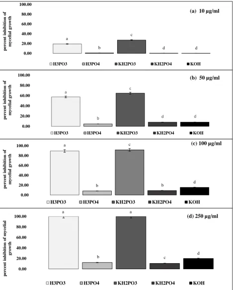
Figure 1 Inhibition of Microdochium nivale mycelial growth on phosphorous acid (H3PO3), phosphoric
444
acid (H3PO4), dihydrogen potassium phosphite (KH2PO3), dihydrogen potassium phosphate (KH2PO4),
445
17
Inhibition of M. nivale mycelial growth on PDA amended with a: 10 μg/ml; b: 50 μg/ml; c: 100 μg/ml; d: 250 447
μg/ml of H3PO3, H2PO4, KH2PO3, KH2PO4 and KOH, presented as % inhibition of growth on unamended PDA.
448
Growth rates calculated from pooled data of each of the four M. nivale isolates, n=6, by measuring the colony 449
radii at four points on each plate, 4 dpi. Bars are 95% confidence intervals. Letters indicate significant 450
differences among compounds, as determined by Tukey HSD at p = 0.05.
451 452
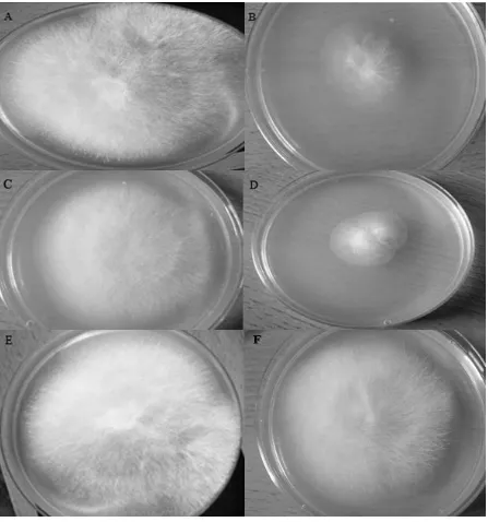
Figure 2Microdochium nivale colonies on amended PDA at 5 days post inoculation. 453
A: unamended control; B: phosphorous acid (H3PO3), 100 μg/ml; C: phosphoric acid (H3PO4), 100 μg/ml; D:
454
dihydrogen potassium phosphite (KH2PO3), 100 μg/ml; E: dihydrogen potassium phosphate (KH2PO4), 100
455
μg/ml F: potassium hydroxide (KOH), 100 μg/ml. 456
457
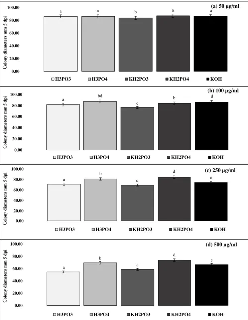
Figure 3Effect of immersion of Microdochium nivale mycelium in solutions of phosphorous acid (H3PO3),
458
phosphoric acid (H3PO4), dihydrogen potassium phosphite (KH2PO3), dihydrogen potassium phosphate
459
(KH2PO4), and potassium hydroxide (KOH).
460
Microdochium nivale colony diameters (mm) 5 days after transfer to unamended PDA, following immersion for 461
10 days in a: 50 μg/ml; b: 100 μg/ml; c: 250 μg/ml; d: 500 μg/ml solutions of H3PO3, H2PO4, KH2PO3, KH2PO4
462
and KOH. Data are mean values, n=6, pooled from four M. nivale isolates. Bars are 95% confidence intervals. 463
Letters indicate significant differences between colony diameters at each compound concentration used, as 464
determined by Tukey HSD at p = 0.05. 465
466
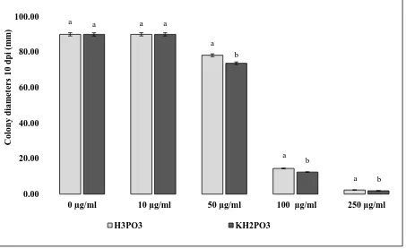
Figure 4Radial growth of Microdochium nivale mycelium 10 days post inoculation on phosphorous acid 467
(H3PO3) anddihydrogen potassium phosphite (KH2PO3) amended PDA.
468
Microdochium nivale colony diameters in mm, 10 days post inoculation, growing on PDA amended with 0 469
(control), 10, 50 100 and 250 μg/mlof H3PO3 and KH2PO3.Colony diameters were determined by measuring the
470
radii at four points on each plate. Bars are 95% confidence intervals. Letters indicate significant differences 471
between compounds at each amendment concentration, as determined by Tukey HSD at p = 0.05. 472
473
Figure 5 Brightfield micrographs of Microdochium nivale hyphal growth in amended PDA. 474
a: unamended control; b: phosphoric acid (H3PO4), 100 μg/ml; c: potassium hydroxide (KOH), 100 μg/ml; d:
475
phosphorous acid (H3PO3), 75 μg/ml; e: phosphorous acid (H3PO3), 100 μg/ml.
476 477
Figure 6 Effect of phosphite on germination of Microdochium nivale conidia. 478
Germination of M. nivale conidia following immersion in solutions of a: 10 μg/ml; b: 50 μg/ml; c: 100 μg/ml; d: 479
250 μg/ml μg/ml concentrations of phosphorous acid (H3PO3), phosphoric acid (H3PO4), dihydrogen potassium
480
phosphite (KH2PO3), dihydrogen potassium phosphate (KH2PO4), and potassium hydroxide (KOH) after
481
incubation at 18° +/- 20 C for 48 h. Data were arcsine transformed prior to analysis and back-transformed for this
482
graph. Bars are 95% confidence intervals. Letters indicate significant differences between compounds as 483
determined by Tukey HSD at p = 0.05. 484
a b c d d 0.00 20.00 40.00 60.00 80.00 100.00 p er ce n t in h ib iti o n o f m y ce li a l g ro w th
(a) 10 µg/ml
H3PO3 H3PO4 KH2PO3 KH2PO4 KOH
a b c d d 0.00 20.00 40.00 60.00 80.00 100.00 p er ce n t in h ib iti o n o f m y ce li a l g ro w th
(b) 50 µg/ml
H3PO3 H3PO4 KH2PO3 KH2PO4 KOH
a b c b d 0.00 20.00 40.00 60.00 80.00 100.00 p er ce n t in h ib iti o n o f m y ce li a l g ro w th
(c) 100 µg/ml
H3PO3 H3PO4 KH2PO3 KH2PO4 KOH
a b a c d 0.00 20.00 40.00 60.00 80.00 100.00 p er ce n t in h ib iti o n o f m y ce li a l g ro w th
(d) 250 µg/ml
H3PO3 H3PO4 KH2PO3 KH2PO4 KOH
Figure 1 Inhibition of Microdochium nivale mycelial growth on phosphorous acid (H3PO3), phosphoric acid (H3PO4), dihydrogen potassium phosphite (KH2PO3), dihydrogen potassium phosphate (KH2PO4), and potassium hydroxide (KOH) amended PDA.
[image:18.595.66.536.73.657.2]Figure 2Microdochium nivale colonies on amended PDA at 5 days post inoculation.
A: unamended control; B: phosphorous acid (H3PO3), 100 μg/ml; C: phosphoric acid (H3PO4), 100 μg/ml; D:
dihydrogen potassium phosphite (KH2PO3), 100 μg/ml; E: dihydrogen potassium phosphate (KH2PO4), 100
a a b a a 0.00 20.00 40.00 60.00 80.00 100.00 C o lo n y d ia m ete rs m m 5 d p i
(a) 50 µg/ml
H3PO3 H3PO4 KH2PO3 KH2PO4 KOH
a bd c b d 0.00 20.00 40.00 60.00 80.00 100.00 C o lo n y d ia m ete rs m m 5 d p i
(b) 100 µg/ml
H3PO3 H3PO4 KH2PO3 KH2PO4 KOH
a b c d e 0.00 20.00 40.00 60.00 80.00 100.00 C o lo n y d ia m ete rs m m 5 d p
i (c) 250 µg/ml
H3PO3 H3PO4 KH2PO3 KH2PO4 KOH
a b c d e 0.00 20.00 40.00 60.00 80.00 100.00 C o lo n y d ia m ete rs m m 5 d p i
(d) 500 µg/ml
[image:20.595.63.563.69.713.2]H3PO3 H3PO4 KH2PO3 KH2PO4 KOH
Figure 3Effect of immersion of Microdochium nivale mycelium in solutions of phosphorous acid (H3PO3), phosphoric
acid (H3PO4), dihydrogen potassium phosphite (KH2PO3), dihydrogen potassium phosphate (KH2PO4), and
potassium hydroxide (KOH).
a a
a
a
a
a a
b
b
b
0.00 20.00 40.00 60.00 80.00 100.00
0 µg/ml 10 µg/ml 50 µg/ml 100 µg/ml 250 µg/ml
Co
lo
ny
dia
m
et
er
s
1
0
dp
i
(m
m
)
H3PO3 KH2PO3
Figure 4 Radial growth of Microdochium nivale mycelium 10 days post inoculation on phosphorous acid (H3PO3) and dihydrogen potassium phosphite (KH2PO3) amended PDA.
Microdochium nivale colony diameters in mm, 10 days post inoculation, growing on PDA amended with 0 (control),
10, 50 100 and 250 μg/mlof H3PO3 and KH2PO3.Colony diameters were determined by measuring the radii at four
[image:21.595.74.527.80.358.2]ab abc bc bc a 0.00 20.00 40.00 60.00 80.00 100.00 Per ce n t co n id ia l g er m in a tio
n (a) 10 µg/ml
H3PO3 H3PO4 KH2PO3 KH2PO4 KOH
a b a b c 0.00 20.00 40.00 60.00 80.00 100.00 Pe rc en t co n id ia l g er m in a tio n
(b) 50 µg/ml
H3PO3 H3PO4 KH2PO3 KH2PO4 KOH
a b a b c 0.00 20.00 40.00 60.00 80.00 100.00 Per ce n t co n id ia l g er m in a tio
n (c) 100 µg/ml
H3PO3 H3PO4 KH2PO3 KH2PO4 KOH
a b a b c 0.00 20.00 40.00 60.00 80.00 100.00 Per ce n t co n id ia l g er m in a tio
n (d) 250 µg/ml
[image:23.595.81.568.71.671.2]H3PO3 H3PO4 KH2PO3 KH2PO4 KOH
Figure 6 Effect of phosphite on germination of Microdochium nivale conidia.
Germination of M. nivale conidia following immersion in solutions of a: 10 μg/ml; b: 50 μg/ml; c: 100 μg/ml; d: 250 μg/ml
μg/ml concentrations of phosphorous acid (H3PO3), phosphoric acid (H3PO4), dihydrogen potassium phosphite (KH2PO3),
dihydrogen potassium phosphate (KH2PO4), and potassium hydroxide (KOH) after incubation at 18° +/- 20 C for 48 h. Data